Review
. 2011 Aug;68(8):415-33.
doi: 10.1002/cm.20527. Epub 2011 Aug 25.

Extracellular inhibitors, repellents, and semaphorin/plexin/MICAL-mediated actin filament disassembly

Affiliations
Review

Extracellular inhibitors, repellents, and semaphorin/plexin/MICAL-mediated actin filament disassembly

Ruei-Jiun Hung et al. Cytoskeleton (Hoboken). 2011 Aug.

Abstract

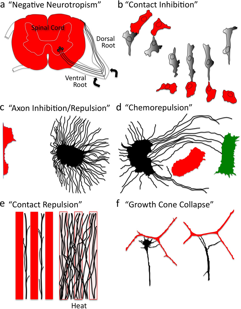
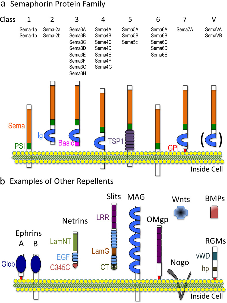
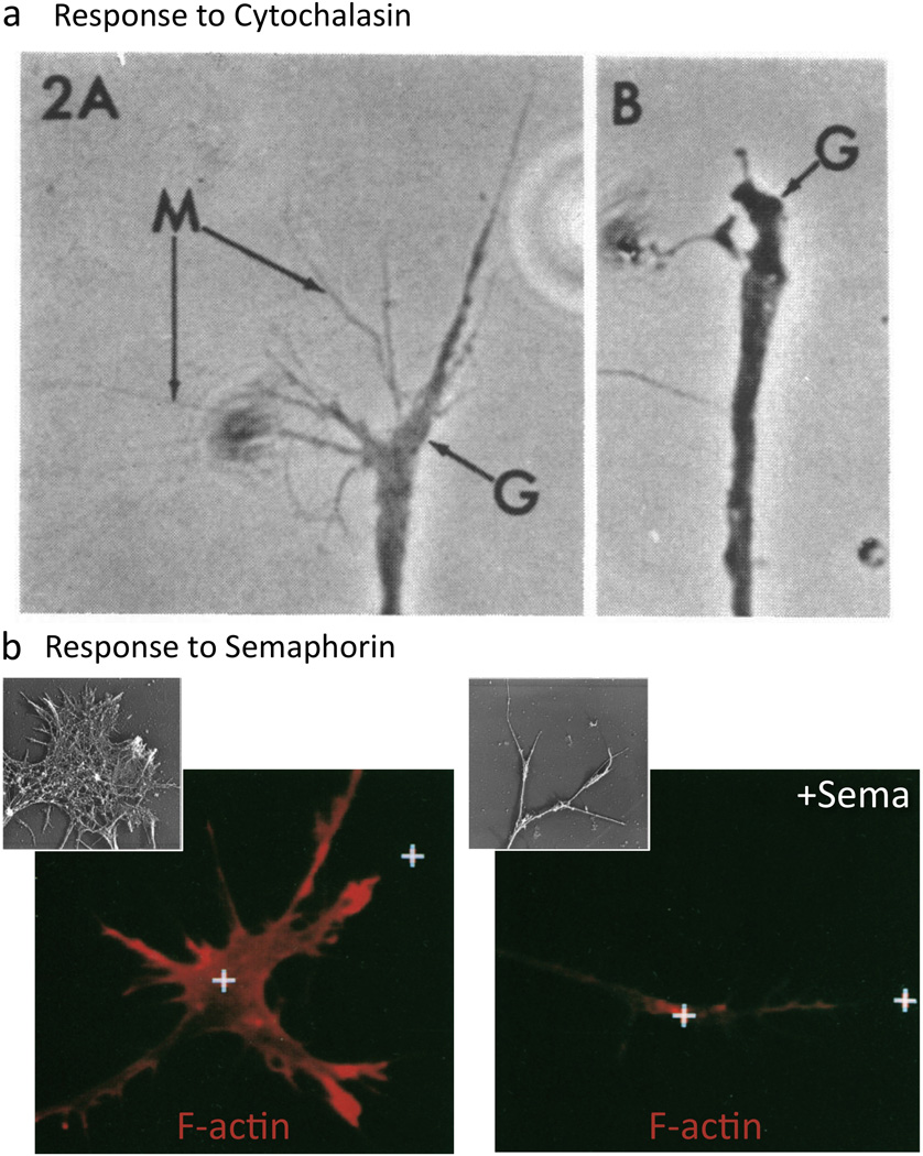
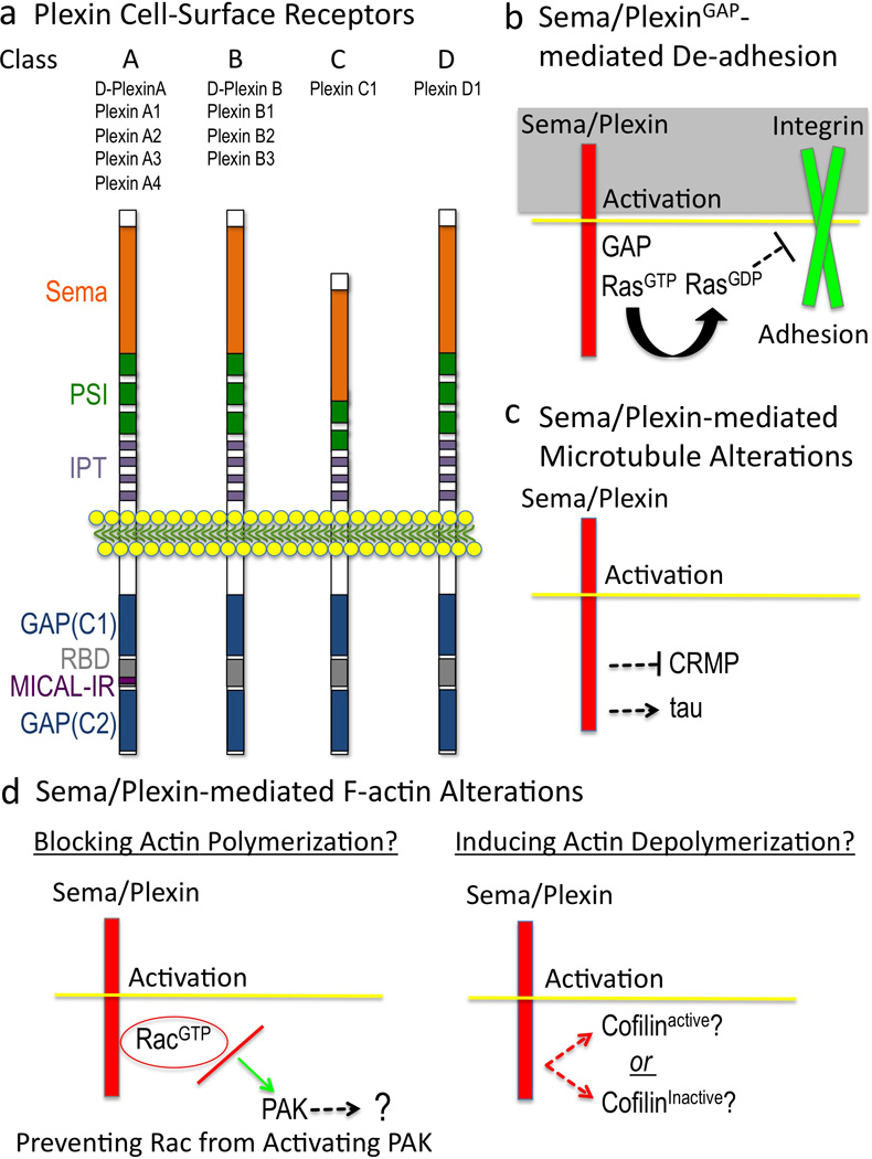
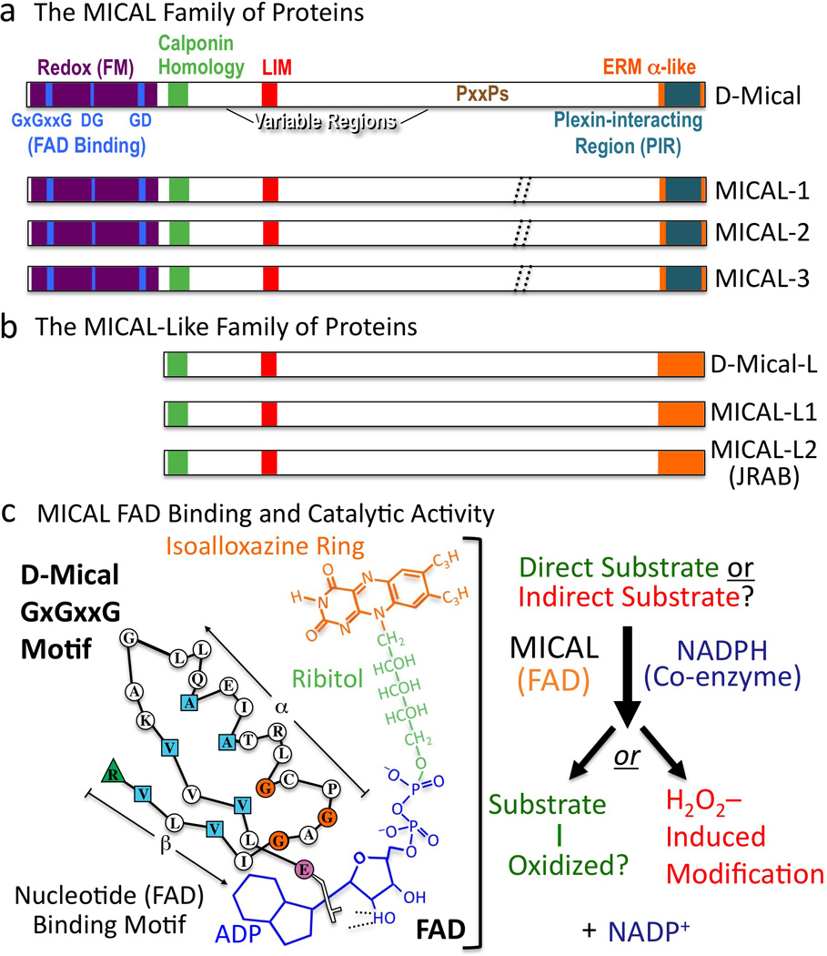
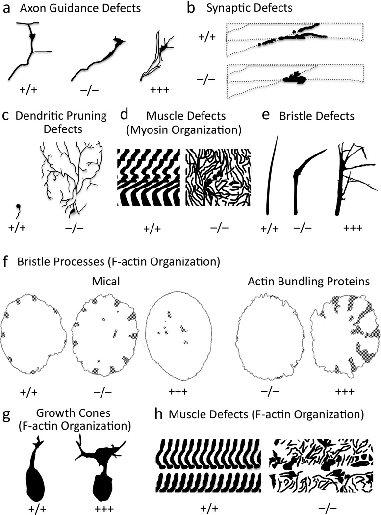
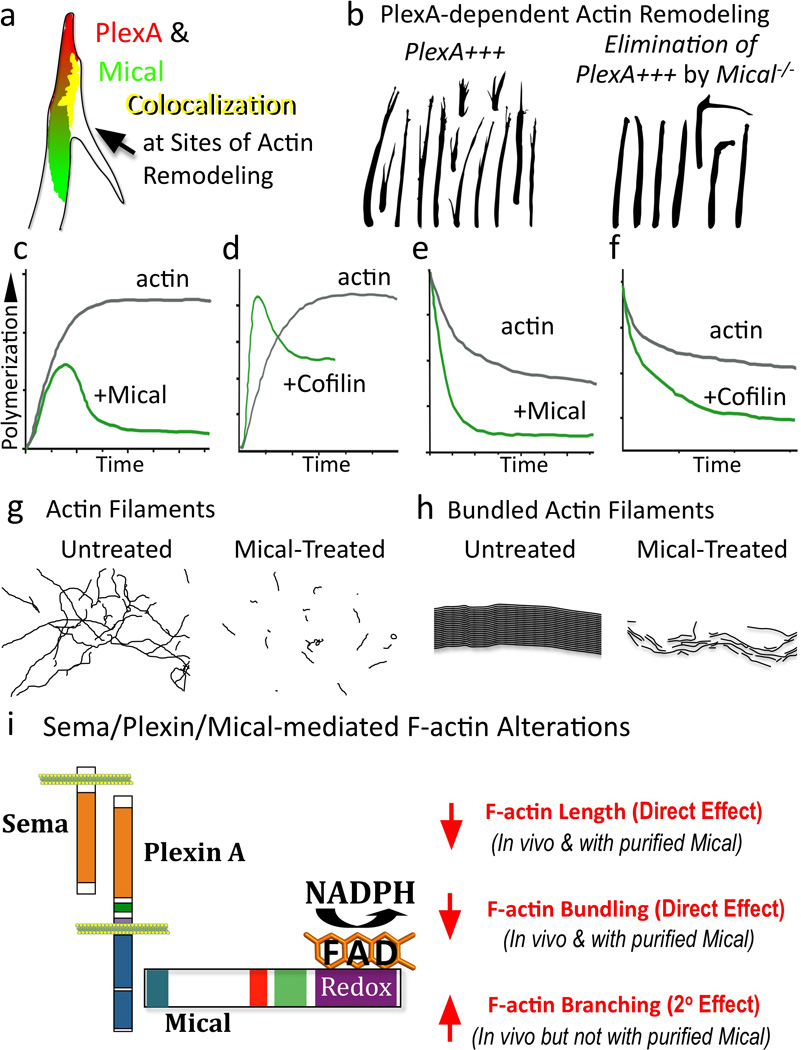
Multiple extracellular signals have been identified that regulate actin dynamics within motile cells, but how these instructive cues present on the cell surface exert their precise effects on the internal actin cytoskeleton is still poorly understood. One particularly interesting class of these cues is a group of extracellular proteins that negatively alter the movement of cells and their processes. Over the years, these types of events have been described using a variety of terms and herein we provide an overview of inhibitory/repulsive cellular phenomena and highlight the largest known protein family of repulsive extracellular cues, the Semaphorins. Specifically, the Semaphorins (Semas) utilize Plexin cell-surface receptors to dramatically collapse the actin cytoskeleton and we summarize what is known of the direct molecular and biochemical mechanisms of Sema-triggered actin filament (F-actin) disassembly. We also discuss new observations from our lab that reveal that the multidomain oxidoreductase (Redox) enzyme Molecule Interacting with CasL (MICAL), an important mediator of Sema/Plexin repulsion, is a novel F-actin disassembly factor. Our results indicate that MICAL triggers Sema/Plexin-mediated reorganization of the F-actin cytoskeleton and suggest a role for specific Redox signaling events in regulating actin dynamics.


Figures

Figure 1
Figure 1. Negative Regulation of Cell Shape, Motility, and Navigation
Various experimental examples of negative cellular behaviors and the terms used to describe them (See also Box 1). (a) Regenerating axons (black) grow extensively on some substrates (ventral and dorsal roots) and not others (spinal cord, red). (Lugaro 1906). (b) Cultured cells retract upon contact and change direction. (Weiss 1958). (c) Neuronal fiber extension (black) is inhibited/repelled upon co-culturing in proximity to a tissue explant (red). (Ebendal 1982). (d) Axons (black) circuitously grow away (red) or towards (green) different tissues explants. (Peterson and Crain 1982). (e) Axons (black) avoid substrates (red), a phenomenon that is abolished upon high temperature treatment (heat); revealing that axons grow on certain substrates not because of “attraction”, but because of avoidance of other substrates. (Walter et al. 1987b). (f) A neuronal growth cone (black) collapses/retracts upon contact with an unlike neuronal fiber (red). (Kapfhammer and Raper 1987a).
Figure 2
Figure 2. Semas and Repulsive/Inhibitory Extracellular Cues
(a) Semas. (b) Other well-known repulsive/inhibitory cues. Domain names are from SMART (http://smart.embl-heidelberg.de) except Glob, globular; hp, hydrophobic. The ( ) in (a) refers to the observation that Ig domains are present in some Class V (viral) Semas.
Figure 3
Figure 3. F-actin Disassembly Underlies Sema-mediated Repulsion
(a) F-actin disassembly collapses growth cones (Yamada et al. 1970). A growth cone (G) exhibits extensive filopodia (M) until cytochalasin treatment (B; 6 minute treatment) disrupts actin polymerization, disassembles F-actin, and induces collapse. (b) Sema treatment results in loss of F-actin. Rhodamine phalloidin staining reveals F-actin present in control (left) and Sema-treated growth cones (right; 5 min treatment). (Fan et al. 1993). (Insets) Rotary shadow EM shows the growth cone F-actin before (left) and after Sema application (right; 30 min). Reprinted with permission of John Wiley & Sons, Inc. (Brown and Bridgman 2009).
Figure 4
Figure 4. Semas Employ Plexins to Direct Repulsion
(a) Plexins. Domain names are from SMART (http://smart.embl-heidelberg.de) except GAP, GTPase activating protein; C1, conserved 1; C2, conserved 2; RBD, Rho GTPase binding domain; MICAL-IR, MICAL interacting region. (b) Sema/Plex-mediated effects on cell adhesion. The current model is that 1) the Plexin GAP is activated by binding of both Sema and a Rac GTPase to the Plexin extracellular and intracellular regions, respectively, 2) the Plexin GAP activity locally enriches for the GDP-bound form of Ras family GTPases, which 3) inactivates (through “inside-out” signaling) integrin-extracellular matrix-mediated adhesion. (c) Sema effects on microtubules. The current model is that Semas “turn-on” tau-mediated microtubule alterations and “turn-off” CRMP-mediated tubulin assembly. (d) Current models suggest that Sema-mediated F-actin alterations occur by limiting actin polymerization (left) and/or by inducing actin depolymerization by regulating the levels of active cofilin (right). A few other actin-associated proteins including myosin II and ERM are involved in Sema-mediated repulsion (Gallo 2008; Mintz et al. 2008; Schlatter et al. 2008), but their effects appear secondary to directly inducing F-actin disassembly (Brown et al. 2009; Gallo 2006; Takamatsu et al. 2010).
Figure 5
Figure 5. MICALs are Multi-domain Cytosolic Redox Enzymes
(a) MICAL family proteins are characterized by their flavoprotein monooxygenase (Redox [FM]), calponin homology, LIM, and ERM alpha-like domains. Variable regions differ in length and continuity among family members (dotted //; Box 3). To conform with Drosophila nomenclature guidelines, lowercase lettering is now used when describing invertebrate MICALs (Mical), and all capitals are used when describing the vertebrate MICALs (and the MICAL family of proteins). (b) MICAL-Like proteins are similar to MICALs in domain organization except they lack the Redox domain. (c) FAD binding and MICAL enzymatic activity. (Left) The MICAL cofactor FAD is composed of ADP (including a pyrophosphate group), ribitol, and an isoalloxazine ring. To bind to the ADP region of FAD, Mical relies on its GxGxxG region where the critical residues are colored (Modified from (Wierenga et al. 1986)). To bind to the pyrophosphate and ribitol moieties of FAD, Mical relies on its DG and GD regions, respectively. (Right) Results indicate that the Redox domain of MICAL binds FAD, consumes NADPH, and generates H2O2. MICAL substrate/s are unknown. Alternatively, MICAL may have no direct substrate and may simply generate H2O2.
Figure 6
Figure 6. MICAL is Necessary and Sufficient to Regulate F-actin Organization In Vivo
(a–e) Mical is required for axon guidance (a), neuronal synapse formation (b), dendrite pruning (c), muscle formation (d), and bristle shape (e). (f-h) Altering Mical levels in vivo generate abnormal F-actin organization in bristles (f), growth cones (g), and muscles (h). In bristles, F-actin organization (stippling) in Mical “knockouts” is similar to overexpression of actin bundling proteins, while Mical overexpression mimics loss of bundling proteins. +/+, wild-type; −/−, “knockout”; +++, overexpression. Modified from (Beuchle et al. 2007; Hung et al. 2010; Kirilly et al. 2009; Tilney et al. 1998; Tilney et al. 1995; Yoon and Terman submitted).
Figure 7
Figure 7. MICAL Disassembles F-actin and Directly Links Sema/Plexin Repulsion and F-actin Collapse
(a–b) Mical colocalizes with and is required by PlexA for F-actin reorganization. (c–f) Mical directly disassembles F-actin in a manner that is distinct from cofilin. (c) Actin polymerization slows-down in the presence of Mical (and its coenzyme NADPH). (d) In the presence of cofilin, polymerization initially speeds-up, a result of the diverse properties of cofilin (e.g., (Andrianantoandro and Pollard 2006; Bernstein and Bamburg 2010)) including that it generates more free ends for polymerization. (e–f) Mical and cofilin affect actin in a similar manner in an actin depolymerization assay. Modified from (Du and Frieden 1998; Hung et al. 2010; Moriyama and Yahara 2002). (g–h) Mical directly decreases F-actin length (g) and the length and width of Fascin-bundled actin filaments (h). (i) Sema/Plexin/Mical/Actin signaling pathway and its effects on F-actin.

References

    1. Abercrombie M. Contact inhibition in tissue culture. In Vitro. 1970;6:128–142. - PubMed
    1. Abercrombie M. The crawling movement of metazoan cells. Proc R Soc Lond B Biol Sci. 1980;207(1167):129–147.
    1. Abercrombie M, Heaysman JE. Observations on the social behaviour of cells in tissue culture. I. Speed of movement of chick heart fibroblasts in relation to their mutual contacts. Exp Cell Res. 1953;5(1):111–131. - PubMed
    1. Aguayo AJ, Bray GM, Perkins CS, Duncan ID. Axon-sheath cell interactions in peripheral and central nervous system transplants. Society for neuroscience symposia: aspects of developmental neurobiology. 1979;4:361–383.
    1. Aizawa H, Wakatsuki S, Ishii A, Moriyama K, Sasaki Y, Ohashi K, Sekine-Aizawa Y, Sehara-Fujisawa A, Mizuno K, Goshima Y, et al. Phosphorylation of cofilin by LIM-kinase is necessary for semaphorin 3A- induced growth cone collapse. Nat Neurosci. 2001;4(4):367–373. - PubMed

Publication types

MeSH terms

LinkOut - more resources