

doi: 10.1021/nl202220q.
Epub 2011 Aug 3.
De novo design of bioactive protein-resembling nanospheres via dendrimer-templated peptide amphiphile assembly
Affiliations
- PMID: 21800917
- PMCID: PMC3223106
- DOI: 10.1021/nl202220q
Item in Clipboard
De novo design of bioactive protein-resembling nanospheres via dendrimer-templated peptide amphiphile assembly
Nano Lett.
.
Abstract
Self-assembling peptide amphiphiles (PAs) have been extensively used in the development of novel biomaterials. Because of their propensity to form cylindrical micelles, their use is limited in applications where small spherical micelles are desired. Here we present a platform method for controlling the self-assembly of biofunctional PAs into spherical 50 nm particles using dendrimers as shape-directing scaffolds. This templating approach results in biocompatible, stable protein-like assemblies displaying peptides with native secondary structure and biofunctionality.
Figures
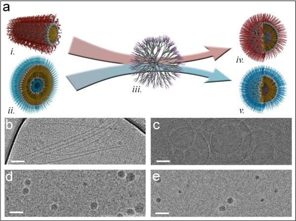
The dendrimers template micelle-forming and vesicle-forming PAs into spherical PRTNs. (a) Schematic of templating process: i. bZip PA cylindrical micelle ii. NLS PA vesicle iii. dendrimer iv. Spherical bZip PRTN v. Spherical NLS PRTN. Cryo-TEM images of self-assemblies: (b) bZip PA cylindrical micelles. (c) NLS PA vesicles. (d) Spherical bZip PRTNs. (e) Spherical NLS PRTNs. Scale bars are 100 nm.
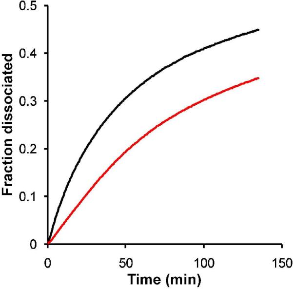
Kinetics experiments show the fraction of aggregates that dissociated over time in the presence of albumin. bZip micelles ( ) dissociate more quickly than bZip PRTNs (
 ).
).
) dissociate more quickly than bZip PRTNs ().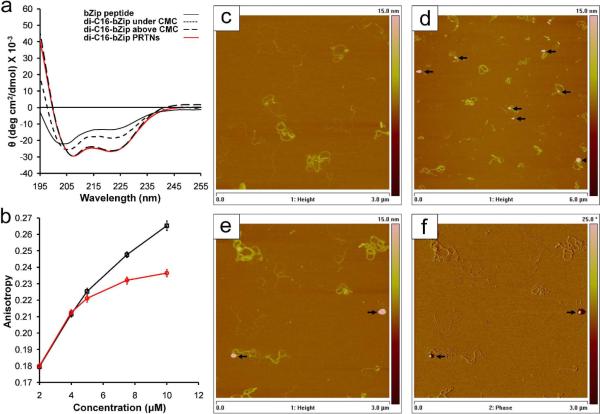
bZip secondary structure and DNA binding. (a) Circular dichroism of bZip peptide, bZip PA below and above the CMC, and bZip PRTNs. The lipidation of the bZip peptide provides a slight increase in helicity over the free peptide, while aggregation into micelles or on the surface of a dendrimer results in drastic increase in α-helicity indicated by the CD minima at 208 nm and 222 nm. (b) An increase in anisotropy corresponds to an increase in binding of bZip PAs (□) and bZip PRTNs ( ) to rhodamine labeled 30bp DNA strands. The bZip PA curve has higher anisotropy at higher concentrations because the DNA strands are bound by long micelles rather than small PTRNs. Data points are the mean of 10 measurements, and error bars are standard deviation. (c) AFM height image of free ctDNA (3 μm frame depicted). (d) AFM height image of bZip PRTNs (spherical objects indicated by black arrows) bound to ctDNA (6 μm frame). PRTNs are not present on the surface but only appear in areas of DNA, indicating they are bound to the DNA and not attached to free mica. (e, f) AFM height and phase image of PRTNs bound to DNA (3 μm frame),where the spherical geometry is clearly seen. Additional AFM images available in the Supplemental Information.
) to rhodamine labeled 30bp DNA strands. The bZip PA curve has higher anisotropy at higher concentrations because the DNA strands are bound by long micelles rather than small PTRNs. Data points are the mean of 10 measurements, and error bars are standard deviation. (c) AFM height image of free ctDNA (3 μm frame depicted). (d) AFM height image of bZip PRTNs (spherical objects indicated by black arrows) bound to ctDNA (6 μm frame). PRTNs are not present on the surface but only appear in areas of DNA, indicating they are bound to the DNA and not attached to free mica. (e, f) AFM height and phase image of PRTNs bound to DNA (3 μm frame),where the spherical geometry is clearly seen. Additional AFM images available in the Supplemental Information.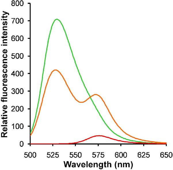
Formation of multifunctional PRTNs. FRET was observed for the co-assembly of 1:1 ratio of bZip:NLS PAs with dendrimer ( ), confirming that the PAs formed “mixed' PRTNs. Fluorescein-tagged bZip PRTNs (
 ) and rhodamine-labeled NLS PRTNs (
 ) were used as controls. These experiments were performed with an excitation wavelength of 475 nm.
), confirming that the PAs formed “mixed' PRTNs. Fluorescein-tagged bZip PRTNs () and rhodamine-labeled NLS PRTNs () were used as controls. These experiments were performed with an excitation wavelength of 475 nm.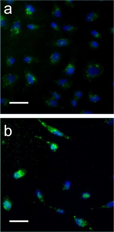
In vitro cell uptake of micelles and PRTNs. The green color in the images represents the fluorescein labeled bZip PAs, while the blue is Hoechst nuclear stain. Micelles and PRTNs are both internalized by HeLa cells. (a) Mixed PRTNs (90% bZip-FAM PA and 10% NLS PA) display punctate fluorescence, suggesting they are trapped in endosomes. The fluorescence is located in the bulk volume of the cell near the nucleus, and not observed throughout the cytoplasm to indicate endosomal escape. Intact PRTNs show low fluorescence, presumably due to fluorescence quenching. (b) Mixed micelles (90% bZip-FAM PA and 10% NLS PA) appear to be internalized via endocytosis as seen through the punctate fluorescence, but also show diffuse fluorescence, indicating PA distribution throughout the cell. The exact mechanisms of internalization are still under investigation. Scale bars are 50 μm.
Similar articles
-
Biomimetic Self-Templated Hierarchical Structures of Collagen-Like Peptide Amphiphiles.Nano Lett. 2015 Oct 14;15(10):7138-45. doi: 10.1021/acs.nanolett.5b03313. Epub 2015 Sep 25. Nano Lett. 2015. PMID: 26392232
-
Probing Peptide Assembly and Interaction via High-Resolution Imaging Techniques: A Mini Review.Int J Mol Sci. 2025 Apr 23;26(9):3998. doi: 10.3390/ijms26093998. Int J Mol Sci. 2025. PMID: 40362238 Free PMC article. Review.
-
Linker chemistry determines secondary structure of p5314-29 in peptide amphiphile micelles.Bioconjug Chem. 2010 Mar 17;21(3):465-75. doi: 10.1021/bc900383m. Epub 2010 Feb 18. Bioconjug Chem. 2010. PMID: 20166676
-
Bionanosphere lithography via hierarchical peptide self-assembly of aromatic triphenylalanine.Small. 2010 Apr 23;6(8):945-51. doi: 10.1002/smll.200902050. Small. 2010. PMID: 20397209
-
Effect of proteins on the synthesis and assembly of calcium phosphate nanomaterials.Nanoscale. 2010 Oct;2(10):1842-8. doi: 10.1039/c0nr00092b. Epub 2010 Aug 2. Nanoscale. 2010. PMID: 20676452 Review.
Cited by
-
The non-peptidic part determines the internalization mechanism and intracellular trafficking of peptide amphiphiles.PLoS One. 2013;8(1):e54611. doi: 10.1371/journal.pone.0054611. Epub 2013 Jan 17. PLoS One. 2013. PMID: 23349939 Free PMC article.
-
Supramolecular guests in solvent driven block copolymer assembly: From internally structured nanoparticles to micelles.Polym Chem. 2013 Oct 7;4(19):5038-5042. doi: 10.1039/C3PY00750B. Polym Chem. 2013. PMID: 25525473 Free PMC article.
-
Effect of Lipidation on the Structure, Oligomerization, and Aggregation of Glucagon-like Peptide 1.Bioconjug Chem. 2025 Mar 19;36(3):401-414. doi: 10.1021/acs.bioconjchem.4c00484. Epub 2025 Jan 22. Bioconjug Chem. 2025. PMID: 39841169 Free PMC article.
-
Vaccine delivery systems and administration routes: Advanced biotechnological techniques to improve the immunization efficacy.Vaccine X. 2024 May 24;19:100500. doi: 10.1016/j.jvacx.2024.100500. eCollection 2024 Aug. Vaccine X. 2024. PMID: 38873639 Free PMC article. Review.
-
Dendrimer-Fullerenol Soft-Condensed Nanoassembly.J Phys Chem C Nanomater Interfaces. 2012 Jul 26;116(29):15775-15781. doi: 10.1021/jp3036692. Epub 2012 Jul 3. J Phys Chem C Nanomater Interfaces. 2012. PMID: 23185644 Free PMC article.
References
-
- Zhang SG. Nat Biotechnol. 2003;21:1171. - PubMed
-
- Cavalli S, Albericio F, Kros A. Chem Soc Rev. 2010;39:241. - PubMed
-
- Versluis F, Marsden HR, Kros A. Chem Soc Rev. 2010;39:3434. - PubMed
-
- Hamley IW. Soft Matter. 2011;7:4122.
-
- Tu RS, Marullo R, Pynn R, Bitton R, Bianco-Peled H, Tirrell MV. Soft Matter. 2010;6:1035.
Publication types
MeSH terms
Substances
Grants and funding
LinkOut - more resources
Full Text Sources
