Review
. 2011:27:653-79.
doi: 10.1146/annurev-cellbio-092910-154026. Epub 2011 Jul 21.

Neurogenesis at the brain-cerebrospinal fluid interface

Affiliations
Review

Neurogenesis at the brain-cerebrospinal fluid interface

Maria K Lehtinen et al. Annu Rev Cell Dev Biol. 2011.

Abstract

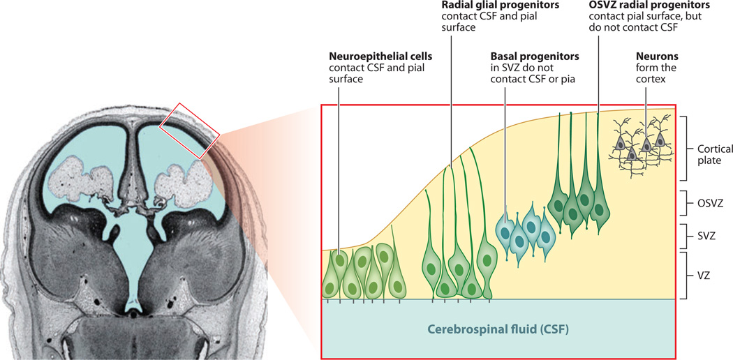
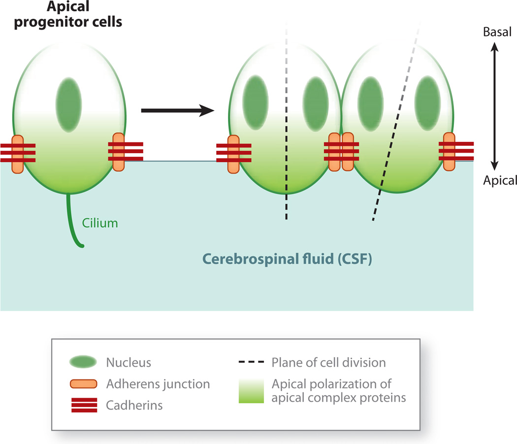
Cerebral cortical progenitor cells can be classified into several different types, and each progenitor type integrates cell-intrinsic and cell-extrinsic cues to regulate neurogenesis. On one hand, cell-intrinsic mechanisms that depend upon appropriate apical-basal polarity are established by adherens junctions and apical complex proteins and are particularly important in progenitors with apical processes contacting the lateral ventricle. The apical protein complexes themselves are concentrated at the ventricular surface, and apical complex proteins regulate mitotic spindle orientation and cell fate. On the other hand, remarkably little is known about how cell-extrinsic cues signal to progenitors and couple with cell-intrinsic mechanisms to instruct neurogenesis. Recent research shows that the cerebrospinal fluid, which contacts apical progenitors at the ventricular surface and bathes the apical complex of these cells, provides growth- and survival-promoting cues for neural progenitor cells in developing and adult brain. This review addresses how the apical-basal polarity of progenitor cells regulates cell fate and allows progenitors to sample diffusible signals distributed by the cerebrospinal fluid. We also review several classes of signaling factors that the cerebrospinal fluid distributes to the developing brain to instruct neurogenesis.


Figures

Figure 1
Figure 1
Neural progenitor cells in the developing mammalian cerebral cortex. Cerebral cortical progenitor cells divide in three principal locations in the developing mammalian brain: the apical ventricular zone (VZ), the inner subventricular zone (SVZ), and the outer subventricular zone (OSVZ). Progenitors in the apical progenitor pool can be divided into two main types of progenitor cells: neuroepithelial cells and radial glial cells. The progenitors in the SVZ constitute the basal progenitor cell pool as well as the most recently discovered class of progenitors, the radial-type progenitors that lie in the OSVZ, which have radial morphology with a long basal process but lack the apical process contacting the ventricle. They are more apparent in mammals with larger brains but are seen in mice as well (Fietz et al. 2010, Hansen et al. 2010, Reillo et al. 2011, Wang et al. 2011). Coronal image of human fetal brain reproduced and adapted from O’Rahilly & Müller (1994), copyright © 1994, Wiley-Liss, Inc.
Figure 2
Figure 2
Apical-basal polarity in cortical progenitor cells. Apical progenitor cells have a distinct apical-basal polarity. Their apical surface contacts the cerebrospinal fluid (CSF) that fills the ventricles, whereas their basal processes extend to and contact the meninges, basal lamina, and vasculature. The adhesion of adjacent progenitor cells to each other is maintained by adherens junctions, which are cadherin-containing contacts between cells. The adherens junctions also define the border of the apical membrane domain that contacts the CSF. The adherens junctions and apical membrane are home to the apical complex proteins, which play an active role in polarizing cellular proteins. The unequal inheritance of the apical membrane and associated proteins appears to regulate whether dividing cells generate pairs of daughter cells with the same, symmetric cell fate (e.g., two progenitor cells) or cells with distinct, asymmetric cell fates (i.e., one progenitor and one neuron). The progenitors of the outer subventricular zone do not appear to show the same expression of apical complex proteins (Fietz et al. 2010).
Figure 3
Figure 3
Cerebrospinal fluid (CSF) flow during embryonic brain development and in adulthood. Schematics of the cerebroventricular system during early human brain development and in the mature adult brain. (a) Upon anterior neural tube closure, the three primary brain vesicles [telencephalic (T), mesencephalic (M), and rhombencephalic (R) vesicles] serve as the rudimentary cerebroventricular system for the developing central nervous system (CNS). Human Carnegie stage (CS) 11 corresponds to approximately embryonic day (E)8.5–9.75 during mouse embryogenesis, and CS13–15 corresponds to approximately E10–11.25. Drawings based on Lowery & Sive 2009. (b) In the mature CNS, CSF generated primarily by the choroid plexus tissues located in each ventricle in the brain fills the ventricles, subarachnoid space, and spinal canal. CSF flows from the lateral ventricles via the foramen of Monro/intraventricular foramen into the mesencephalic/third ventricle, and then via the aqueduct of Sylvius/cerebral aqueduct into the hindbrain/fourth ventricle. The CSF then continues through the foramina of Magendie/median apertures and Luschka/lateral apertures into the spinal canal and subarachnoid space, and is finally resorbed into the venous system via arachnoid villi. An adult human circulates approximately 150 ml of CSF within the cerebroventricular system. The CSF is estimated to turn over approximately three to four times per day, so a healthy CNS produces close to 500 ml of CSF daily.
Figure 4
Figure 4
The cerebrospinal fluid (CSF, blue) distributes diffusible factors during brain development. (a) IGF2 and other factors (purple spheres) are secreted by the choroid plexus and delivered by the CSF to targets on the apical ventricular surface of the developing cerebral cortex (Lehtinen et al. 2011). CSF and CSF-FGF2 have been shown to support proliferation of midbrain progenitor cells (Martin et al. 2006, 2009). Whether similar rules apply to CSF-borne retinoic acid, Wnt, and Bmp signaling, as well as other as yet uncharacterized signals, remains to be elucidated. (b) Sonic hedgehog (Shh) secreted by the hindbrain/fourth ventricle choroid plexus signals in an autocrine manner to instruct choroid plexus development. CSF-Shh also signals in a paracrine manner to stimulate the proliferation of cerebellar granule neuron precursors located in the external granule cell layer (Huang et al. 2009, 2010).

References

LITERATURE CITED

    1. Aaku-Saraste E, Oback B, Hellwig A, Huttner WB. Neuroepithelial cells downregulate their plasma membrane polarity prior to neural tube closure and neurogenesis. Mech. Dev. 1997;69:71–81. - PubMed
    1. Agnati LF, Zoli M, Stromberg I, Fuxe K. Intercellular communication in the brain: wiring versus volume transmission. Neuroscience. 1995;69:711–726. - PubMed
    1. Angevine JB, Jr., Bodian D, Coulombre AJ, Edds MVJr, Hamburger V, et al. Embryonic vertebrate central nervous system: revised terminology. Anat. Rec. 1970;166:257–261. - PubMed
    1. Awatramani R, Soriano P, Rodriguez C, Mai JJ, Dymecki SM. Cryptic boundaries in roof plate and choroid plexus identified by intersectional gene activation. Nat. Genet. 2003;35:70–75. - PubMed
    1. Baker J, Liu JP, Robertson EJ, Efstratiadis A. Role of insulin-like growth factors in embryonic and postnatal growth. Cell. 1993;75:73–82. - PubMed

RELATED RESOURCES

    1. He S, Nahada D, Morrison SJ. Mechanisms of stem cell self-renewal. Annu. Rev. Cell Dev. Biol. 2009;25:377–406. - PubMed
    1. Jackson Lab. Hydrocephalus in laboratory mice. JAX® NOTES. 2003:490. http://jaxmice.jax.org/jaxnotes/archive/490f.html.
    1. Nachury MV, Seeley ES, Jin H. Trafficking to the ciliary membrane: how to get across the periciliary diffusion barrier? Annu. Rev. Cell Dev. Biol. 2010;26:59–87. - PMC - PubMed
    1. Sundberg JP, Woolcott BL, Cunlifee-Beamer T, Brown KS, Bronson R. Spontaneous hydrocephalus in inbred strains of mice. JAX® NOTES. 1991:445. http://jaxmice.jax.org/jaxnotes/archive/445a.html.

Publication types

MeSH terms

LinkOut - more resources