Comparative Study
. 2012 Jun;16(6):1245-59.
doi: 10.1111/j.1582-4934.2011.01400.x.

In vitro analyses of the anti-fibrotic effect of SPARC silencing in human Tenon's fibroblasts: comparisons with mitomycin C

Affiliations
Comparative Study

In vitro analyses of the anti-fibrotic effect of SPARC silencing in human Tenon's fibroblasts: comparisons with mitomycin C

Li-Fong Seet et al. J Cell Mol Med. 2012 Jun.

Abstract

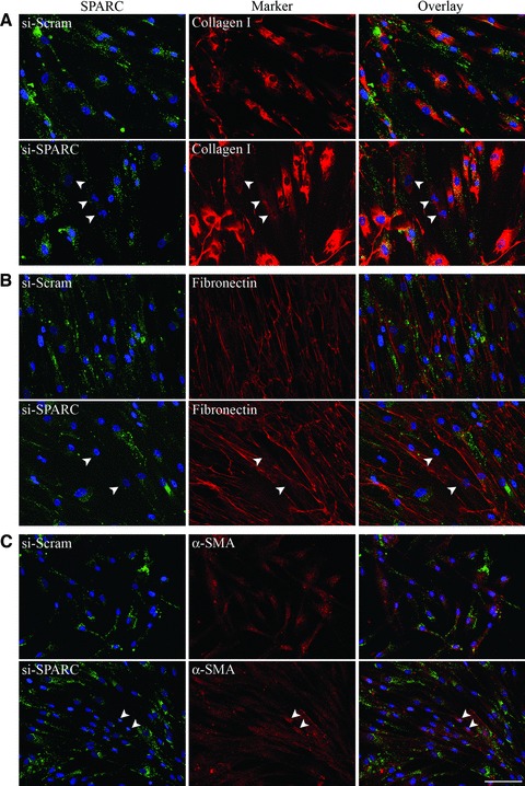
Failure of glaucoma filtration surgery (GFS) is commonly attributed to scarring at the surgical site. The human Tenon's fibroblasts (HTFs) are considered the major cell type contributing to the fibrotic response. We previously showed that SPARC (secreted protein, acidic, rich in cysteine) knockout mice had improved surgical success in a murine model of GFS. To understand the mechanisms of SPARC deficiency in delaying subconjunctival fibrosis, we used the gene silencing approach to reduce SPARC expression in HTFs and examined parameters important for wound repair and fibrosis. Mitomycin C-treated HTFs were used for comparison. We demonstrate that SPARC-silenced HTFs showed normal proliferation and negligible cellular necrosis but were impaired in motility and collagen gel contraction. The expression of pro-fibrotic genes including collagen I, MMP-2, MMP-9, MMP-14, IL-8, MCP-1 and TGF-β(2) were also reduced. Importantly, TGF-β(2) failed to induce significant collagen I and fibronectin expressions in the SPARC-silenced HTFs. Together, these data demonstrate that SPARC knockdown in HTFs modulates fibroblast functions important for wound fibrosis and is therefore a promising strategy in the development of anti-scarring therapeutics.


Figures

Fig 1
Fig 1
Silencing of HTFs with si-SPARC was effective for at least 7 days. (A) HTFs were transfected with si-Scram (control) or si-SPARC and the mRNA level of SPARC was determined by quantitative real-time PCR after 1 or 7 days post-transfection. The β-actin transcript was used for normalization. Values are shown as fold expression relative to control for each time-point analysed. (B) HTFs were transfected as in (A) and the protein level of SPARC was determined by Western blotting with SPARC antibody after 7 days. Immunoblotting with β-tubulin was carried out to determine the loading levels in each sample.
Fig 2
Fig 2
Silencing of SPARC did not affect the cell proliferation capacity of HTFs. (A) HTFs were treated with 0.4 mg/ml MMC for 1 min. and washed before being plated on 96-well plates for real-time analysis of cell proliferation using the RTCA SP instrument. Untreated HTFs were similarly analysed to serve as controls. The cell index profiles of untreated cells (dotted line) and MMC-treated cells (solid line) reflected the logarithmic growth phase and response to treatment respectively for up to 6 days. The cell index values for MMC-treated cells did not demonstrate a logarithmic growth phase that was measured for the control cells, indicating that the MMC-treated cells did not proliferate. (B) HTFs were transfected with either si-Scram (control) or si-SPARC and plated the next day on 96-well plates for real-time analysis of cell proliferation as in (A). The cell index values for HTFs silenced for SPARC demonstrated a similar logarithmic growth phase as si-Scram–transfected control cells, indicating that knockdown of SPARC had no influence on the proliferative capacity of HTFs.
Fig 3
Fig 3
Silencing of SPARC protected HTFs from necrosis. (A) HTFs, treated as indicated for 72 hrs, were harvested for both adherent and non-adherent cells and analysed for apoptosis and late apoptosis/necrosis. The representative scatterplots are shown. Apoptotic cells are enclosed within the blue perimeter and the mean percentage of apoptotic cells is indicated. Necrotic cells are shown within the green perimeter and the mean percentage of necrotic/late apoptotic is indicated. Data are presented as mean ± S.D. of the averages of three independent experiments, each performed in triplicate. (B) Cells were treated as in (A) and analysed by TUNEL staining (green). Nuclei were visualized by DAPI staining (blue). Cells positive for TUNEL staining are indicated by arrowheads; scale bar: 100 μm.
Fig 4
Fig 4
Silencing of SPARC impaired HTF motility. (A) Plated HTFs were wounded with a pipette tip. After wounding, cell culture medium was replaced with fresh medium and wound closure was monitored by microscopy and photographed at the indicated times post-wounding. Triplicates were performed. A representative micrograph for each condition is shown. Vertical lines outline the wound edge. (B) Quantitative evaluation of HTF migration shown in (A). Values are expressed as percentages of the original wound widths. si-SPARC–transfected HTFs demonstrated significantly slower cell migration into the wound relative to si-Scram–transfected HTFs at 23 hrs (*P ∇ 0.0004) and 31 hrs (**P ∇ 0.0009) post-wounding. Data are presented as mean ± S.D. (n ∇ 3).
Fig 5
Fig 5
Silencing of SPARC reduced the contractility of HTFs but to a lesser extent compared to MMC treatment. (A) Graphical representations of the contraction of free-floating collagen lattices seeded with control (untreated) or MMC-treated (left panel) and si-Scram- or si-SPARC–transfected HTFs (right panel) are shown. Data are presented as mean percentage of the initial gel size ± S.D. The data shown are from one experiment but is representative of three independent experiments, each performed in triplicate. (B) MMP-2 activity was reduced in medium conditioned by HTFs knocked down for SPARC seeded in collagen gels. Left panel: MMP-2 activity in medium conditioned by untreated or MMC-treated HTFs seeded in collagen gels for 5 days was analysed by gelatin zymography. Proteolytic activities corresponding to the molecular weights of MMP-9 (92 kD) and pro- and active MMP-2 (72 and 66 kD, respectively) are indicated by arrowheads. Right panel: MMP-2 activity in medium conditioned by si-Scram- or si-SPARC–transfected HTFs seeded in collagen gels for 5 days was similarly analysed. (C) MMP-14 expression was reduced in SPARC-silenced HTFs. Left panel: HTFs, control or treated with MMC, were seeded in collagen gels for 5 days before being lysed and equal amounts of protein subjected to immunoblotting with antibodies specific for MMP-14 and GAPDH (loading control). Right panel, similarly seeded si-Scram- or si-SPARC–transfected HTFs were subjected to the same immunoblot analysis for MMP-14 expression.
Fig 6
Fig 6
Collagen I and fibronectin protein expressions were not induced in SPARC knockdown HTFs in response to TGF-β2 but α-SMA expression remained inducible. (A) HTFs were treated with MMC (left panel) or transfected with siRNAs (right panel), with or without TGF-β2, for 72 hrs before being analysed for collagen I protein abundance by immunoblotting for collagen Iα1 and GAPDH (loading control). The data shown are representative of three independent experiments. (B) Densitometric analysis of immunoblots from three independent experiments represented in (A). Data are presented as mean fold induction ± S.D. relative to their respective controls (untreated: left panel; si-Scram–transfected HTFs: right panel) from three independent experiments. The GAPDH level was used for normalization. The P value for each comparison is indicated above the bars. (C) HTFs were subjected to the indicated treatments as in (A) and analysed for fibronectin expression. The data shown are representative of three independent experiments. (D) Densitometric analysis of immunoblots from three independent experiments represented in (C). (E) HTFs were subjected to the indicated treatments as in (A) and analysed for α-SMA protein expression. The data shown are representative of three independent experiments. (F) Densitometric analysis of immunoblots from three independent experiments represented in (E).
Fig 7
Fig 7
Silencing of SPARC reduced collagen I in HTFs. (A) HTFs were transfected with either si-Scram or si-SPARC for 72 hrs and then simultaneously immunostained for SPARC (green) and collagen I (red). Nuclei were visualized by DAPI staining (blue). Cells expressing reduced levels of SPARC from transfection with si-SPARC also showed reduced collagen I expression (arrowheads). Overlapping staining is shown in the overlay. (B) HTFs transfected as in (A) were visualized for SPARC (green) and fibronectin (red) expressions simultaneously. Cells expressing reduced levels of SPARC from transfection with si-SPARC did not show obvious changes in fibronectin expression (arrowheads). (C) HTFs transfected as in (A) were visualized for SPARC (green) and α-SMA (red) expressions simultaneously. Cells expressing reduced levels of SPARC from transfection with si-SPARC showed slightly increased α-SMA staining (arrowheads). Scale bar: 100 μm.
Fig 8
Fig 8
Silencing of SPARC reduced MMP-2 activity. (A) MMP-2 activity was reduced in medium conditioned by HTFs knocked down for SPARC. Five μg of protein in the respective culture medium was analysed by gelatin zymography. Left panel: MMP-2 activity in medium conditioned by control (untreated) or MMC-treated HTFs cultured for 3 days was analysed. Proteolytic activities corresponding to MMP-9 and active MMP-2 are indicated by arrowheads. Right panel: MMP-2 activity in medium conditioned by si-Scram- or si-SPARC–transfected HTFs for 3 days was similarly analysed. The presented data are representative of three independent experiments. (B) Densitometric analysis of MMP-2 activity from three independent experiments represented in (A). Values are expressed as mean fold MMP-2 activity ± S.D. relative to that in control (untreated) HTFs (left panel; P ∇ 0.54) or si-Scram–transfected HTFs (right panel; *P ∇ 0.019). Data are calculated based on the fold difference from three independent experiments.

Similar articles

Cited by

References

    1. Quigley HA, Broman AT. The number of people with glaucoma worldwide in 2010 and 2020. Br J Ophthalmol. 2006;90:262–7. - PMC - PubMed
    1. Heijl A, Leske MC, Bengtsson B. Reduction of intraocular pressure and glaucoma progression: results from the early manifest glaucoma trial. Arch Ophthalmol. 2002;120:1268–78. - PubMed
    1. Kass MA, Heuer DK, Higginbotham EJ, et al. The ocular hypertension treatment study: a randomized trial determines that topical ocular hypotensive medication delays or prevents the onset of primary open-angle glaucoma. Arch Ophthalmol. 2002;120:701–13. - PubMed
    1. Hitchings RA, Grierson I. Clinico pathological correlation in eyes with failed fistulizing surgery. Trans Ophthalmol Soc UK. 1983;103:84–8. - PubMed
    1. Skuta GL, Parrish RK. Wound healing in glaucoma filtering surgery. Surv Ophthalmol. 1987;32:149–70. - PubMed

Publication types

MeSH terms