Characterization of antigenic variants of hepatitis C virus in immune evasion
- PMID: 21801418
- PMCID: PMC3158126
- DOI: 10.1186/1743-422X-8-377
Characterization of antigenic variants of hepatitis C virus in immune evasion
Abstract
Background: Antigenic variation is an effective way by which viruses evade host immune defense leading to viral persistence. Little is known about the inhibitory mechanisms of viral variants on CD4 T cell functions.
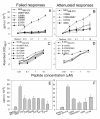
Results: Using sythetic peptides of a HLA-DRB1*15-restricted CD4 epitope derived from the non-structural (NS) 3 protein of hepatitis C virus (HCV) and its antigenic variants and the peripheral blood mononuclear cells (PBMC) from six HLA-DRB1*15-positive patients chronically infected with HCV and 3 healthy subjects, the in vitro immune responses and the phenotypes of CD4+CD25+ cells of chronic HCV infection were investigated. The variants resulting from single or double amino acid substitutions at the center of the core region of the Th1 peptide not only induce failed T cell activation but also simultaneously up-regulate inhibitory IL-10, CD25-TGF-β+ Th3 and CD4+IL-10+ Tr1 cells. In contrast, other variants promote differentiation of CD25+TGF-β+ Th3 suppressors that attenuate T cell proliferation.
Conclusions: Naturally occuring HCV antigenic mutants of a CD4 epitope can shift a protective peripheral Th1 immune response into an inhibitory Th3 and/or Tr1 response. The modulation of antigenic variants on CD4 response is efficient and extensive, and is likely critical in viral persistence in HCV infection.
Figures


Similar articles
-
Modulation of the peripheral T-Cell response by CD4 mutants of hepatitis C virus: transition from a Th1 to a Th2 response.Hum Immunol. 2003 Jul;64(7):662-73. doi: 10.1016/s0198-8859(03)00070-3. Hum Immunol. 2003. PMID: 12826368
-
Hypervariable region 1 variants act as TCR antagonists for hepatitis C virus-specific CD4+ T cells.J Immunol. 1999 Jul 15;163(2):650-8. J Immunol. 1999. PMID: 10395654
-
Impaired effector function of hepatitis C virus-specific CD8+ T cells in chronic hepatitis C virus infection.J Immunol. 2002 Sep 15;169(6):3447-58. doi: 10.4049/jimmunol.169.6.3447. J Immunol. 2002. PMID: 12218168
-
[Immunopathogenesis of viral hepatitis C. Immunological markers of the disease progression].Zh Mikrobiol Epidemiol Immunobiol. 2007 Nov-Dec;(6):75-84. Zh Mikrobiol Epidemiol Immunobiol. 2007. PMID: 18277544 Review. Russian.
-
Immunobiology of hepatitis C virus (HCV) infection: the role of CD4 T cells in HCV infection.Immunol Rev. 2000 Apr;174:90-7. doi: 10.1034/j.1600-0528.2002.017403.x. Immunol Rev. 2000. PMID: 10807509 Review.
Cited by
-
Control of the inflammatory response mechanisms mediated by natural and induced regulatory T-cells in HCV-, HTLV-1-, and EBV-associated cancers.Mediators Inflamm. 2014;2014:564296. doi: 10.1155/2014/564296. Epub 2014 Nov 30. Mediators Inflamm. 2014. PMID: 25525301 Free PMC article. Review.
-
Designing and Development of a DNA Vaccine Based On Structural Proteins of Hepatitis C Virus.Iran J Pathol. 2016 Summer;11(3):222-230. Iran J Pathol. 2016. PMID: 27799971 Free PMC article.
-
Hepatitis C Virus: Insights Into Its History, Treatment, Challenges, and Future Directions.Cureus. 2023 Aug 22;15(8):e43924. doi: 10.7759/cureus.43924. eCollection 2023 Aug. Cureus. 2023. PMID: 37614826 Free PMC article. Review.
-
Genetic and epidemiological insights into the emergence of peste des petits ruminants virus (PPRV) across Asia and Africa.Sci Rep. 2014 Nov 13;4:7040. doi: 10.1038/srep07040. Sci Rep. 2014. PMID: 25391314 Free PMC article.
-
Intracellular Pathogens: Host Immunity and Microbial Persistence Strategies.J Immunol Res. 2019 Apr 14;2019:1356540. doi: 10.1155/2019/1356540. eCollection 2019. J Immunol Res. 2019. PMID: 31111075 Free PMC article. Review.
References
Publication types
MeSH terms
Substances
Grants and funding
LinkOut - more resources
Full Text Sources
Research Materials
Miscellaneous

