. 2011 Jul 31:10:95.
doi: 10.1186/1476-4598-10-95.

Essential role of caspase-8 in p53/p73-dependent apoptosis induced by etoposide in head and neck carcinoma cells

Affiliations

Essential role of caspase-8 in p53/p73-dependent apoptosis induced by etoposide in head and neck carcinoma cells

Juan Liu et al. Mol Cancer. .

Abstract

Background: Caspase-8 is a key upstream mediator in death receptor-mediated apoptosis and also participates in mitochondria-mediated apoptosis via cleavage of proapoptotic Bid. However, the role of caspase-8 in p53- and p73-dependent apoptosis induced by genotoxic drugs remains unclear. We recently reported that the reconstitution of procaspase-8 is sufficient for sensitizing cisplatin- but not etoposide-induced apoptosis, in chemoresistant and caspase-8 deficient HOC313 head and neck squamous cell carcinoma (HNSCC) cells.

Results: We show that p53/p73-dependent caspase-8 activation is required for sensitizing etoposide-induced apoptosis by utilizing HOC313 cells carrying a temperature-sensitive p53G285K mutant. Restoration of wild-type p53 function under the permissive conditions, together with etoposide treatment, led to substantial transcriptional activation of proapoptotic Noxa and PUMA, but failed to induce apoptosis. In addition to p53 restoration, caspase-8 reconstitution was needed for sensitization to etoposide-induced apoptosis, mitochondria depolarization, and cleavage of the procaspases-3, and -9. In etoposide-sensitive Ca9-22 cells carrying a temperature-insensitive mutant p53, siRNA-based p73 knockdown blocked etoposide-induced apoptosis and procaspase-8 cleavage. However, induction of p73 protein and up-regulation of Noxa and PUMA, although observed in Ca9-22 cells, were hardly detected in etoposide-treated HOC313 cells under non-permissive conditions, suggesting a contribution of p73 reduction to etoposide resistance in HOC313 cells. Finally, the caspase-9 inhibitor Ac-LEHD-CHO or caspase-9 siRNA blocked etoposide-induced caspase-8 activation, Bid cleavage, and apoptosis in both cell lines, indicating that p53/p73-dependent caspase-8 activation lies downstream of mitochondria.

Conclusions: we conclude that p53 and p73 can act as upstream regulators of caspase-8, and that caspase-8 is an essential mediator of the p53/p73-dependent apoptosis induced by etoposide in HNSCC cells. Our data suggest the importance of caspase-8-mediated positive feedback amplification in the p53/p73-dependent apoptosis induced by etoposide in HNSCC cells.


Figures

Figure 1
Figure 1
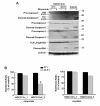
The restoration of wild-type p53 function induced p53-target gene expression in etoposide-treated HOC313 cells. A, cells treated with cisplatin (10 μg/ml) or etoposide (100 μg/ml) were incubated at 32.5°C or 37°C for 6 h; the amount of mRNA encoding PUMA, Noxa and p21 was measured using real-time RT-PCR. n > 3 for all conditions. Bars, SD. B, p53 phosphorylation and p21 expression in drug-treated cells. Cells were treated with drugs as in A for 24 hours followed by Western blot analysis with the indicated antibodies. β-actin was used as a loading control.
Figure 2
Figure 2
Both p53 restoration and caspase-8 reconstitution are needed to sensitize etoposide-induced apoptosis in HOC313 cells. A, effects of temperature downshift on the chemosensitivity of HOC313 and Ca9-22 cells. Cells were treated with increasing doses of cisplatin or etoposide at 32.5°C (solid circle) or 37°C (open triangle) for 24 h. Cell viability was measured by WST-1 assay. The results are the means ± SD from three independent experiments. Bars, SD. The asterisk indicates a significant difference (p < 0.05) between 32.5°C and 37°C (t-test). B, effects of temperature downshift on the DNA profile of drug-treated HOC313 and HOC313/c8_3 cells. Cells treated with cisplatin (10 μg/ml) or etoposide (100 μg/ml) were incubated at 32.5°C or 37°C for 24 h, followed by LSC analysis. The horizontal and vertical axes represent the DNA content and cell number, respectively. The apoptotic cell population is indicated as a percentage of the sub-G1 fraction.
Figure 3
Figure 3
Both p53 and caspase-8 functions are needed for etoposide-induced caspase activation in HOC313 cells. A, effects of temperature downshift on the cleavage of the procaspases and PARP in drug-treated HOC313 cells and HOC313/c8_3 cells. The cells treated with drugs as in Figure 2B were subjected to Western blotting for PARP, and the caspases-8, -9 and -3. β-actin was used as a loading control. B, effects of temperature downshift on caspase 3/7 activities in drug-treated HOC313 cells and HOC313/c8_3 cells. The cells treated with drugs as in Figure 2B for 12 h were subjected to Caspase-Glo 3/7 assay. Results are expressed as fold induction compared with untreated control. The bars show the SD. The asterisk indicates a significant difference (p < 0.05) between 32.5°C and 37°C in etoposide-treated HOC313/c8_3 cells (t-test). C, the caspase-8 (C360S) mutant did not induced apoptosis in etoposide-treated HOC313 cells at 32.5°C. HOC313 cells were transiently transfected with the indicated expression vectors by electroporation. Twenty-four hours after transfection, cells were treated with etoposide (100 μg/ml) at 32.5°C. DNA profiles were analyzed 40 h after drug treatment as in Figure 2B (left panel). Caspase-8 expression was analyzed by Western blot using anti-FLAG antibody 24 h after drug treatment (right panel).
Figure 4
Figure 4
p73 is responsible for etoposide-induced caspase-8 activation and apoptosis in drug-sensitive Ca9-22 cells. A, the induction of endogenous p73 following drug treatment. Cells treated with the drugs as in Figure 2B were subjected to Western blotting with p73, caspase-8 or caspase-9 antibodies. B, C and D, Ca9-22 cells were transfected with either p73 or control siRNA. At 48 h after siRNA transfection, cells were treated with cisplatin (5 μg/ml) or etoposide (50 μg/ml). DNA profiles were analyzed by LSC 30 h after drug treatment (B, upper panel). The horizontal and vertical axes represent the DNA content and cell number, respectively. Apoptotic cell population is indicated as a percentage of the sub-G1 fraction. p73 expression was analyzed by Western blot 30 h after drug treatment (B, lower panel). Caspase 3/7 activity was analyzed at the indicated time after drug treatment (C). Results are expressed as fold induction compared with untreated control. The bars represent the SD. The asterisk indicates a significant difference (p < 0.05) between p73 siRNA-1 and control siRNA treatment cells (t-test). The cleavage of procaspases was analyzed by western blot with caspases-3 and -8 antibodies 24 h after drug treatment (D). E and F, Ca9-22 cells were transfected with either caspase-8 or control siRNA. At 48 h after siRNA transfection, cells were treated with etoposide (100 μg/ml). DNA profiles were analyzed by LSC 24 h after drug treatment as in B (E). The expression and cleavage of procaspases and Bid was analyzed by western blot with caspases-3, -8, -9, and Bid antibodies 24 h after drug treatment (F). β-actin was used as a loading control.
Figure 5
Figure 5
Caspase-9 is functionally relevant in etoposide-induced caspase-8 activation and apoptosis. A and B, effects of caspase inhibitors on caspase-8 activation and the induction of apoptosis by etoposide in HNSCC cell lines. HOC313/c8_3 cells and Ca9-22 cells were incubated at 32.5°C and 37°C respectively with the indicated caspase inhibitors (80 μM) for 1.5 h, followed by treatment with cisplatin (10 μg/ml) or etoposide (100 μg/ml) for 24 h. The DNA profiles were analyzed by LSC as in Figure 2B (A). Caspase-8 activity was analyzed by Caspase-Glo 8 assay (B). Results are expressed as fold induction compared with untreated control. The bars represent the SD. The asterisk indicates a significant difference (p < 0.05) between the presence and absence of Ac-LEHD-CHO in etoposide-treated cells (t-test).
Figure 6
Figure 6
Caspase-9 is functionally relevant in etoposide-induced caspase-8 activation and apoptosis. A and B, cells were transfected with either caspase-9 or control siRNA. At 48 h after siRNA transfection, cells were treated with cisplatin (10 μg/ml) or etoposide (100 μg/ml). Caspase-9 expression was analyzed by Western blot 24 h after drug treatment (A). The DNA profiles were analyzed by LSC 24 h after drug treatment (B). The horizontal and vertical axes represent the DNA content and cell number, respectively. Apoptotic cell population is indicated as a percentage of the sub-G1 fraction. The cleavage of procaspase-9 and Bid was analyzed by Western blot with caspases-9 and Bid antibodies 24 h after drug treatment (C). β-actin was used as a loading control.
Figure 7
Figure 7
Caspase-8 reconstitution activates the p53-mediated mitochondrial pathway induced by etoposide. A, cleavage of procaspase-9 and Bid were analyzed by Western blot with caspase-9 and Bid antibodies 24 h after drug treatment. Exo and endo represent exogenous and endogenous, respectively. B, effects of drug treatment on the mitochondrial membrane potential (ΔΨm) in HOC313/v_1 and HOC313/c8_3 cells at 32.5°C or 37°C. Cells treated with cisplatin (10 μg/ml) or etoposide (100 μg/ml) for 10 h were subjected to JC-1 assay as described in Materials and methods. To measure ΔΨm, the ratio of red to green fluorescence of JC-1 was calculated for each sample and compared with the untreated control JC-1 fluorescence ratio at each temperature. Results are the means ± SD from three independent experiments. The bars represent the SD. The asterisk indicates a significant difference (p < 0.05) between 32.5°C and 37°C in etoposide-treated HOC313/c8_3 cells (t-test).
Figure 8
Figure 8
Model of the drug-specific activation of apoptotic pathways in HNSCC cells. Caspase-8 plays critical roles in inducing both death receptor- and mitochondria-mediated apoptosis in HNSCC cells. The caspase-8-mediated signal amplification of the mitochondrial pathway is important for determining the sensitivity to etoposide-induced and p53/p73-mediated apoptosis (A). Caspase-8 also mediates cisplatin-induced and death receptor-mediated apoptosis (B) [42].

Similar articles

Cited by

References

    1. Danial NN, Korsmeyer SJ. Cell death: critical control points. Cell. 2004;116:205–219. doi: 10.1016/S0092-8674(04)00046-7. - DOI - PubMed
    1. Boatright KM, Salvesen GS. Mechanisms of caspase activation. Curr Opin Cell Biol. 2003;15:725–731. doi: 10.1016/j.ceb.2003.10.009. - DOI - PubMed
    1. Ashkenazi A, Dixit VM. Death receptors: signaling and modulation. Science. 1998;281:1305–1308. - PubMed
    1. Harris MH, Thompson CB. The role of the Bcl-2 family in the regulation of outer mitochondrial membrane permeability. Cell Death Differ. 2000;7:1182–1191. doi: 10.1038/sj.cdd.4400781. - DOI - PubMed
    1. Houghton JA, Harwood FG, Tillman DM. Thymineless death in colon carcinoma cells is mediated via fas signaling. Proc Natl Acad Sci USA. 1997;94:8144–8149. doi: 10.1073/pnas.94.15.8144. - DOI - PMC - PubMed

Publication types

MeSH terms