

Induction of apoptotic change in the rat hippocampus caused by ferric nitrilotriacetate
- PMID: 21801493
- PMCID: PMC6837460
- DOI: 10.1179/174329211X13049558293597
Induction of apoptotic change in the rat hippocampus caused by ferric nitrilotriacetate
Abstract
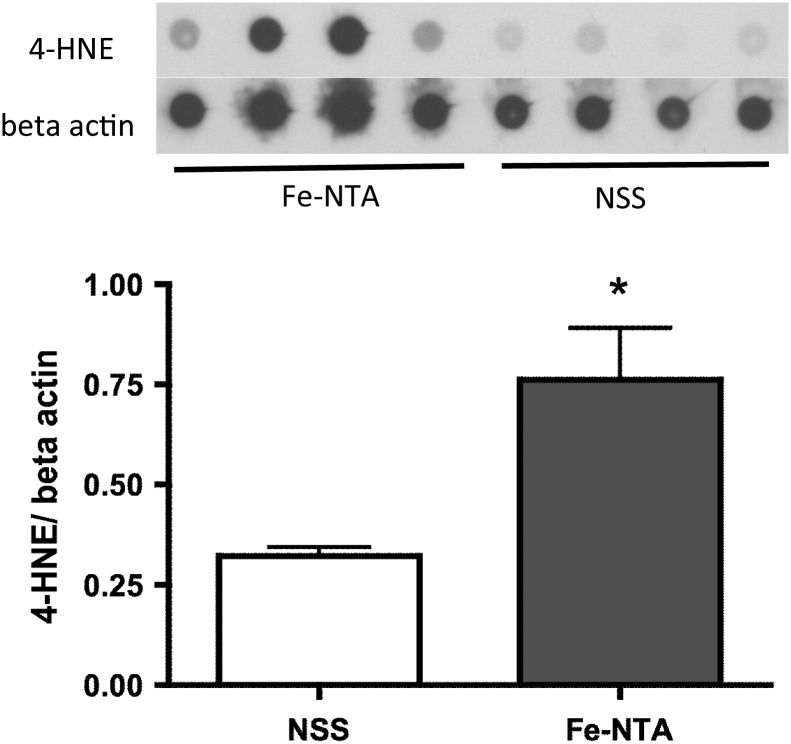
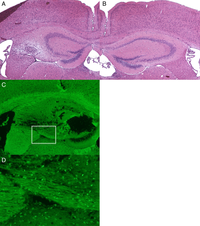
Iron, a source of oxidative stress, plays a major role in the pathology of neurodegenerative disease. In Alzheimer's disease, the hippocampus is vulnerable to oxidative stress, leading to impairment in memory formation. In our previous study, a brain oxidative reaction was induced after intraperitoneal injection of ferric nitrilotriacetate (Fe-NTA). However, since only a small amount of iron reached the brain in the previous study, Fe-NTA was administered into the hippocampus using an osmotic pump in this study. After continuous injection of Fe-NTA for 2 weeks, a high level of apoptotic change was induced in the hippocampus, in accordance with the iron localization. After injection for 4 weeks, the hippocampus was totally destroyed. A small amount of iron infiltrated into the cerebral cortex and the striatum, and deposition was observed at the choroid plexus and ependymal cells. However, no apoptotic reaction or clear tissue injury was observed in these areas. In addition, muscarinic acetylcholine receptors (M1, M2, and M4) were decreased in both the cortex and hippocampus while it increased in the striatum. Thus, the hippocampus is likely vulnerable to oxidative stress from Fe-NTA, and the oxidative stress is considered to bring the disturbance in the muscarinic acetylcholine receptors.
Figures


Similar articles
-
Oxidative changes in the rat brain by intraperitoneal injection of ferric nitrilotriacetate.Redox Rep. 2009;14(3):109-14. doi: 10.1179/135100009X392575. Redox Rep. 2009. PMID: 19490752
-
Attenuation of iron-nitrilotriacetate (Fe-NTA)-mediated renal oxidative stress, toxicity and hyperproliferative response by the prophylactic treatment of rats with garlic oil.Food Chem Toxicol. 1998 Jun;36(6):485-95. doi: 10.1016/s0278-6915(98)00008-8. Food Chem Toxicol. 1998. PMID: 9674956
-
Oxidation and turnover of renal metallothioneins after an injection of ferric nitrilotriacetate.Chem Biol Interact. 2012 Jan 5;195(1):61-7. doi: 10.1016/j.cbi.2011.09.006. Epub 2011 Oct 4. Chem Biol Interact. 2012. PMID: 22001350
-
Ferric nitrilotriacetate promotes N-diethylnitrosamine-induced renal tumorigenesis in the rat: implications for the involvement of oxidative stress.Carcinogenesis. 1998 Jun;19(6):1133-9. doi: 10.1093/carcin/19.6.1133. Carcinogenesis. 1998. PMID: 9667754
-
Differential role of hydrogen peroxide and organic hydroperoxides in augmenting ferric nitrilotriacetate (Fe-NTA)-mediated DNA damage: implications for carcinogenesis.Teratog Carcinog Mutagen. 2003;Suppl 1:13-21. doi: 10.1002/tcm.10045. Teratog Carcinog Mutagen. 2003. PMID: 12616593
References
-
- Schipper HM. Heme oxygenase-1 in Alzheimer disease: a tribute to Moussa Youdim. J Neural Transm 2010;118:381–7. - PubMed
-
- Levenson CW, Tassabehji NM. Iron and ageing: an introduction to iron regulatory mechanisms. Ageing Res Rev 2004;3:251–63. - PubMed
-
- Altamura S, Muckenthaler MU. Iron toxicity in diseases of aging: Alzheimer's disease, Parkinson's disease and atherosclerosis. J Alzheimers Dis 2009;16:879–95. - PubMed
-
- Seabrook GR, Rosahl TW. Transgenic animals relevant to Alzheimer's disease. Neuropharmacology 1999;38:1–17. - PubMed
MeSH terms
Substances
LinkOut - more resources
Full Text Sources