. 2011 Nov;141(5):1915-26.e1-14.
doi: 10.1053/j.gastro.2011.07.033. Epub 2011 Jul 27.

Dendritic cells promote pancreatic viability in mice with acute pancreatitis

Affiliations

Dendritic cells promote pancreatic viability in mice with acute pancreatitis

Andrea S Bedrosian et al. Gastroenterology. 2011 Nov.

Abstract

Background & aims: The cellular mediators of acute pancreatitis are incompletely understood. Dendritic cells (DCs) can promote or suppress inflammation, depending on their subtype and context. We investigated the roles of DC in development of acute pancreatitis.

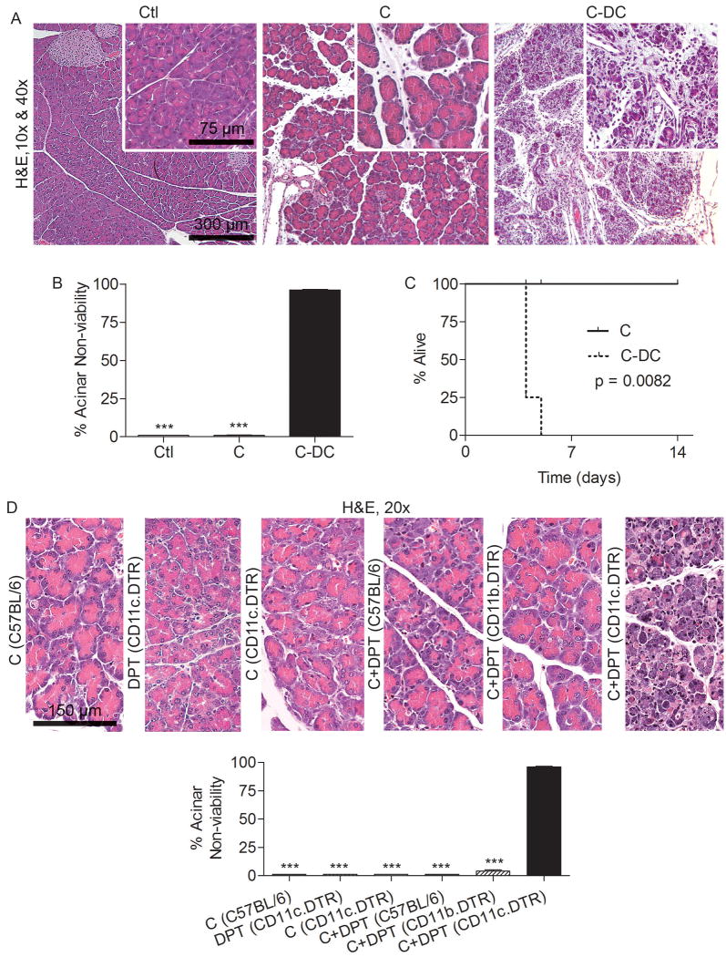
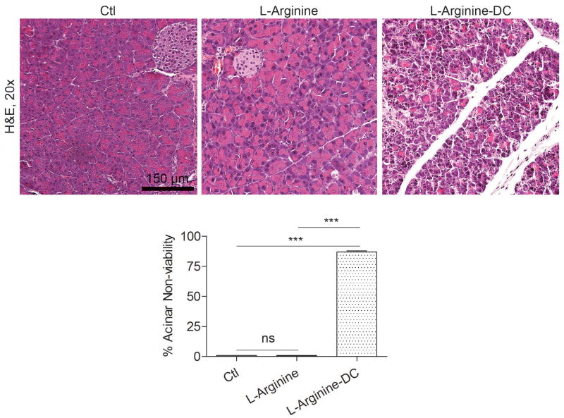
Methods: Acute pancreatitis was induced in CD11c.DTR mice using caerulein or L-arginine; DCs were depleted by administration of diphtheria toxin. Survival was analyzed using Kaplan-Meier method.

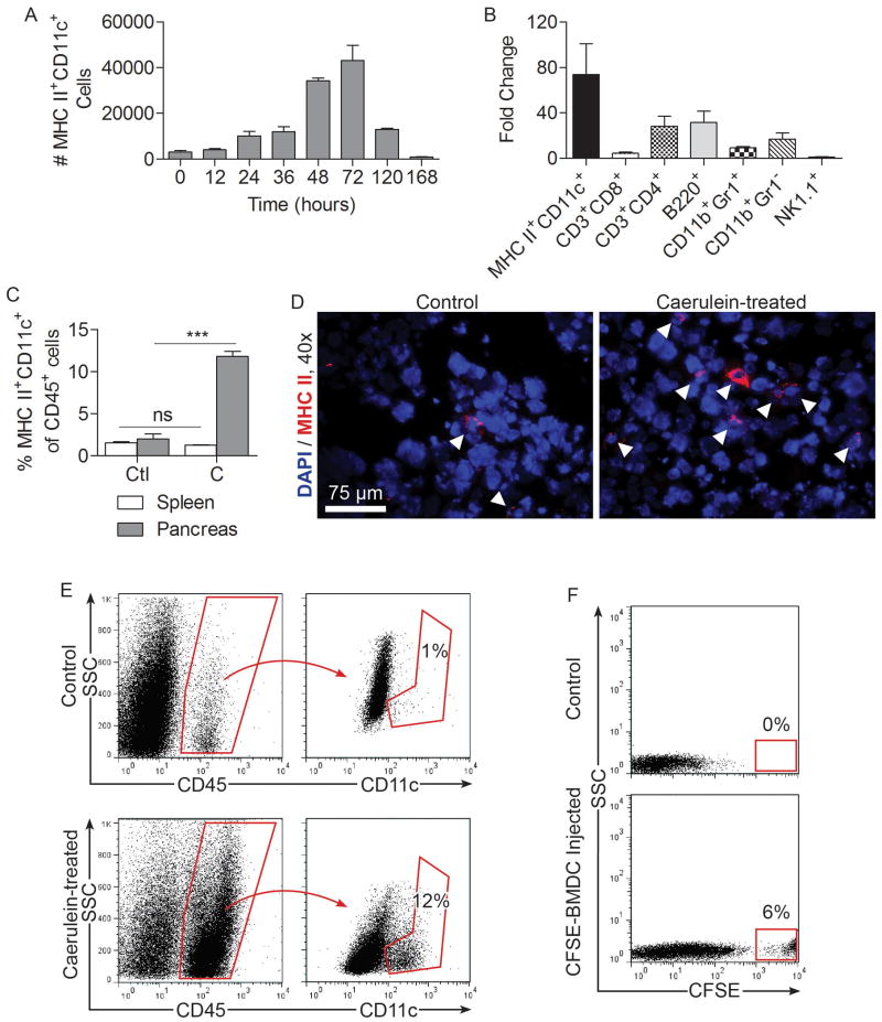
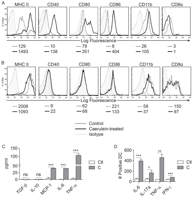
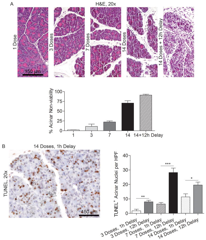
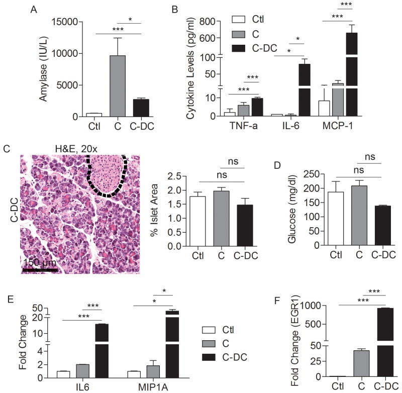
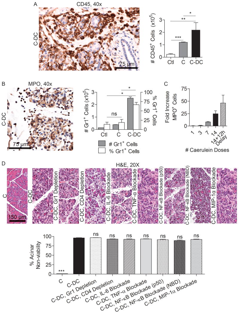
Results: Numbers of major histocompatibility complex II(+)CD11c(+) DCs increased 100-fold in pancreata of mice with acute pancreatitis to account for nearly 15% of intrapancreatic leukocytes. Intrapancreatic DCs acquired a distinct immune phenotype in mice with acute pancreatitis; they expressed higher levels of major histocompatibility complex II and CD86 and increased production of interleukin-6, membrane cofactor protein-1, and tumor necrosis factor-α. However, rather than inducing an organ-destructive inflammatory process, DCs were required for pancreatic viability; the exocrine pancreas died in mice that were depleted of DCs and challenged with caerulein or L-arginine. All mice with pancreatitis that were depleted of DCs died from acinar cell death within 4 days. Depletion of DCs from mice with pancreatitis resulted in neutrophil infiltration and increased levels of systemic markers of inflammation. However, the organ necrosis associated with depletion of DCs did not require infiltrating neutrophils, activation of nuclear factor-κB, or signaling by mitogen-activated protein kinase or tumor necrosis factor-α.

Conclusions: DCs are required for pancreatic viability in mice with acute pancreatitis and might protect organs against cell stress.


Figures

Figure 1
Figure 1. Intra-pancreatic DC expand in acute pancreatitis
(A) Mice were challenged with caerulein at hourly intervals seven times daily for two days. The number of intra-pancreatic DC was determined at various time points starting from immediately before the first injection. (B) Pancreas-infiltrating leukocytes from caerulein treated mice were characterized at 48 hours after initiation of caerulein treatment and their numbers compared with saline-treated controls. (C) The fraction of DC among intra-pancreatic and splenic CD45+ leukocytes was compared in saline (Ctl) and caerulein (C) treated animals. (D) Frozen pancreatic sections were stained using antibodies directed against MHC II and visualized by immunofluorescence. (E) Intra-pancreatic CD45+CD11c+ cells in control and caerulein-treated mice were measured at 48 hours by flow cytometry. (F) The number of intra-pancreatic CFSE+ cells was determined at 48 hours after injection of saline or CFSE-labeled BMDC. Experiments were repeated more than three times using 2-5 mice per data point (***p<0.001).
Figure 2
Figure 2. Intra-pancreatic DC are mature and pro-inflammatory in acute pancreatitis
(A) Pancreatic and (B) splenic DC were assayed for surface marker expression by flow cytometry. Median fluorescence index is indicated below each histogram. (C, D) Intra-pancreatic DC production of various inflammatory mediators was measured (C) in cell culture supernatant and (D) using intracellular cytokine analysis. Data are representative of experiments repeated three times and performed in triplicate using 2-5 mice per group (*p<0.05, **p<0.01, ***p<0.001).
Figure 3
Figure 3. DC depletion in acute pancreatitis results in pancreatic necrosis
(A-C) Mice were challenged for two days with caerulein after DC depletion or mock depletion. (A) Pancreata were analyzed using H&E. (B) The percentage of non-viable acinar cells was determined by examining 10 HPF per mouse (mean 5 mice per group). (C) Cohorts of mice (4-5 per group) were not sacrificed but instead analyzed for survival according to the Kaplan-Meier method. (D) Diffuse acinar cell death was specific to DC depletion and not mouse strain or DPT treatment. Notably, treatment of CD11b.DTR mice with caerulein and DPT to deplete macrophages did not result in exacerbated injury. Mouse strain is indicated in parentheses preceded by treatment (***p<0.001).
Figure 4
Figure 4. DC depletion in L-arginine induced acute pancreatitis results in severe injury
Mice were challenged with two doses of L-arginine after DC depletion or mock depletion (3 mice per group). Percent organ necrosis was calculated as above by examining 10 high powered fields (HPF) per pancreas (***p<0.001).
Figure 5
Figure 5. Time course of pancreatic injury
(A) Mice were depleted of DC and treated with various doses of caerulein over 1-2 days. Mice were sacrificed either one hour after the final dose or after a 12 hour delay and pancreata examined by H&E staining (average of 3 mice per data point). (B) Mice were treated with 1, 3, 7, or 14 doses of caerulein and sacrificed either one hour or 12 hours after the final treatment. Formalin-fixed, paraffin-embedded pancreata were then examined by TUNEL staining (*p<0.05; **p<0.01; ***p<0.001).
Figure 6
Figure 6. Effects of DC depletion in pancreatitis
Mice were challenged for two days with caerulein after DC depletion or mock depletion. Serum levels of (A) amylase and (B) TNF-α, IL-6, and MCP-1 were measured at 48 hours. (C) The fraction of pancreatic area occupied by islets and (D) serum levels of glucose were also measured. (E, F) RNA was harvested from whole pancreata and analyzed for expression of (E) IL6, MIP1A, and (F) EGR1. Assays were performed in triplicate and repeated three times (*p<0.05; ***p<0.001).
Figure 7
Figure 7. Additional cellular depletion or cytokine blockade does not mitigate the effects of DC depletion on pancreas viability
Pancreata were harvested from C-DC mice and controls and (A) the number of intra-pancreatic CD45+ leukocytes and (B) the number and fraction of pancreas infiltrating neutrophils were examined by both immunohistochemistry and flow cytometry. (C) The time course of neutrophil recruitment was determined by immunohistochemistry. Mice were sacrificed either one hour after the final dose of caerulein or after a 12 hour delay. (D) Acinar death in C-DC mice was studied in mice concurrently depleted of neutrophils, CD4+ T cells, mice with blockaded NF-κB signaling, or treated with neutralizing antibodies targeting IL-6, MIP-1α, or TNF-α (mean 4 mice/group; *p<0.05; **p<0.01; ***p<0.001).

Similar articles

Cited by

References

    1. Fagenholz PJ, Fernandez-del Castillo C, Harris NS, et al. Direct medical costs of acute pancreatitis hospitalizations in the United States. Pancreas. 2007;35:302–7. - PubMed
    1. Bradley EL, 3rd, Dexter ND. Management of severe acute pancreatitis: a surgical odyssey. Ann Surg. 2010;251:6–17. - PubMed
    1. Hegyi P, Rakonczay Z, Jr, Sari R, et al. L-arginine-induced experimental pancreatitis. World J Gastroenterol. 2004;10:2003–9. - PMC - PubMed
    1. Willemer S, Elsasser HP, Adler G. Hormone-induced pancreatitis. Eur Surg Res. 1992;24 1:29–39. - PubMed
    1. Glasbrenner B, Adler G. Pathophysiology of acute pancreatitis. Hepatogastroenterology. 1993;40:517–21. - PubMed

Publication types

MeSH terms