

Statistical association of basal cell keratins with metastasis-inducing proteins in a prognostically unfavorable group of sporadic breast cancers
- PMID: 21801876
- PMCID: PMC3157230
- DOI: 10.1016/j.ajpath.2011.04.022
Statistical association of basal cell keratins with metastasis-inducing proteins in a prognostically unfavorable group of sporadic breast cancers
Abstract
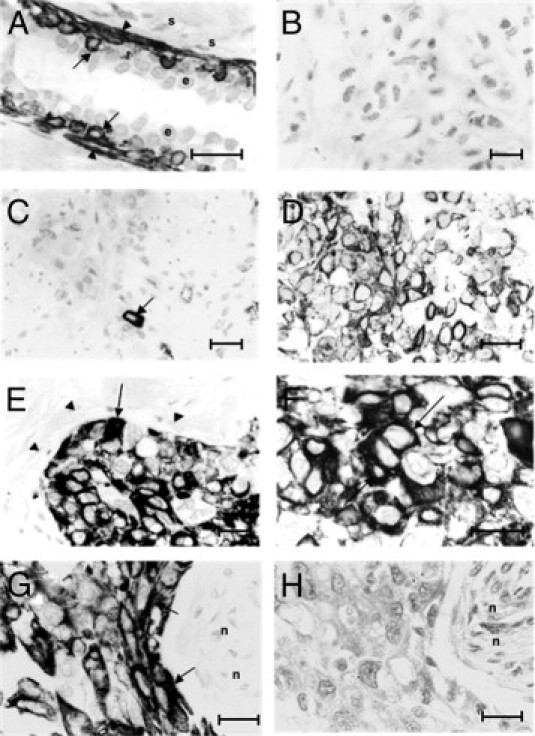
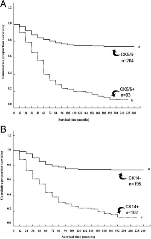
Two subgroups of invasive breast carcinomas have been identified with a poor prognosis in different patient cohorts: the basal-like category and the subgroup containing proteins capable of inducing metastasis in experimental rodents, the metastasis-inducing proteins (MIPs). Here we identify by immunohistochemical staining for cytokeratin CK5/6 or CK14 the basal-like subgroup in a set of 297 primary invasive breast carcinomas in which the staining profile for the MIPs S100A4, osteopontin, anterior gradient-2, and S100P has already been established. Monoclonal antibodies to CK5/6 or CK14 specifically stain 31% to 34% of the primary carcinomas. These positively stained tumors are highly significantly associated with premature death of the patient (Wilcoxon statistics, P < 0.0001), the increased relative risk being approximately 5.6-fold. Positive staining for either cytokeratin is very significantly associated with that for each of the four MIPs separately and with loss of staining for the Fanconi anemia protein FANCD2 (corrected Fisher's exact test, P < 0.0007). There is no significant correlation with the remaining tumor variables tested, including staining for the estrogen receptor α, progesterone receptor, and c-erbB-2. These results show that the basal cytokeratin-like carcinomas contain many of the MIPs and that these may arise by their selection for tumors with an inherent deficiency in the FANC/BRCA pathway of DNA repair.
Copyright © 2011 American Society for Investigative Pathology. Published by Elsevier Inc. All rights reserved.
Figures
References
-
- Wellings S.R., Jensen H.M., Marcum R.G. An atlas of subgross pathology of the human breast with special reference to possible precancerous lesions. J Natl Cancer Inst. 1975;55:231–273. - PubMed
-
- Page D.L., Anderson T.J. Churchill Livingstone; Edinburgh: 1988. Diagnostic Histopathology of the Breast.
-
- Sorlie T., Tibshirani R., Parker J., Hastie T., Marron J.S., Nobel A., Deng S., Johnsen H., Pesich R., Geisler S., Demeter J., Perou C.M., Lønning P.E., Brown P.O., Børresen-Dale A.L., Botstein D. Repeated observation of breast tumor subtypes in independent gene expression data sets. Proc Natl Acad Sci USA. 2003;100:8418–8423. - PMC - PubMed
-
- Moll R., Franke W.W., Schiller D.L., Geiger B., Krepler R. The catalog of human cytokeratins: patterns of expression in normal epithelia, tumors and cultured cells. Cell. 1982;31:11–24. - PubMed
-
- Dairkee S.H., Mayall B.H., Smith H.S., Hackett A.J. Monoclonal marker that predicts early recurrence of breast cancer. Lancet. 1987;1(8531):514. - PubMed
Publication types
MeSH terms
Substances
Grants and funding
LinkOut - more resources
Full Text Sources
Other Literature Sources
Medical
Research Materials
Miscellaneous
