. 2011 Aug 16;21(2):301-14.
doi: 10.1016/j.devcel.2011.06.033. Epub 2011 Jul 28.

Semaphorin-PlexinD1 signaling limits angiogenic potential via the VEGF decoy receptor sFlt1

Affiliations

Semaphorin-PlexinD1 signaling limits angiogenic potential via the VEGF decoy receptor sFlt1

Tomasz Zygmunt et al. Dev Cell. .

Abstract

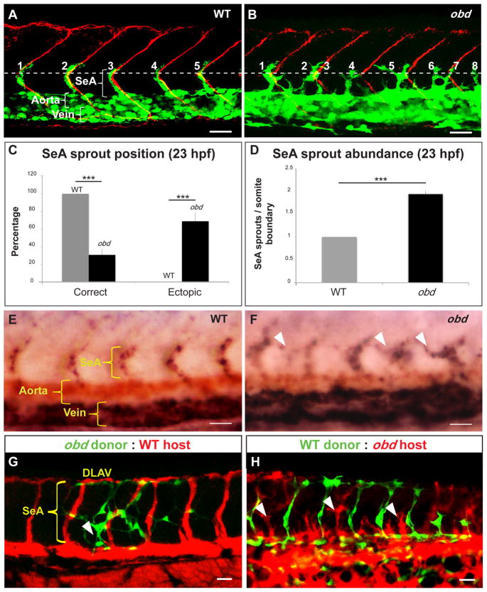
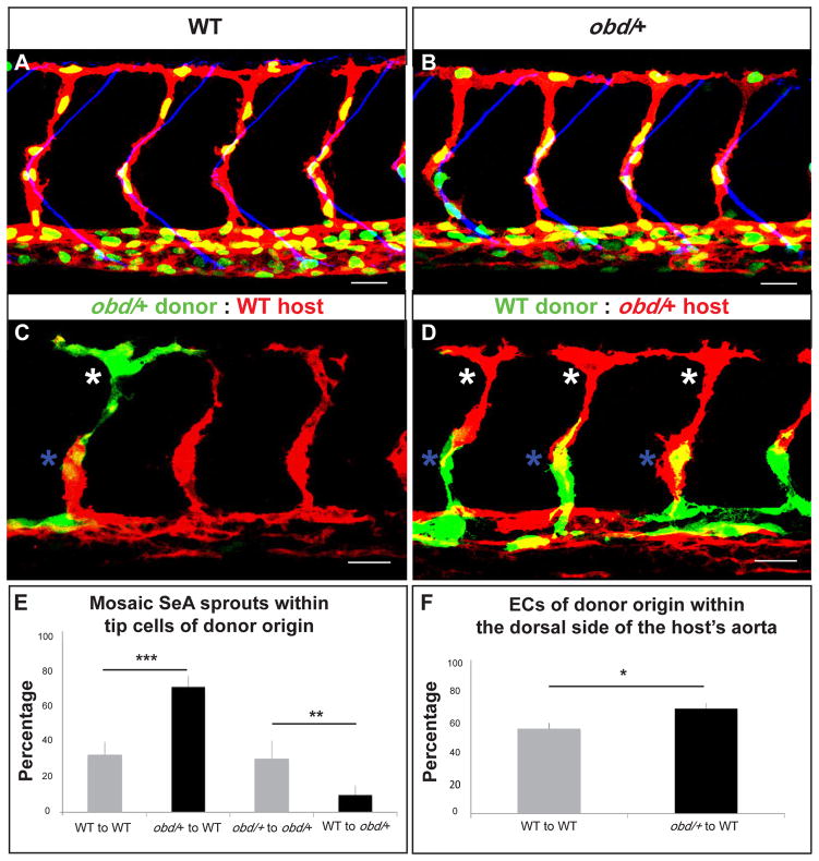
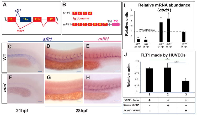
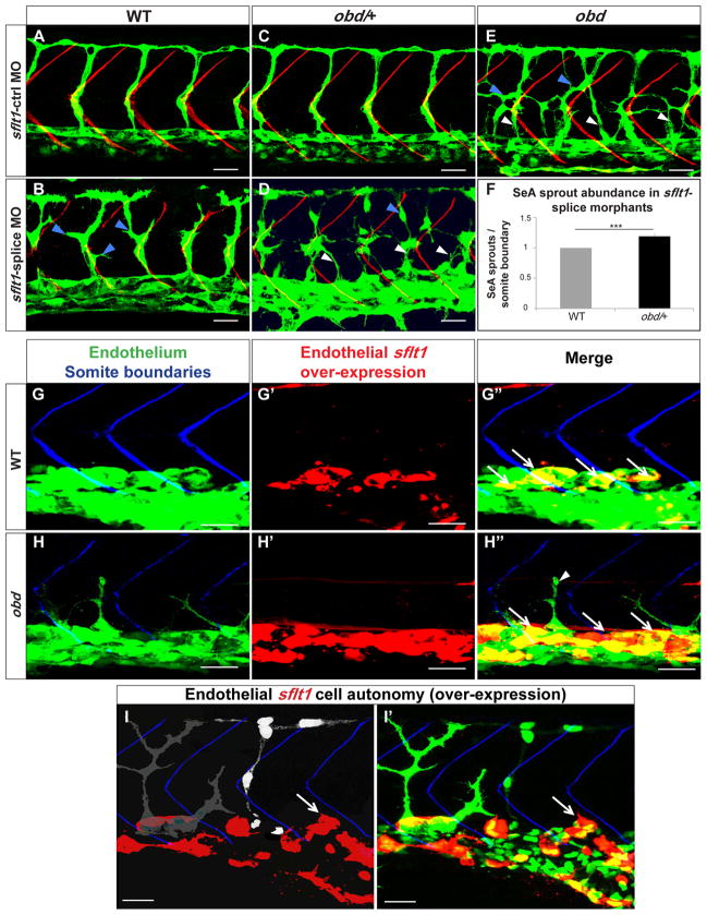
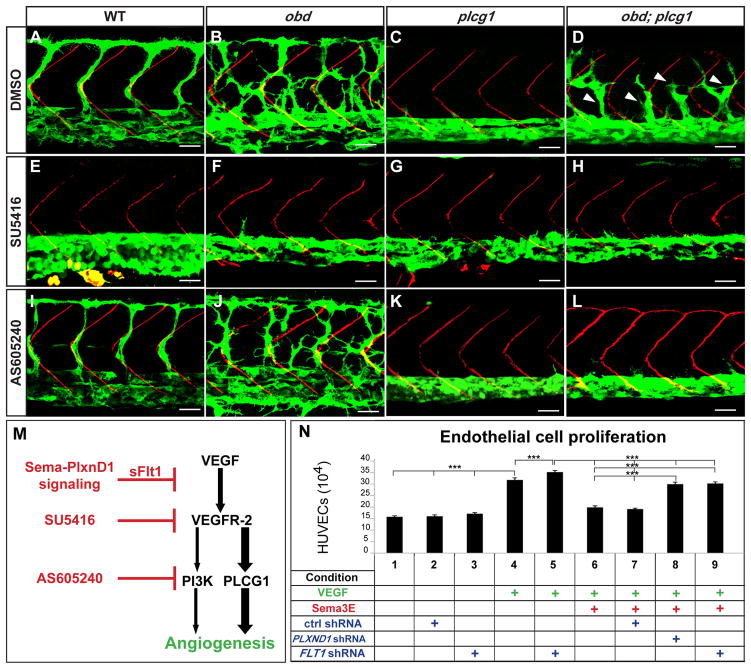
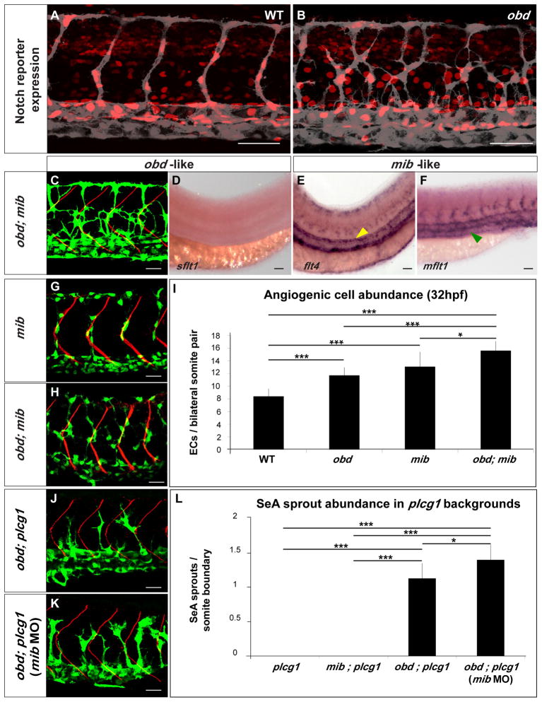
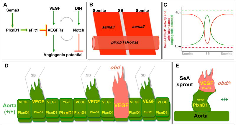
Sprouting angiogenesis expands the embryonic vasculature enabling survival and homeostasis. Yet how the angiogenic capacity to form sprouts is allocated among endothelial cells (ECs) to guarantee the reproducible anatomy of stereotypical vascular beds remains unclear. Here we show that Sema-PlxnD1 signaling, previously implicated in sprout guidance, represses angiogenic potential to ensure the proper abundance and stereotypical distribution of the trunk's segmental arteries (SeAs). We find that Sema-PlxnD1 signaling exerts this effect by antagonizing the proangiogenic activity of vascular endothelial growth factor (VEGF). Specifically, Sema-PlxnD1 signaling ensures the proper endothelial abundance of soluble flt1 (sflt1), an alternatively spliced form of the VEGF receptor Flt1 encoding a potent secreted decoy. Hence, Sema-PlxnD1 signaling regulates distinct but related aspects of angiogenesis: the spatial allocation of angiogenic capacity within a primary vessel and sprout guidance.


Conflict of interest statement

The authors declare no competing financial interests.

Figures

Figure 1
Figure 1. Sema-PlxnD1 signaling is cell autonomously required within the endothelium for proper SeA sprout abundance and distribution
(A–B) 23 hpf vasculatures, green. SBs, red. Horizontal myoseptum (HM), white dotted line. SeA sprouts, numbered. (A) WT. (B) obd. SeA sprout position (C) and abundance (D) in 23 hpf WT and obd. (E–F) WISH, 28 hpf trunks. Ectopic SeA sprouts, white arrowheads. Riboprobes: flt4 (blue), cdh5 (red). WT (E). obd (F). (G–H) 32 hpf chimeric vasculatures with ECs of donor (green) and host (red) origin. Examples of ectopic SeA sprouts, white arrowheads. (C–D) n = 8 WT, 12 obd. Error bars, s.e.m. ***p < 0.001. (E–F) n = 10/10 WT, 10/10 obd. (A–B, E–H) Anterior, left; dorsal, up. Scale bars, 30 μm. See Figure S1.
Figure 2
Figure 2. ECs with less Sema-PlxnD1 signaling tend to become tip cells and occupy the aorta’s dorsal side
(A–B) 32 hpf vasculatures. EC nuclei (green), membranes (red). SBs, blue. (A) WT. (B) obd/+. (C–D) 28 hpf vasculatures with ECs of donor (green) and host (red) origin. Asterisks: Tip cells (white), stalk cells (blue). (E) Percentage of mosaic SeAs with tip cells of donor origin in homogenotypic (grey bars) and heterogenotypic (black bars) chimeras. (F) Percentage of ECs of donor origin found within the dorsal side of the host’s arterial tree in homogenotypic (grey bar) and heterogenotypic (black bar) chimeras. (E–F) *p < 0.05, **p < 0.01, ***p < 0.001. Error bars, s.e.m. (E) n = 27 WT to obd/+, n = 18 obd/+ to obd/+, n = 38 obd/+ to WT, n = 34 WT to WT. Error bars, s.e.m. (F) n = 24 WT to WT, n = 32 obd/+ to WT. (A–D) Anterior, left; dorsal, up. Scale bars, 30 μm. See Figure S2 and Movie S1.
Figure 3
Figure 3. Sema-PlxnD1 signaling ensures the proper endothelial abundance of sflt1
(A) Alternative flt1 splicing yields sflt1 and mflt1 isoforms with unique eleventh exons. Exons, colored boxes. Introns, black lines. (B) sflt1 encodes a soluble 474 aa protein. mflt1 encodes a 1,273 aa transmembrane protein. Protein domains: Immunoglobulin (Ig, red numbered boxes), transmembrane (TM, grey box), tyrosine kinase (TK, pink box). (C–H) WISH, embryo trunks (genotypes and ages indicated) hybridized with sflt1 (C–D, F–G) and mflt1 (E, H) riboprobes (blue). (I) qPCR measurements. Relative mRNA abundance of sflt1, mflt1 and YFP (from Tg(flt1:YFP)hu4624/+) in 28 hpf obd/+ (WT level = 1, dashed line). Error bars, coefficient of variance *p < 0.05. (J) ELISA-based quantification of FLT1 prepared from cell extracts of HUVECs treated with both VEGF and Sema3E and the control or PLXND1-targeting shRNAs. Error bars, s.e.m. ***p < 0.001. (C–H) n = 10 embryos per riboprobe, stage and genotype. Pictures of representative examples of stainings observed (10/10 embryos in each category). Anterior, left; dorsal, up. Scale bars, 50 μm. See Figure S3.
Figure 4
Figure 4. plxnD1 and sflt1 interact genetically, sflt1 limits SeA angiogenesis cell autonomously
(A–I) 32 hpf trunk vasculatures, green. (A–E) SBs, red. White arrowheads, ectopic SeA sprouts. Blue arrowheads, ectopic SeA branching. (A, C, E) Embryos treated with 20 ng of sflt1-ctrl MO: WT (A), obd/+ (C), obd (E). Embryos treated with 20 ng of sflt1-splice MO: WT (B), obd/+ (D). (F) 23 hpf SeA sprout abundance in WT (left, grey bar) and obd/+ (right, black bar) sflt1-splice morphants. n = 20 WT and n = 19 obd/+. Error bars, s.e.m. ***p < 0.001. (G–I′) SBs, blue. GAL4FF/UAS-mediated endothelial-specific sflt1 over-expression, red. White arrows, missing SeA sprouts. (G–H″) Endothelial sflt1 over-expression inhibits SeA sprouting. WT (G-G″). obd (H-H″), note lack of sflt1 over-expression (red) in remaining SeA sprout (white arrowhead). (I-I′) Mosaic vasculature with ECs from both obd donor and WT host. Endothelial-specific and mosaic sflt1 and DsRed co-expression restricted to the WT endothelium (red, I-I′). obd ECs express cytosolically-targeted EGFP (grey in I; green in I′). WT ECs express nuclear-targeted EGFP (white in I; green in I′). obd and WT ECs without sflt1 over-expression (DsRed-) from SeA sprouts even next to sflt1 over-expressing WT ECs (DsRed+). WT ECs over-expressing sflt1 (DsRed+) fail to form SeA sprouts (white arrows, I-I′). (G-H″) n = 30 embryos with over-expression per genotype, all showing suppression of SeA sprouting. Anterior, left; dorsal, up. Scale bars, 30 μm. See Figure S4.
Figure 5
Figure 5. Enhanced VEGF signaling causes obd’s exacerbated SeA angiogenesis
A–L, 32 hpf trunk vasculatures. WT, obd, plcg1 and obd; plcg1 treated with DMSO, SU5416 (VEGFR inhibitor) or AS605240 (PI3K inhibitor). Genotypes, top; treatments, left. Endothelium, green. SBs, red. White arrowheads, recovered SeA sprouts in obd; plcg1. Anterior, left; dorsal, up. Scale bars, 30 μm. n = 18 embryos per genotype and treatment. Pictures show representative phenotypes (18/18 embryos per category). (M) Diagram of the VEGF cascade and steps inhibited by sflt1 and drugs used in (E–L). (N) HUVEC proliferation in response to combinations of VEGF, Sema3E and shRNAs (control, PLXND1 and FLT1). ***p < 0.001. Error bars, s.e.m. See Figure S5.
Figure 6
Figure 6. Notch signaling loss does not phenocopy obd
(A–B) Expression of Notch’s activity nuclear reporter Tg(Tp1bglob:hmgb1-mCherry)jh11 (red) in the endothelium (grey) of WT (A) and obd (B). (C–F) obd; mib. (C) Endothelium, green. SBs, red. (D–F) WISH with sflt1, flt4 and mflt1 riboprobes, as indicated. Double mutant phenotypes classed as obd-like (C–D) or mib-like (E–F) based on the mutant they resemble most. Note lack of sflt (as in Fig. 3G) and ectopic aortic flt4 (yellow arrowhead; as in Fig. S6A) and venous mflt1 stainings (green arrowhead, as in Fig. S6B). (G–I) Angiogenic cell abundance within the trunk’s arterial tree of WT, obd, mib (G) and obd; mib (H) in Tg(fli1:nEGFP)y7 embryos. (G–H) EC nuclei, green. SBs, red. (I) Quantification; n= 10 per genotype. (J–L) SeA sprout abundance in plcg, mib; plcg, obd; plcg (J) and obd; plcg embryos injected with 10 ng of mib MO (mib MO) (K). (J–K) Endothelium, green. SBs, red. (L) n = 8, 7, 11 and 9 for plcg, mib; plcg, obd; plcg and obd; plcg (mib MO), respectively. Scale bars: 50 μm (A–B, D–F), 30 μm (C, G–H, J–K). (I, L) *p <0.05, ***p < 0.001. Error bars: s.e.m. (A–F, G–H, J–K) Anterior, left; dorsal, up. Trunk images and quantifications: 32 hpf (A–C, G–L), 28 hpf (D–F). See Figure S6.
Figure 7
Figure 7. Model for how Sema3-PlxnD1 signaling restricts angiogenic potential along the aorta and limits angiogenic responses within SeA sprouts
(A) Sema3-PlxnD1 signaling inhibits VEGF’s pro-angiogenic effects via sFlt1, limiting angiogenic potential. The complex cross-regulation (grey lines) between the VEGF and Notch cascades implies Sema-PlxnD1 signaling impacts Notch activity indirectly. (B) Somitic sema3s (dark red) and endothelial plxnD1 (light red) expression precedes SeA sprouting (SB, grey) (Roos et al., 1999; Torres-Vazquez et al., 2004; Yee et al., 1999). (C) WT aortic Sema-PlxnD1 signaling levels (red solid line) are highest in ECs next to the somites and lowest in ECs next to SBs, where angiogenic potential (green solid line) is highest. obd lacks Sema-PlxnD1 activity and thus sflt1 abundance is greatly reduced (red dotted line), leading to uniformly enhanced angiogenic potential levels (green dotted line) that yield too many and ectopic SeA sprouts. (D–E) VEGF signaling and angiogenic responses are cell autonomously enhanced by loss (obd) or decreased (obd/+) endothelial plxnD1 activity, as exemplified by obd to WT (D) and obd/+ to WT (E) chimeras. VEGF signaling and PlxnD1 activity levels are indicated by font size.

Comment in

References

    1. Acevedo LM, Cheresh DA. Suppressing NFAT increases VEGF signaling in hemangiomas. Cancer Cell. 2008;14:429–430. - PubMed
    1. Ahn DG, Ruvinsky I, Oates AC, Silver LM, Ho RK. tbx20, a new vertebrate T-box gene expressed in the cranial motor neurons and developing cardiovascular structures in zebrafish. Mech Dev. 2000;95:253–258. - PubMed
    1. Ambati BK, Nozaki M, Singh N, Takeda A, Jani PD, Suthar T, Albuquerque RJ, Richter E, Sakurai E, Newcomb MT, et al. Corneal avascularity is due to soluble VEGF receptor-1. Nature. 2006;443:993–997. - PMC - PubMed
    1. Aref S, El Sherbiny M, Goda T, Fouda M, Al Askalany H, Abdalla D. Soluble VEGF/sFLt1 ratio is an independent predictor of AML patient out come. Hematology. 2005;10:131–134. - PubMed
    1. Banu N, Teichman J, Dunlap-Brown M, Villegas G, Tufro A. Semaphorin 3C regulates endothelial cell function by increasing integrin activity. FASEB J. 2006;20:2150–2152. - PubMed

Publication types

MeSH terms

Associated data