. 2011 Aug 5;9(2):113-8.
doi: 10.1016/j.stem.2011.07.002. Epub 2011 Jul 28.

Direct reprogramming of adult human fibroblasts to functional neurons under defined conditions

Affiliations

Direct reprogramming of adult human fibroblasts to functional neurons under defined conditions

Rajesh Ambasudhan et al. Cell Stem Cell. .

Abstract

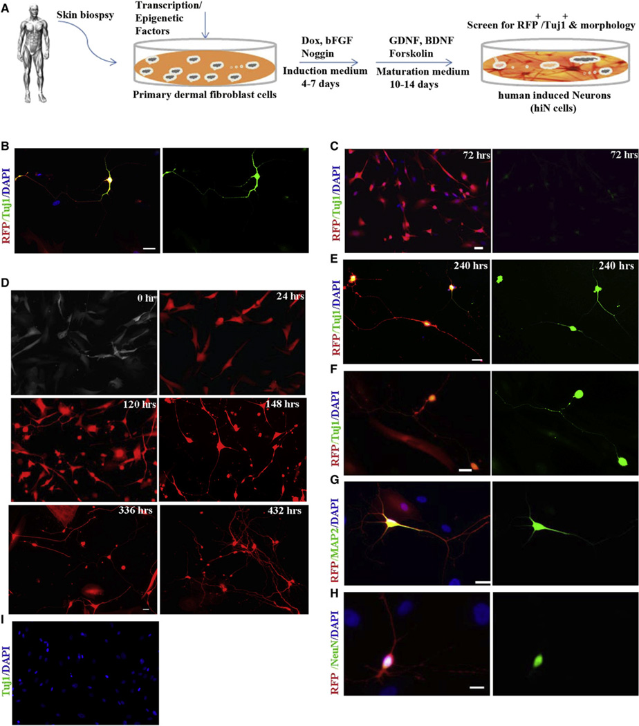
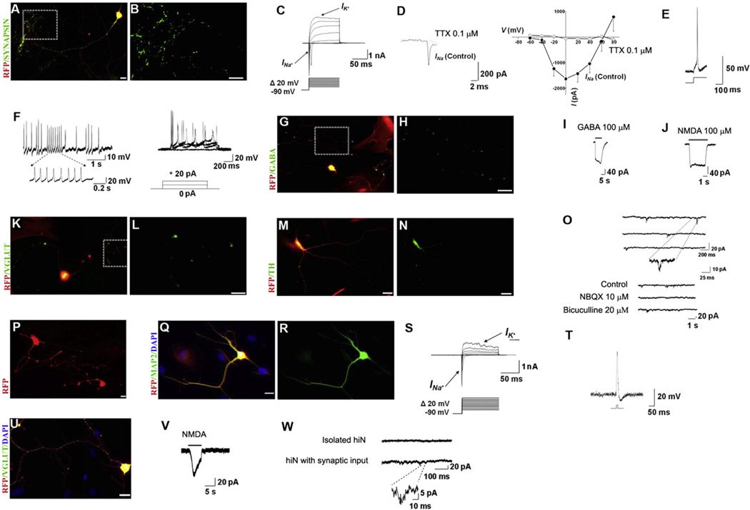
Human induced pluripotent stem cells (hiPSCs) have been generated by reprogramming a number of different somatic cell types using a variety of approaches. In addition, direct reprogramming of mature cells from one lineage to another has emerged recently as an alternative strategy for generating cell types of interest. Here we show that a combination of a microRNA (miR-124) and two transcription factors (MYT1L and BRN2) is sufficient to directly reprogram postnatal and adult human primary dermal fibroblasts (mesoderm) to functional neurons (ectoderm) under precisely defined conditions. These human induced neurons (hiNs) exhibit typical neuronal morphology and marker gene expression, fire action potentials, and produce functional synapses between each other. Our findings have major implications for cell-replacement strategies in neurodegenerative diseases, disease modeling, and neural developmental studies.


Figures

Figure 1
Figure 1. Conversion of Postnatal Human Dermal Fibroblasts to hiN Cells using Defined Factors under Defined Conditions
(A) Schematic showing the experimental protocol. (B) Tuj1-stained hiN cells 18 days after infection of BJ cells with the 12F pool. (C) Within 3 days of infection, 3F (miBM)-transduced fibroblasts exhibited notable morphological changes and weak immunoreactivity with Tuj1 antibody. (D) Time-lapse live images of RFP+ BJ cells infected with miBM showed gradual changes leading to neuronal-like morphology. (E) 3F-infected BJ cells were Tuj1+ and exhibited characteristic neuronal morphology when stained 240 hr (10 days) after infection. (F–H) By day 18, hiN cells expressed, in addition to Tuj1 (F), mature neuronal markers, including MAP2 (G) and NeuN (H). (I) Control cultures infected with BM transgenes along with nonspecific scrambled RNA did not generate hiN cells or show immunoreactivity to Tuj1 antibody. Red: RFP; green: Tuj1 (B, C, E, F, and I), MAP2 (G), or NeuN (H); blue: DAPI. Scale: 20 µm. See also Figure S1 and Table S1.
Figure 2
Figure 2. hiN Cells from Postnatal and Adult Human Fibroblasts Show Functional Maturation and Synaptic Properties
(A and B) hiN cells, assessed 25 days postinfection, stained positively for synapsin-1. (C) Representative traces of whole-cell currents recorded in voltage-clamp mode. Cells were hyperpolarized to −90 mV for 300 ms before depolarizing pulses were applied to elicit Na+ and K+ currents. (D) The inward currents could be blocked by Na+ channel blocker tetrodotoxin (TTX). CsCl was present in the patch electrode-filling solution to suppress K+ currents. Representative current traces recorded in the presence of TTX are shown at left, and the current-voltage (I/V) relationship, at right (mean + SEM, n = 13). (E and F) Traces of evoked (E) and spontaneous (F, left panel) action potentials recorded in current-clamp mode on day 25. In other hiN cells, repetitive trains of evoked action potentials were observed after transgene inactivation (F, right panel). (G and H) hiN cells expressing GABA. (I) GABA-evoked current from hiN cell at a holding potential of −80 mV. (J–L) Other hiN cells responded to application of exogenous NMDA in the nominal absence of extracellular Mg2+ (J) and expressed glutamate transporter VGLUT (K and L). (M and N) hiN cell staining for tyrosine hydroxylase (TH) on day 25. (O) Top panel: Representative traces of mEPSCs recorded at a holding potential of −80 mV in hiN cells on day 30 in culture, indicating functional synapse formation. The insert demonstrates the rapid kinetics of these synaptic currents. Bottom panel: Spontaneous synaptic currents were reversibly inhibited by addition of 10 µM NBQX (to block excitatory AMPA-type glutamate receptors), but not by 20 µM bicuculline (to block inhibitory GABA receptors). (P) Representative image of live aHDF-converted hiN cells on day 18 postinfection exhibiting typical neuronal morphology and RFP fluorescence. (Q and R) aHDF-converted hiN cells displayed mature neuronal marker MAP2 when fixed and immunostained 18 days after 3F (miBM) infection. (S) Representative traces of whole-cell currents in voltage-clamp mode from aHDF-converted hiN cells 25 days postinfection. Cells were hyperpolarized to −90 mV for 300 ms before depolarizing pulses were applied to elicit Na+ and K+ currents. (T) Representative action potential recorded in current-clamp mode. (U) Day 25 aHDF-converted hiN cell displaying immunoreactivity for VGLUT antibody. (V) Patch-clamp recording showing response of day 25 aHDF-converted hiN cell to application of exogenous NMDA in the nominal absence of Mg2+ in the bath solution. (W) Spontaneous synaptic currents from an aHDF hiN cell plated at high density (lower trace), reflecting mEPSCs given the composition of the intracellular and bath solutions used in the recording (see Experimental Procedures). The magnified trace shows the rapid kinetics of an mEPSC. When plated at lower density to isolate the cells, aHDF hiNs were synaptically silent (upper trace). Red: RFP; green: synapsin (B), GABA (H), VGLUT (L), TH (N), MAP2 (R), and VGLUT (U); blue: DAPI stained nuclei (Q and U). Boxed areas in the left-hand panels (A, G, and K) are shown at higher magnification in the adjacent right-hand panels (B, H, and L). Large red cells in (G), (K), (M), and (Q) are infected fibroblasts expressing RFP that have not undergone conversion to hiN cells. (A), (G), (K), (M), (Q), and (U) are merged images. Scale bars: 20 µm. See also Figure S2 and Table S2.

Comment in

References

    1. Caiazzo M, Dell’anno MT, Dvoretskova E, Lazarevic D, Taverna S, Leo D, Sotnikova TD, Menegon A, Roncaglia P, Colciago G, et al. Direct generation of functional dopaminergic neurons from mouse and human fibroblasts. Nature. 2011 - PubMed
    1. Campbell KH, McWhir J, Ritchie WA, Wilmut I. Sheep cloned by nuclear transfer from a cultured cell line. Nature. 1996;380:64–66. - PubMed
    1. Cao X, Pfaff SL, Gage FH. A functional study of miR-124 in the developing neural tube. Genes Dev. 2007;21:531–536. - PMC - PubMed
    1. Cheng LC, Pastrana E, Tavazoie M, Doetsch F. miR-124 regulates adult neurogenesis in the subventricular zone stem cell niche. Nat. Neurosci. 2009;12:399–408. - PMC - PubMed
    1. Cobaleda C, Jochum W, Busslinger M. Conversion of mature B cells into T cells by dedifferentiation to uncommitted progenitors. Nature. 2007;449:473–477. - PubMed

Publication types