. 2011 Dec;8(12):1837-46.
doi: 10.1016/j.hrthm.2011.07.032. Epub 2011 Jul 28.

Robotic catheter ablation of left ventricular tachycardia: initial experience

Affiliations

Robotic catheter ablation of left ventricular tachycardia: initial experience

Miguel Valderrábano et al. Heart Rhythm. 2011 Dec.

Abstract

Background: Catheter ablation of ventricular tachycardia (VT) can be technically challenging due to difficulty with catheter positioning in the left ventricle (LV) and achieving stable contact. The Hansen Sensei Robotic system (HRS) has been used in atrial fibrillation but its utility in VT is unclear.

Objective: The purpose of this study was to test the technical feasibility of robotic catheter ablation of LV ventricular tachycardia (VT) using the HRS.

Methods: Twenty-three patients underwent LV VT mapping and ablation with the HRS via a transseptal, transmitral valve approach. Nineteen patients underwent substrate mapping and ablation (18 had ischemic cardiomyopathy, 1 had an apical variant of hypertrophic cardiomyopathy). Four patients had focal VT requiring LV VT mapping and ablation. Procedural endpoints included substrate modification by endocardial scar border ablation and elimination of late potentials, or elimination of inducible focal VT.

Results: Mapping and ablation were entirely robotic without requiring manual catheter manipulation in all patients and reaching all LV regions with stable contact. Fluoroscopy time of the LV procedure was 22.2 ± 11.2 minutes. Radiofrequency time was 33 ± 21 minutes. Total procedural times were 231 ± 76 minutes. Complications included a left groin hematoma (opposite to the HRS sheath), 1 pericardial effusion without tamponade that was drained successfully, and transient right ventricular failure in a patient with previous left ventricular assist device. At 13.4 ± 6.7 months of follow-up (range 1-19 months), recurrence of VT occurred in 3 of 23 patients.

Conclusion: Our initial experience suggests that the HRS allows successful mapping and ablation of LV VT.


Figures

Figure 1
Figure 1
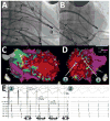
Mapping and ablation of anteroapical scar-related VT. A and B show right anterior oblique and left anterior oblique fluoroscopic views of the robotic catheter system reaching the LV apex (in particular, the lateral aspect of the apex) where the LV VT substrate was found. An epicardial sheath is also present. C and D show corresponding 3D maps of bipolar endocardial voltage amplitudes, demonstrating a large scar. Sites 1 and 2 correspond to the exit site of the VT circuit and its middiastolic location, respectively. E on the left shows pacing from site 1, with concealed entrainment and post-pacing interval (PPI) identical to the VT cycle length (TCL), and stimulus-to-QRS delay identical to signal-to-QRS (S-QRS). E, on the right, shows a middiastolic potential at site 2 during VT.
Figure 2
Figure 2
Mapping and ablation of inferior scar-related VT. A and B show the robotic catheter position in left and right anterior oblique fluoroscopic views, respectively. The septal aspect of the mid-inferior wall is being mapped. C, shows the 3D map of the inferior scar, with sites 1 and 2 at the border zone and in the center of the scar, respectively. D shows, on the left, the 12 lead surface electrogram of the VT. On D, mid panel, pacing from the edge of the scar (site 1) shows near-identical pace-map (numbers refer to percent QRS matching) with short stimulus-to-QRS delay (86 ms) that matched the delay from signal-to-QRS during VT at that site (right panel). These data were consistent with site 1 being the exit site of the scar in the VT circuit. E shows pacing from the center of the scar (site 2), with near-identical pace-maps, but long (162 ms) stimulus-to-QRS delay, that matched the delay from signal-to-QRS during VT at that site (mid panel). During paced rhythm, a post-systolic signal was present (right panel).
Figure 3
Figure 3
Mapping and ablation of a septal scar. Stable catheter positioning allowed real-time monitoring of lesion creation on ICE. A and B show the robotic catheter position in left and right anterior oblique fluoroscopic views, respectively. The septal aspect of the apex was being mapped. C and D show ICE images before and 15 seconds after application of radiofrequency. The catheter tip is in the distal septum, where increased echogenicity is seen at the ablation site. E and F show 3D maps of the large septal and apical scar and the ablation lesions, respectively.
Figure 4
Figure 4
Mapping and ablation of a basal lateral scar. A and B show the robotic catheter position in left and right anterior oblique fluoroscopic views, respectively. The mid lateral wall is being mapped. C shows an ICE image showing catheter contact and lesion formation in the LV lateral wall. D shows the 3D map of the lateral scar and the ablation lesions created.
Figure 5
Figure 5
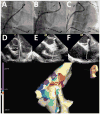
Mapping and ablation of an inferior wall scar in the presence of an LVAD. A, B, and C show the robotic catheter position in the right (A) and left (B and C) anterior oblique fluoroscopic views, with the catheter tip around the LVAD inflow cannula. D shows an ICE image of the LVAD cannula and the ablation catheter. E shows the 3D scar maps and ablation lesions.
Figure 6
Figure 6
Mapping and ablation of an apical scar in the setting of end-stage apical hypertrophy. A, B and C show the robotic catheter position in the right anterior oblique fluoroscopic view, with the catheter tip manipulated around the apical aneurysm. C shows the robotic sheath retracted to allow additional manual catheter manipulation. D through G show ICE images of the apical aneurysm, the catheter tip in different positions, and real-time monitoring of an ablation lesion just proximal to the apical aneurysm. H and I show the 3D scar maps and ablation lesions.
Figure 7
Figure 7
Mapping and ablation of subaortic PVCs. A shows the robotic catheter at the earliest mapped site in the right ventricular outflow tract in a right anterior oblique projection. B and C show the robotic catheter at the earliest mapped site of the LVOT in right and left anterior oblique fluoroscopic views, respectively. D, E, and F show ICE images with the robotic catheter in the right (D) and left (E and F) outflow tract. E and F show frames in diastole (E) and systole (F), to highlight the subaortic location of the catheter tip. The right and left earliest sites were identical in timing, and ablation was delivered from both sites. G shows the 3-dimensional map and the ablation lesions.

References

    1. Reddy VY, Reynolds MR, Neuzil P, et al. Prophylactic catheter ablation for the prevention of defibrillator therapy. N Engl J Med. 2007;357:2657–2665. - PMC - PubMed
    1. Stevenson WG, Wilber DJ, Natale A, et al. Irrigated radiofrequency catheter ablation guided by electroanatomic mapping for recurrent ventricular tachycardia after myocardial infarction: the multicenter thermocool ventricular tachycardia ablation trial. Circulation. 2008;118:2773–2782. - PubMed
    1. Kuck KH, Schaumann A, Eckardt L, et al. Catheter ablation of stable ventricular tachycardia before defibrillator implantation in patients with coronary heart disease (VTACH): a multicentre randomised controlled trial. Lancet. 2010;375:31–40. - PubMed
    1. Aliot EM, Stevenson WG, Almendral-Garrote JM, et al. EHRA/HRS Expert Consensus on Catheter Ablation of Ventricular Arrhythmias: developed in a partnership with the European Heart Rhythm Association (EHRA), a Registered Branch of the European Society of Cardiology (ESC), and the Heart Rhythm Society (HRS); in collaboration with the American College of Cardiology (ACC) and the American Heart Association (AHA) Heart Rhythm. 2009;6:886–933. - PubMed
    1. Wazni OM, Barrett C, Martin DO, et al. Experience with the hansen robotic system for atrial fibrillation ablation--lessons learned and techniques modified: Hansen in the real world. J Cardiovasc Electrophysiol. 2009;20:1193–1196. - PubMed

Publication types