Review
. 2011 Oct;95(2):104-32.
doi: 10.1016/j.pneurobio.2011.07.003. Epub 2011 Jul 23.

GABAergic synchronization in the limbic system and its role in the generation of epileptiform activity

Affiliations
Review

GABAergic synchronization in the limbic system and its role in the generation of epileptiform activity

Massimo Avoli et al. Prog Neurobiol. 2011 Oct.

Abstract

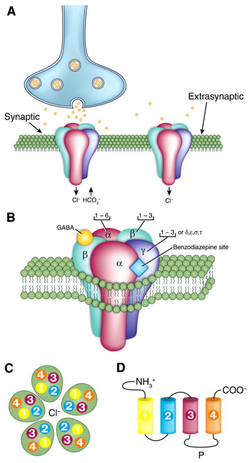
GABA is the main inhibitory neurotransmitter in the adult forebrain, where it activates ionotropic type A and metabotropic type B receptors. Early studies have shown that GABA(A) receptor-mediated inhibition controls neuronal excitability and thus the occurrence of seizures. However, more complex, and at times unexpected, mechanisms of GABAergic signaling have been identified during epileptiform discharges over the last few years. Here, we will review experimental data that point at the paradoxical role played by GABA(A) receptor-mediated mechanisms in synchronizing neuronal networks, and in particular those of limbic structures such as the hippocampus, the entorhinal and perirhinal cortices, or the amygdala. After having summarized the fundamental characteristics of GABA(A) receptor-mediated mechanisms, we will analyze their role in the generation of network oscillations and their contribution to epileptiform synchronization. Whether and how GABA(A) receptors influence the interaction between limbic networks leading to ictogenesis will be also reviewed. Finally, we will consider the role of altered inhibition in the human epileptic brain along with the ability of GABA(A) receptor-mediated conductances to generate synchronous depolarizing events that may lead to ictogenesis in human epileptic disorders as well.


Figures

Fig. 1
Fig. 1. Molecular structure of the ionotropic GABAA receptor
(A) Localization of GABAA receptors in the postsynaptic neuron membrane. Both a synaptic and an extra- or peri-synaptic receptor are shown. (B) Structure of the GABAA receptor and its subunit composition; the heteropentameric, Cl-permeable channel is made of five subunits that come from seven subunit subfamilies (α, β, γ, δ, ε, θ and π). Note that receptors composed of α (1–3) subunits together with β and γ subunits are presumably synaptically localized, whereas those containing α5, β an γ receptors are located at extrasynaptic sites. Both types of GABAA modulated by benzodiazepine, while extrasynaptically localized receptors composed of α (4 or 6), β and δ are benzodiazepine insensitive. Note also that binding of GABA occurs at the interface between α and β subunits while the benzodiazepine binding occurs at the interface between α (1, 2, 3 or 5) and γ subunits. (C) Top view of the GABAA receptor. Note that each subunit comprises four hydrophobic transmembrane domains (TM1–TM4) with TM2 providing the lining of the Cl pore. (D) Unfolded view of the transmembrane domains (TM1–TM4). Note that: (i) the extracellular amino terminus is the site of GABA binding, and also contains binding sites for psychoactive drugs, such as benzodiazepines; (ii) the large intracellular loop between TM3 and TM4 (P) is the site for various protein interactions as well as for various post-translational modifications that modulate receptor activity. Panels in this figure were drawn according to information obtained from Bormann (2000) and Jacob et al. (2008).
Fig. 2
Fig. 2. Unitary inhibitory field potentials recorded from the rat hippocampal CA3 area
(A) Simultaneous intracellular (intra) and extracellular (extra) recordings obtained from the CA3 area in a brain slice that was superfused with ionotropic glutamatergic receptor antagonists; the extracellular electrode was at ~100 μm from the pyramidal cell. Note that many spontaneously occurring intracellular and extracellular events are correlated as well as that their shapes closely resemble the signals recorded following a weak stimulus (triangle) delivered in the stratum pyramidale at 200 μm from the recorded pyramidal cell. (B) Reconstruction of the dendritic and axonal arbour of a biocytin-filled presumptive interneuron. Axon terminals were largely confined to the CA3 stratum pyramidale (st. pyr.) with some in stratum oriens (st. o.), and were distributed over about 1 mm along CA3 st. pyr. with their density falling with distance. Extracellular recordings shown in C were obtained from sites E1-3. (C) Single action potentials generated by a presumptive interneuron evoke field IPSPs in two of three extracellular recording sites. Thirty responses recorded from electrodes positioned at E1–E3 are shown below. Note that intracellular action potentials elicited extracellular field potentials at two of three recording sites in stratum pyramidale while no extracellular event was detected by a third electrode located at a site which was not innervated by this cell. Modified from Bazelot et al. (2010) with permission.
Fig. 3
Fig. 3. Cortical post-synaptic GABA receptor-mediated potentials
(A) Intracellular potentials recorded from a regularly firing neuron in the rodent perirhinal cortex in response to single-shock stimuli delivered in the amygdala in an in vitro slice preparation. Responses were recorded during application of control medium with a K-acetate-filled microelectrode at resting membrane potential (−70 mV), and at depolarizing and hyperpolarizing levels set by intracellular injection of steady current. (B) Intracellular responses generated by a neocortical pyramidal cell in an in vitro slice preparation following single-shock stimuli delivered in medium containing glutamatergic receptor antagonists; these responses were recorded with a K-acetate-filled microelectrode at resting membrane potential (−75 mV) and at depolarized and hyperpolarized potentials. Note in A and B that the hyperpolarizing response comprises an early (GABA type A) and a late (GABA type B) component. (C) Intracellular response recorded from a hippocampal neuron following a train of 40 stimuli delivered at 100 Hz; this recording was performed in medium containing glutamatergic receptor antagonists with a patch microelectrode (from Kaila et al., 1997). (D) Spontaneous (a) and stimulus-induced (b) intracellular potentials recorded from a pyramidal cell in a hippocampal slice that was superfused with medium containing 4AP. Note that both here and in C the intracellular responses consist of an early hyperpolarizing component followed by a long-lasting depolarization. (E) Superimposed intracellular potentials recorded from a cortical neuron in an in vitro slice preparation during 4AP application in response to single-shock stimuli. Responses were recorded with a K-acetate-filled microelectrode at different membrane potentials set by intracellular injection of current pulses as indicated by the current trace (below).
Fig. 4
Fig. 4. Fast oscillation generated by carbachol (CCh) perfusion in the entorhinal cortex (EC) of the isolated in vitro guinea pig brain preparation
(A) Extracellular recording of fast activity induced by CCh (100 μM) is blocked by local co-perfusion with bicuculline methiodide (BMI, 50 μM). The peak activity at 22 Hz induced by CCh is shown in the spectrogram on the right (thick line). Frequency content of the signals in control conditions and during CCh is illustrated in the right panel. (B and C) Intracellular recordings performed in a principal neuron (stellate cell in B) and a putative interneuron (in C) during carbachol-induced gamma oscillations. In the inset in panel C, the typical fast firing that characterizes interneurons is shown. The simultaneous field potential recording is shown in B. (D) Extracellular laminar profile of gamma activity recorded with a 16-channel silicon probe and simultaneous recording from a pyramidal EC neuron. (E) Correlation between oscillations of a stellate EC cell (upper traces) and superimposed simultaneous field potential recordings (lower traces). The intracellular signals recorded at different membrane potentials (left of each trace) and synchronized with reference to the simultaneous extracellular recording demonstrate sequences of PSPs with different reversal potentials. A component of the oscillation shows a reversal membrane potential around −60 mV, close to the values that characterize GABAA receptor-mediated reversal potentials.
Fig. 5
Fig. 5. Interictal- and ictal-like discharges analyzed in two in vitro brain preparations
A and B panels illustrate the synchronous field discharges recorded in an in vitro brain slice preparation from the entorhinal cortex (EC) and the dentate gyrus (DG) during application of the GABAA receptor antagonist picrotoxin (50 μM) (A) or 4AP (50 μM) (B). Note that only short-lasting interictal discharges occur in the presence of picrotoxin while during 4AP both interictal and ictal events are generated. (C) Epileptiform activity induced in the isolated guinea pig brain preparation by a brief (3 min) arterial perfusion with medium containing the GABAA receptor antagonist bicuculline methiodide (50 μM). Recordings were performed in the piriform cortex (PC), in the lateral and medial entorhinal cortex (l-EC and m-EC) and in the CA1 region of the hippocampus (Hip). Note that seizure-like activity is mainly present in CA1 and medial entorhinal cortex. The late bursting phase of the seizure is shown in the middle and right group of traces.
Fig. 6
Fig. 6. Characteristics of the fast, CA3-driven interictal discharges
(A) Diagramatic drawing of an extended brain slice including the hippocampus, the entorhinal and perirhinal cortices as well as the insular cortex. (B) Interictal and ictal discharges are recorded from a brain slice similar to that shown in A during application of 4AP. Note that two type of interictal events can be identified; the first type (multiple arrows) is recorded in this experiment in the CA3 area only, while the second type (asteriks) is seen in all limbic areas. Note also that the second type of interictal discharge occurs at a slower pace than the CA3-driven discharge. (C) CA3-driven interictal activity can occur simultaneously in several limbic areas in the presence of 4AP (note in the expanded sample the different onset latencies). (D) Changes induced by intracellular injections of hyperpolarizing current on the amplitude of the fast interictal events recorded from a CA3 pyramidal cell during 4AP application; note that at resting membrane potential (−67 mV) the interictal discharge is associated with an action potential burst that rides on a depolarization that increases in amplitude when the membrane potential is made more negative; this characteristic suggests that the interictal depolarizations (also termed paroxysmal depolarizing shifts) are largely contributed by synaptic currents
Fig. 7
Fig. 7. Slow interictal discharges recorded from the hippocampus and from the entorhinal-perirhinal cortices in the presence of 4AP
(A) Field potential recordings obtained in vitro from the medial and lateral aspects of the entorhinal cortex (MEC and LEC, respectively) and from the perirhinal cortex (PC) demonstrate the occurrence of interictal and ictal discharges; note in the expanded panels (a–d) that these slow interictal discharges do not have a fixed site of initiation. (B) Intracellular characteristics of the slow interictal discharges recorded from hippocampal pyramidal cells; in a, both field and intracellular signals were recorded. Note in all examples the presence of a long-lasting depolarization that in a follows a single action potential, in b appears to be initiated by an action potential burst, and in c arises from a clear hyperpolarizing event during which ectopic action potentials occur. (C and D) Simultaneous field potential and “sharp” intracellular recordings obtained from the rat entorhinal cortex in the presence of 4AP. Note that both the rate of occurrence and the duration of these interictal discharges are quite diverse in spite of an identical pharmacological procedure and of the similar in vitro brain preparation. (E) Local applications of the GABAA receptor antagonist bicuculline methiodide (BMI) to the CA1 stratum radiatum blocks the long-lasting depolarization and unmasks a long-lasting hyperpolarization. Intracellular recordings in all samples (B–E) were obtained by employing K-acetate-filled “sharp” microelectrodes.
Fig. 8
Fig. 8. 4AP-induced, slow interictal discharges continue to occur during blockade of ionotropic glutamatergic receptors
(A) Effects induced by concomitant application of NMDA (CPP) and non-NMDA (CNQX) glutamatergic receptor antagonists on the 4AP-induced epileptiform discharges recorded from a combined brain slice that included the entorhinal (EC) and perirhinal (PC) cortices as well as the insular cortex (IC) (see Fig. 4A for detail on this preparation); note that during CPP + CNQX application spontaneous field events continue to occur as well as that this glutamatergic-independent synchronous activity is seen in different areas while fast, CA3-driven interictal activity is readily abolished. (B) Histogram of the latencies of the glutamatergic-independent events recorded from perirhinal and insular cortices; note that these field potentials occur without a preferential site of initiation. (C) Intracellular recording obtained with a K-acetate filled microelectrode from a perirhinlal cortex principal cell during application of medium containing 4AP + CPP + CNQX demonstrates a sequence of hyperpolarizing-depolarizing potentials that change in amplitude during injection of steady hyperpolarizing and depolarizing current (RMP = −66 mV); note that the early hyperpolarizing component inverts in polarity between −66 and −85 mV suggesting the contribution of Cl conductances. (D) Glutamatergic independent interictal spikes – which were recorded from a combined brain slice that comprised the entorhinal and perirhinal cortices as well as the insular cortex (see Fig. 4A) during application of medium containing 4AP + CPP + CNQX – are abolished by the application of the GABAA receptor antagonist picrotoxin.
Fig. 9
Fig. 9. Seizure-like activity and glutamatergic independent interictal-like events recorded in the in vitro, isolated guinea pig brain during arterial perfusion with 4AP
(A) Schematic representation of of the recording electrodes that were positioned in the medial entorhinal cortex (mEC) and in the hippocampal CA1 field is shown in the top panel. Seizure-like discharges along with interictal spikes induced by 4AP are illustrated in the upper panel; note that one interictal-like spike is shown at fast speed in the right panel. In the lower part only interictal spikes but not seizure-like events are recorded during concomitant application of 4AP and the non-NMDA glutamatergic receptor antagonist CNQX; also in this case interictal-like spike is illustrated in the right panel at high speed. Note the similar shape of the interictal spike under these two conditions. (B) Amplitude of spikes (upper graph) and duration of the slow component that follows the spike (lower panel) recorded during arterial perfusion of 4AP and during co-perfusion of 4AP + CNQX. (C) Simultaneous intracellular (upper traces) and extracellular (lower traces) recordings obtained from two different experiments from the medial entorhinal cortex during arterial perfusion of 4AP + CNQX. Note in both cases that these glutamatergic independent events are characterized by a pronounced hyperpolarization as well as that action potentials are evident during the hyperpolarizing component of the discharge thus suggesting that they were ectopically generated. Resting membrane potentials (dotted lines) were −65 mV and −60 mV for the neurons on the left and right, respectively, in C.
Fig. 10
Fig. 10. Field potential and [K+]o features of the interictal discharges induced by 4AP in the rat entorhinal cortex
(A) Two examples of slow interictal spikes obtained during application of 4AP containing medium, and recorded with simultaneous field (upper trace) and K+ selective microelectrodes. Note that these interictal events are associated with transient increases in [K+]o from a baseline of 3.2 mM up to approx. 5.3 mM, and that, in the left sample, the [K+]o remains slightly elevated during the period of field oscillations that follow the initial negative field transient. Note also in the right sample the close relation between field amplitude and transient increases in [K+]o. (B) Field events and similar concomitant elevations in [K+]o (see Table I) can be recorded during application of medium containing 4AP and glutamatergic receptor antagonists (Control). This pattern of spontaneous activity is depressed by application of the μ-opioid receptor agonist DAGO. In this experiment field potential and [K+]o were recorded simultaneously by two electrodes (indicated as 1 and 2) that were positioned in the deep layers of the entorhinal cortex, approx. 1 mm apart.
Fig. 11
Fig. 11. Glutamatergic-independent interictal spikes and spreading depression like-episodes are contributed by GABAA receptor-mediated conductances
(A) Under control condition (i.e., in the presence of 4AP, CNQX, CPP and TEA) a spreading depression-like event (a), a prolonged field-potential discharge (b) and a negative-going field potential (c) are recorded with simultaneous extracellular (Field) and “sharp” intracellular (Intra) microelectrodes from the CA3 stratum radiatum and a CA3 pyramidal cell, respectively. Note that a large decrease in membrane input resistance, monitored by continuous injection of brief (100 ms; −0.3 nA) hyperpolarizing current pulses, characterizes all three types of synchronous activity (RMP of the CA3 neuron = −75 mV). (B) Effects induced by the GABAA receptor antagonist bicuculline methiodide (BMI) on the pattern spontaneous activity shown in A; note that this pharmacological procedure abolishes all types of synchronous events.
Fig. 12
Fig. 12. Relationship between slow interictal spikes and ictal discharges recorded from different limbic structures during 4AP application
(A) Simultaneous extracellular (Field) and “sharp” intracellular (−68 mV) recordings obtained from the rat perirhinal cortex during 4AP application in an in vitro brain slice preparation. Both slow interictal spikes and an ictal discharges are illustrated. Note that the ictal discharge appears to be initiated by an interictal spike (arrow). (B) Ictal discharge recorded with simultaneous “sharp” intracellular (−69 mV) and extracellular (Field) recordings in the lateral amygdala in an in vitro slice preparation during 4AP application. Note in the expanded panel that a long-lasting depolarization, which is associated with only two small amplitude “spikes”, characterizes the ictal discharge onset as well as that the corresponding field recording shows a spike-slow wave pattern. (C) Electrophysiological features of the long-lasting depolarization occurring at ictal discharge onset in an entorhinal cortex neuron recorded with K-acetate-filled microelectrode during 4AP application. RMP of this neuron = −70 mV. Note that when the membrane is hyperpolarized by intracellular injection of steady negative current (−80 mV trace) both long-lasting depolarization and ictal depolarization increase in amplitude as compared with the RMP sample; on the contrary, when the neuronal membrane is depolarized by intracellular injection of steady positive current (−55 mV) the amplitude of the sustained ictal depolarization decreases while the initial long-lasting depolarization becomes hyperpolarizing as compared with the recording obtained at RMP; the time occupied by this initial long-lasting depolarization is indicated by the continuous line on top of the −55 mV trace. (D) Simultaneous field and [K+]o recordings obtained from the rat entorhinal cortex at seizure onset during 4AP application. Note that the slow interictal spikes are associated with transient increases in [K+]o, while a sustained elevation is seen during the tonic phase of the ictal discharge; note that the ictal discharge is initiated by an increase in [K+]o that is larger than what observed during the isolated slow interictal discharges.
Fig. 13
Fig. 13. Seizure-like activity in the isolated guinea pig brain induced by arterial perfusion of bicuculline methiodide
“Sharp” intracellular (intra) and extracellular (extra) recordings, which were obtained from the medial entorhinal cortex, are illustrated at different time scale in A and B. Note in A that the ictal discharge (i) rises from a very slow depolarization that leads to sustained action potential firing during the “tonic phase” and then (ii) progresses to the “clonic phase” that is characterized by rhythmic bursting. Note in B that the very slow depolarization is associated to fast oscillatory activity at 20–30 Hz in both intracellular and field recordings; moreover, the intracellular recording reveals that these fast oscillations consist of IPSPs that are in phase with the extracellular oscillation. The peak frequencies of the oscillations recorded extra- and intracellularly are shown in the FFT plots in the inserts. The asterisks point to pre-ictal spikes that correlate at intracellular level with a hyperpolarizing potential (presumably an IPSP). Resting membrane potentials (dotted lines) were −63 mV.
Fig. 14
Fig. 14. Blockade or reduction of GABAA receptor signaling abolishes ictal discharges in limbic areas
(A) Effects induced by bath application of picrotoxin in a combined hippocampus-entorhinal cortex slice after Schaffer collateral cut. Note that picrotoxin abolishes both the slow interictal events and the ictal discharges seen under control conditions (i.e., during application of 4AP only), and discloses a robust pattern of intercital discharges. Note also the inversion in polarity of the discharges during picrotoxin application. The expanded panels show the onset of the ictal discharge seen under control conditions (i.e., 4AP) and of an interictal event recorded during additional application of picrotoxin. (B) Epileptiform activity in the anterior cingulate cortex is modulated by mu-opioid receptors. Note that ictal activity induced by 4AP (Control) no longer persists after bath-application of the μ-opioid agonist DAGO; this effect is reversed by the opioid antagonist naloxone. Field signals were obtained from different areas of the cingulate cortex.
Fig. 15
Fig. 15. Inhibitory networks control propagation of activity from perirhinal to entorhinal cortex
(A) Schematic drawing of the position of the stimulation and recording electrodes in the isolated guinea pig brain preparation are shown in the top part. Laminar profiles of the responses recorded in the entorhinal cortex (EC) with a 16-channel linear slicon probe (100 μM interelectrode spacing) to the lateral olfactory tract (LOT) and to the perirhinal cortex (PRC) are shown in the bottom part. Note that LOT stimulation induced a field response in EC, while PRC stimulation did not evoke a field response in the EC. (B) Ventral view of the guinea pig brain and photomicrograph of the imaging area with the stimulation electrode in PRC (blue dot) and the bicuculline (BIC) injection site (red dot). (C) Optical imaging of the signals generated by voltage-sensitive dye (2-ANEPS) in the entorhinal cortex following perirhinal cortex stimulation under control conditions and during local injection of 1 mM bicuculline (BIC) in area 35. In the right panels, stripe images representing the changes in optical signal over time along a line positioned across the rhinal sulcus between the perirhinal and entorhinal cortices are shown. In control conditions, PRC-evoked responses do not propagate to the entorhinal cortex (left). The same stimulus applied after BIC injection induced both a stronger activation of the PRC and a spread of signal into the entorhinal cortex across the rhinal sulcus.
Fig. 16
Fig. 16. GABAA receptor-mediated inhibition in subiculum controls the spread of epileptiform discharges from the hippocampus to entorhinal cortex
Under control conditions (4AP only in the superfusing medium) independent fast and slow interictal discharges are recorded with field microelectrodes from the CA3 and entorhinal cortex (EC), respectively, while hyperpolarizing IPSPs are generated by a subicular cells (−60 mV) analyzed intracellularly with a K-acetate-filled “sharp” microelectrode. During application of picrotoxin, both amplitude and rate of occurrence of the spontaneous IPSPs in the subicular cell decrease in amplitude and frequency of occurrence while an initial ictal discharge occurs in the EC (transition panel). Later, IPSPs disappear in the subiculum and CA3-driven interictal discharge spread to subiculum and EC (late panel). Expanded traces are shown in the lower insterts.
Fig. 17
Fig. 17. Changes induced by Schaffer collateral cut on the 4AP-induced epileptiform activity
Synchronous activity was recorded durring application of 4AP with field potential and [K+]o selective electrodes in the entorhinal cortex and dentate gyrus, and with a field electrode in the CA3 area before and after cutting the Schaffer collateral. Note in A that under control conditions intercital discharges are recorded in EC and DG; note also that the slow (presumptive GABAergic) spikes (asterisks) are accompanied by larger increases in [K+]o than CA3 driven interictal events. Cutting the Schaffer collateral prevents CA3-driven interictal activity to propagate to the entorhinal cortex and uncovers ictal discharge. Note that under these conditions the slow interictal event eirther occurs in isolation or shortly precedes the ictal discharge onset. (B) Histogram showing the changes in rate of occurrence of the slow interictal events before and after Schaffer collateral cut (n = 8 experiments; *p < 0.05). (C) Quantitative summary of the changes in duration of the slow interictal events recorded in dentate gyrus and entorhinal cortex before and after Schaffer collateral cut (n = 7 experiments; p < 0.05). (D) Histogram showing the effects of Schaffer collateral cut on the increases in [K+]o associated with the slow interictal discharge and recorded in entorhinal cortex and dentate (*p < 0.05).
Fig. 18
Fig. 18
Interplay between principal cells, interneurons and [K+]o during different EEG states. Drawings summarizing the role of GABAA receptor signaling subsequent to GABA released by interneurons during normal and epileptic conditions. The hypothetical EEG, the total amounts of firing generated by principal cell and by interconnected interneuron networks, as well as the [K+]o are shown. In the epileptic condition, interictal, preictal and ictal discharges are considered. Note that an increase in interneuron network activity (and consequent GABA release) occurs in all three cases, while a decrease in principal cell network activity (secondary to activation of GABAA receptors) can result during the interictal discharges as well as during the pre-ictal spike as indicated by the experimental data shown in Fig. 12C and Fig. 13. The preictal mechanism proposed by Ziburkus et al. (2006), Derchansky et al. (2008) and Huberfeld et al. (2011) in which field spikes leading to an ictal discharge are mainly glutamatergic and capable of activating interneurons secondarily is hihglighted by the dotted blue lines. Note that the dotted yellow line indicates the possible decrease in interneuron firing secondary do depolarization block as reported by Ziburkus et al. (2006).

Similar articles

Cited by

References

    1. Aicardi J. Epilepsy in children. In: Aicardi J, editor. International Review of Child Neurology Series. Raven Press; New York: 1986. p. 413.
    1. Achilles K, Okabe A, Ikeda M, Shimizu-Okabe C, Yamada J, Fukuda A, Luhmann HJ, Kilb W. Kinetic properties of Cl uptake mediated by Na+-dependent K+-2Cl cotransport in immature rat neocortical neurons. J Neurosci. 2007;27:8616–8627. - PMC - PubMed
    1. Alakuijala A, Alakuijala J, Pasternack M. Evidence for a functional role of GABA-C receptors in the rat mature hippocampus. Eur J Neurosci. 2006;23:514–520. - PubMed
    1. Alger BE, Nicoll RA. Pharmacological evidence for two kinds of GABA receptor on rat hippocampal pyramidal cells studied in vitro. J Physiol. 1982;328:125–141. - PMC - PubMed
    1. Alonso-Nanclares L, Garbelli R, Sola RG, Pastor J, Tassi L, Sreafico R, De Felipe J. Microanatomy of the dysplastic neocortex from epileptic patients. Brain. 2005;128:158–173. - PubMed

Publication types

Grants and funding