. 2011 Nov 1;82(9):1090-9.
doi: 10.1016/j.bcp.2011.07.082. Epub 2011 Jul 23.

Growth differentiation factor 15 (GDF15)-mediated HER2 phosphorylation reduces trastuzumab sensitivity of HER2-overexpressing breast cancer cells

Affiliations

Growth differentiation factor 15 (GDF15)-mediated HER2 phosphorylation reduces trastuzumab sensitivity of HER2-overexpressing breast cancer cells

Jayashree P Joshi et al. Biochem Pharmacol. .

Abstract

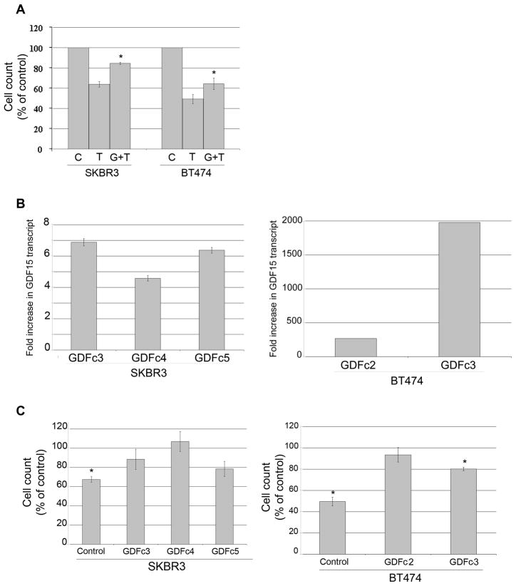
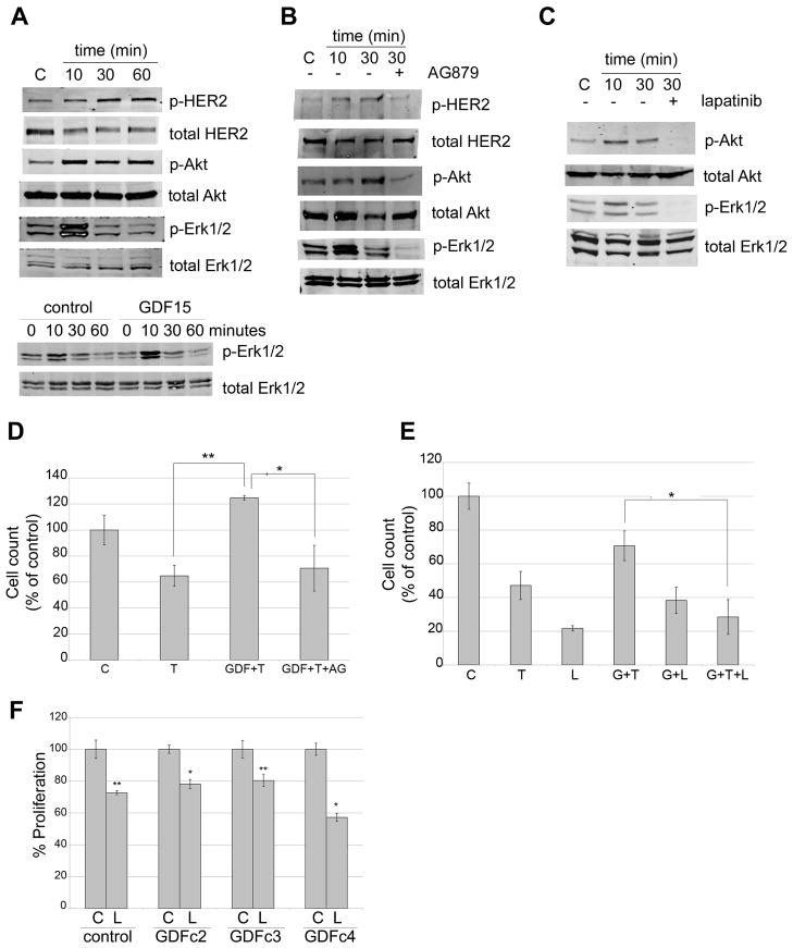
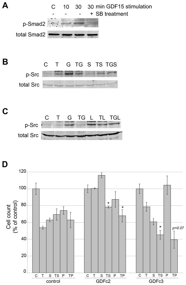
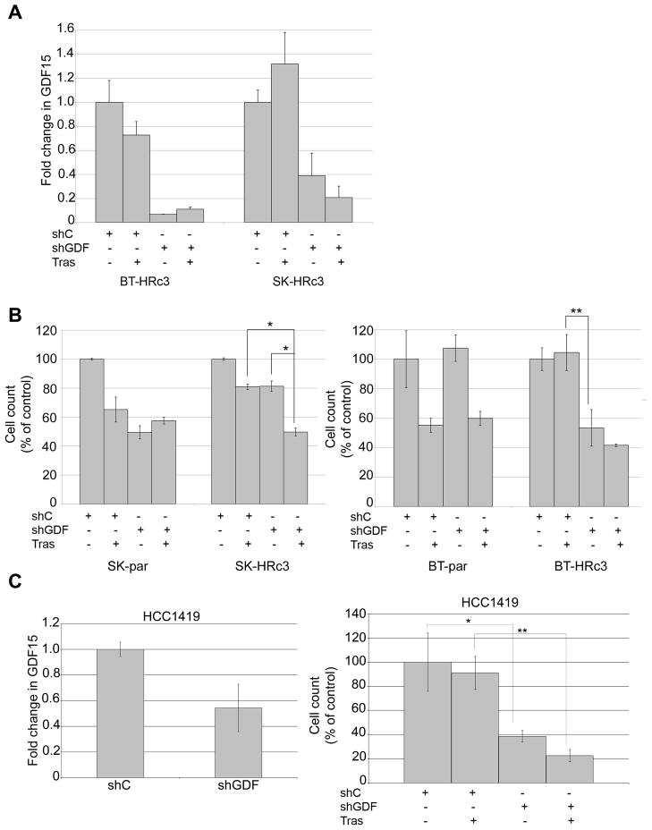
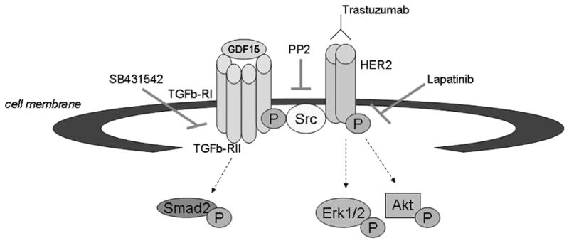
Resistance to the anti-HER2 monoclonal antibody trastuzumab is a major problem in the treatment of HER2-overexpressing metastatic breast cancer. Growth differentiation factor 15 (GDF15), which is structurally similar to TGF beta, has been reported to stimulate phosphorylation of HER2. We tested the hypothesis that GDF15-mediated phosphorylation of HER2 reduces the sensitivity of HER2-overexpressing breast cancer cell lines to trastuzumab. Gene microarray analysis, real-time PCR, and ELISA were used to assess GDF15 expression. Growth inhibition and proliferation assays in response to pharmacologic inhibitors of HER2, TGF beta receptor, or Src were performed on cells stimulated with recombinant human GDF15 or stable GDF15 transfectants. Western blotting was performed to determine effects of GDF15 on HER2 signaling. Cells were infected with lentiviral GDF15 shRNA plasmid to determine effects of GDF15 knockdown on cell survival in response to trastuzumab. Cells with acquired or primary trastuzumab resistance showed increased GDF15 expression. Exposure of trastuzumab-sensitive cells to recombinant human GDF15 or stable transfection of a GDF15 expression plasmid inhibited trastuzumab-mediated growth inhibition. HER2 tyrosine kinase inhibition abrogated GDF15-mediated Akt and Erk1/2 phosphorylation and blocked GDF15-mediated trastuzumab resistance. Pharmacologic inhibition of TGF beta receptor blocked GDF15-mediated phosphorylation of Src. Further, TGF beta receptor inhibition or Src inhibition blocked GDF15-mediated trastuzumab resistance. Finally, lentiviral GDF15 shRNA increased trastuzumab sensitivity in cells with acquired or primary trastuzumab resistance. These results support GDF15-mediated activation of TGF beta receptor-Src-HER2 signaling crosstalk as a novel mechanism of trastuzumab resistance.


Figures

Figure 1
Figure 1. HER2-overexpressing trastuzumab-resistant breast cancer cells express increased levels of GDF15
(A) Total RNA was extracted from BT474 parental (Par), resistant clone 2 (c2) and clone 3 (c3), and SKBR3 parental (Par) and resistant clone 3 (c3) cells. RNA was converted to cDNA and analyzed by real-time PCR for GDF15 transcript level. Results are reported as fold increase in GDF15 transcript level versus parental counterpart. Values were normalized to RPLPO housekeeping ribosomal gene transcript levels as internal control. Each sample was run in triplicate per experiment, and 3 independent experiments were performed on separate occasions to ensure reproducibility. Values represent the average of the 3 separate experiments; error bars represent standard error between the triplicate experiments. P-values were calculated by student’s t-test for each resistant line versus the parental line; *p<0.05, **p<0.005. GDF15 levels were also determined by (B) ELISA using total protein lysates excluding media, or (C) ELISA using cell culture media only. Error bars represent standard deviation between triplicates. P-values were calculated by student’s t-test for each resistant line versus the parental line; *p<0.05, **p<0.005. Results were confirmed on three separate occasions. Acquired resistant cells showed statistically significant increase in both the endogenous and secreted forms of GDF15 versus sensitive lines. (D) Secreted GDF15 was measured by ELISA in the cell culture media of HCC1419, HCC1954, MDA-MB-453, and MDA-MB-361 cells, which have primary resistance to trastuzumab, versus BT474 and SKBR3 trastuzumab-sensitive cells. Error bars represent standard deviation between triplicates. P-values were calculated by student’s t-test for each resistant line versus BT474; **p<0.005. Results were confirmed on three separate occasions. Cells with primary trastuzumab resistance showed higher levels of secreted GDF15 versus trastuzumab-sensitive cells.
Figure 2
Figure 2. Increased GDF15 expression reduces trastuzumab-mediated growth inhibition
(A) SKBR3 and BT474 cells were treated with vehicle control (C), 20 μg/mL trastuzumab (T), or pre-treated with 10 ng/mL GDF15 for 48h followed by 20 μg/mL trastuzumab plus 10 ng/mL GDF15 for an additional 72 h (G+T); media plus treatments were changed each day. Cells were counted by trypan blue exclusion; cell count is shown as a percentage of the control group per cell line. Each sample was run in triplicate cultures per experiment; experiments were performed on 3 independent occasions for reproducibility. Error bars represent standard error between the 3 independent experiments. P-values were determined by student’s t-test for GDF15 plus trastuzumab (G+T) group versus trastuzumab alone (T); *p<0.05. Stimulation with recombinant human GDF15 significantly reduced trastuzumab-mediated growth inhibition. (B) SKBR3 and BT474 clones stably transfected with pCMV empty vector control or pCMV-myc-GDF15 were analyzed by real-time PCR for GDF15 transcript level. Results are reported as fold increase in GDF15 transcript level versus stable control clone per line. Values were normalized to RPLPO housekeeping ribosomal gene transcript levels as internal control. Error bars represent standard deviation between triplicates. (C) Stable clones were treated with 20 μg/mL trastuzumab (Tr) for 72h. Cells were counted by trypan blue exclusion; cell count is shown as a percentage of control untreated cells (C) per clone. Error bars represent standard error between duplicate experiments, each run in triplicate. P-values were determined by student’s t-test for trastuzumab-treated cells versus untreated cells per clone; *p<0.05. Trastuzumab significantly reduced growth of stable control clones; no significant response to trastuzumab was measured in stable GDF15-overexpressing SKBR3 clones. Stable GDF15 BT474 clone 3 showed statistically significant reduction in growth, but less than control BT474 clone cells.
Figure 3
Figure 3. GDF15-mediated activation of HER2 signaling reduces trastuzumab sensitivity
(A) upper panel: BT474 cells were treated with 2 ng/mL recombinant human GDF15 for 10, 30, or 60 min or with the corresponding volume of vehicle control for 60 min. Total protein lysates were Western blotted for phosphorylated and total HER2, Akt, and Erk1/2. lower panel: BT474 cells were treated with 2 ng/mL vehicle control for 0, 10, 30, or 60 min, or with 2 ng/mL recombinant human GDF15 for 0, 10, 30, or 60 min. Total protein lysates were Western blotted for phosphorylated and total Erk1/2. (B) BT474 cells were treated with vehicle control (HCl-BSA plus DMSO), 2 ng/mL GDF15 for 10 or 30 min, or 2 ng/mL GDF15 plus 5 μM AG879 for 30 min. Total protein lysates were Western blotted for phosphorylated and total HER2, Akt, and Erk1/2. (C) BT474 cells were treated with vehicle control (HCl-BSA plus DMSO), 2 ng/mL GDF15 for 10 or 30 min, or 2 ng/mL GDF15 plus 1 μM lapatinib for 30 min. Total protein lysates were Western blotted for phosphorylated and total Akt and Erk1/2. (D) BT474 cells were treated with vehicle control (C), 20 μg/mL trastuzumab (T), pre-treated with 2 ng/mL GDF15 for 48h followed by 20 μg/mL trastuzumab plus 2 ng/mL GDF15 for an additional 72 h (GDF+T), or pre-treated with 2 ng/mL GDF15 for 48h followed by 20 μg/mL trastuzumab plus 2 ng/mL GDF15 plus 5 μM AG879 for an additional 72 h (GDF+T+AG); media plus treatments were changed each day. Cells were counted by trypan blue exclusion; cell count is shown as a percentage of the control group per cell line. Error bars represent standard error between duplicate experiments, each run in triplicate. . P-values were determined by student’s t-test for GDF+T versus T alone, and for GDF+T+AG versus GDF+T; *p<0.05, **p<0.005. (E) BT474 cells were treated with vehicle control (C), 20 μg/mL trastuzumab (T), 1 μM lapatinib (L), pre-treated with 2 ng/mL GDF15 for 48h followed by 20 μg/mL trastuzumab plus 2 ng/mL GDF15 for an additional 72 h (G+T), pre-treated with 2 ng/mL GDF15 for 48h followed by 1 μM lapatinib plus 2 ng/mL GDF15 for an additional 72 h (G+L), or pre-treated with 2 ng/mL GDF15 for 48h followed by 20 μg/mL trastuzumab plus 1 μM lapatinib plus 2 ng/mL GDF15 for an additional 72 h (G+T+L); media plus treatments were changed each day. Cells were counted by trypan blue exclusion; cell count is shown as a percentage of the control group per cell line. Error bars represent standard deviation between triplicates. P-values were determined by student’s t-test for G+T+L versus G+T; *p<0.05. (F) Stable SKBR3 control and GDF15 clones were treated with 100 nM lapatinib (Lap) or corresponding dose of DMSO control (C) for 48h. Proliferation was measured by MTS assay, and is shown as a percentage of control per clone. Error bars represent standard error between duplicate experiments, each run in triplicate. . P-values were determined by student’s t-test for lapatinib-treated cells versus control per clone; *p<0.05, **p<0.005. Stable GDF15-overexpressing clones retained sensitivity to lapatinib.
Figure 4
Figure 4. TGF beta receptor-dependent Src phosphorylation contributes to GDF15-mediated resistance
(A) BT474 cells were treated with vehicle control (HCl-BSA plus DMSO), 2 ng/mL GDF15 for 10 or 30 min, or 2 ng/mL GDF15 plus 5 μM SB431542 for 30 min. Total protein lysates were Western blotted for phosphorylated and total Smad2. (B) BT474 cells were treated with vehicle control (C) (HCl-BSA plus DMSO), 20 μg/mL trastuzumab (T), 2 ng/mL GDF15 (G), trastuzumab plus GDF15 (TG), 5 μM SB431542 (S), trastuzumab plus SB431542 (TS), or trastuzumab plus GDF15 plus SB431542 (TGS) for 30 min. Total protein lysates were Western blotted for phosphorylated and total Src. (C) BT474 cells were treated with vehicle control (C) (HCl-BSA plus DMSO), 20 μg/mL trastuzumab (T), 2 ng/mL GDF15 (G), trastuzumab plus GDF15 (TG), 1 μM lapatinib (L), trastuzumab plus lapatinib (TL), or trastuzumab plus GDF15 plus lapatinib (TGL) for 30 min. Total protein lysates were Western blotted for phosphorylated and total Src. (D) BT474 control and GDF15 stable clones 2 and 3 were treated with vehicle control (C) (HCl-BSA plus DMSO), 20 μg/mL trastuzumab (T), 5 μM SB431542 (S), trastuzumab plus SB431542 (TS), 1 μM PP2 (P), or trastuzumab plus PP2 (TP) for 72 h; media plus treatments were changed each day. Cells were counted by trypan blue exclusion; cell count is shown as a percentage of the control group per cell line. Error bars represent standard deviation between triplicates. P-values were determined by student’s t-test for each group versus T alone; *p<0.05.
Figure 5
Figure 5. GDF15 knockdown increases trastuzumab sensitivity in cells with acquired or primary trastuzumab resistance
(A) BT-HRc3 and SK-HRc3 cells were infected with lentiviral control shRNA (shC) or GDF15 shRNA (shGDF). After 72 h, 20 μg/mL trastuzumab (Tras) was added to cultures for an additional 72 h or cells were left untreated. Cell culture media was examined using GDF15-specific ELISA to confirm knockdown of GDF15. Error bars represent standard deviation between triplicates. (B) BT-parental, BT-HRc3, SK-parental, and SK-HRc3 cells were infected with lentiviral control shRNA (shC) or GDF15 shRNA (shGDF). After 72 h, 20 μg/mL trastuzumab (Tras) was added to cultures for an additional 72 h or cells were left untreated. Cells were then counted by trypan blue exclusion; growth is shown as a percentage of shC-infected cells per line. Treatments were done in triplicate, with error bars representing standard deviation between replicates. Results were confirmed in duplicate experiments. P-values were determined by student’s t-test; *p<0.05, **p<0.005. (C) HCC1419 cells were infected with lentiviral control shRNA (shC) or GDF15 shRNA (shGDF); knockdown of GDF15 was confirmed by real-time PCR. Results are reported as fold change in GDF15 transcript level versus control shRNA. Values were normalized to RPLPO housekeeping ribosomal gene transcript levels as internal control. Error bars represent standard deviation between triplicates. Cells were infected with control shRNA or GDF15 shRNA, and after 72 h, treated with 20 μg/mL trastuzumab (Tras) for an additional 72 h or left untreated. Cells were counted by trypan blue exclusion; growth is shown as a percentage of shC-infected cells. Treatments were done in triplicate, with error bars representing standard deviation between replicates. Results were confirmed in duplicate experiments. P-values were determined by student’s t-test; *p<0.05, **p<0.005.
Figure 6
Figure 6. Proposed mechanism of GDF15-mediated trastuzumab resistance
We propose that GDF15 activates TGF beta receptor-Src-HER2 signaling crosstalk as a novel mechanism of trastuzumab resistance. GDF15 appears to activate TGF beta receptor, as measured by phosphorylation of the TGF beta receptor substrate Smad2. GDF15 activates Src in a TGF beta receptor-dependent manner, which subsequently induces phosphorylation of HER2 and abrogates the growth inhibitory effects of the HER2-targeted antibody trastuzumab. Inhibition of the HER2 kinase by lapatinib restores sensitivity to trastuzumab in models of GDF15-mediated trastuzumab resistance.

Similar articles

Cited by

References

    1. Slamon DJ, Clark GM, Wong SG, Levin WJ, Ullrich A, McGuire WL. Human breast cancer: correlation of relapse and survival with amplification of the HER-2/neu oncogene. Science. 1987;235:177–82. - PubMed
    1. Baselga J, Tripathy D, Mendelsohn J, Baughman S, Benz CC, Dantis L, et al. Phase II study of weekly intravenous recombinant humanized anti-p185HER2 monoclonal antibody in patients with HER2/neu-overexpressing metastatic breast cancer. J Clin Oncol. 1996;14:737–44. - PubMed
    1. Cobleigh MA, Vogel CL, Tripathy D, Robert NJ, Scholl S, Fehrenbacher L, et al. Multinational study of the efficacy and safety of humanized anti-HER2 monoclonal antibody in women who have HER2-overexpressing metastatic breast cancer that has progressed after chemotherapy for metastatic disease. J Clin Oncol. 1999;17:2639–48. - PubMed
    1. Slamon DJ, Leyland-Jones B, Shak S, Fuchs H, Paton V, Bajamonde A, et al. Use of chemotherapy plus a monoclonal antibody against HER2 for metastatic breast cancer that overexpresses HER2. N Engl J Med. 2001;344:783–92. - PubMed
    1. Esteva FJ, Valero V, Booser D, Guerra LT, Murray JL, Pusztai L, et al. Phase II study of weekly docetaxel and trastuzumab for patients with HER-2-overexpressing metastatic breast cancer. J Clin Oncol. 2002;20:1800–8. - PubMed

Publication types

MeSH terms

Substances