

Protein S-nitrosylation: role for nitric oxide signaling in neuronal death
- PMID: 21803124
- PMCID: PMC3251724
- DOI: 10.1016/j.bbagen.2011.07.010
Protein S-nitrosylation: role for nitric oxide signaling in neuronal death
Abstract
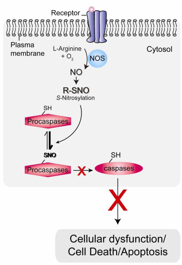
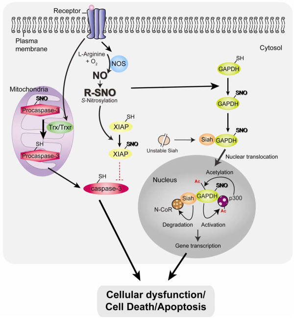
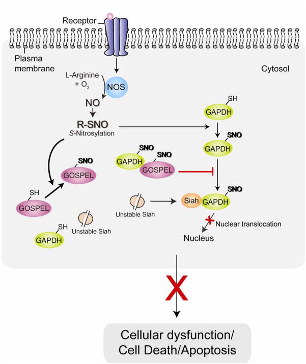
Background: One of the signaling mechanisms mediated by nitric oxide (NO) is through S-nitrosylation, the reversible redox-based modification of cysteine residues, on target proteins that regulate a myriad of physiological and pathophysiological processes. In particular, an increasing number of studies have identified important roles for S-nitrosylation in regulating cell death.
Scope of review: The present review focuses on different targets and functional consequences associated with nitric oxide and protein S-nitrosylation during neuronal cell death.
Major conclusions: S-Nitrosylation exhibits double-edged effects dependent on the levels, spatiotemporal distribution, and origins of NO in the brain: in general Snitrosylation resulting from the basal low level of NO in cells exerts anti-cell death effects, whereas S-nitrosylation elicited by induced NO upon stressed conditions is implicated in pro-cell death effects.
General significance: Dysregulated protein S-nitrosylation is implicated in the pathogenesis of several diseases including degenerative diseases of the central nervous system (CNS). Elucidating specific targets of S-nitrosylation as well as their regulatory mechanisms may aid in the development of therapeutic intervention in a wide range of brain diseases.
Copyright © 2011 Elsevier B.V. All rights reserved.
Figures
References
-
- Mattson MP. Neuronal life-and-death signaling, apoptosis, and neurodegenerative disorders. Antioxid Redox Signal. 2006;8:1997–2006. - PubMed
-
- Cotter TG. Apoptosis and cancer: the genesis of a research field. Nat Rev Cancer. 2009;9:501–507. - PubMed
-
- Nakamura T, Lipton SA. Cell death: protein misfolding and neurodegenerative diseases. Apoptosis. 2009;14:455–468. - PubMed
-
- Zhivotovsky B, Orrenius S. Cell death mechanisms: cross-talk and role in disease. Exp Cell Res. 2010;316:1374–1383. - PubMed
Publication types
MeSH terms
Substances
Grants and funding
LinkOut - more resources
Full Text Sources
Medical
Research Materials
