

Self-modulation of primary motor cortex activity with motor and motor imagery tasks using real-time fMRI-based neurofeedback
- PMID: 21803163
- PMCID: PMC3222744
- DOI: 10.1016/j.neuroimage.2011.07.035
Self-modulation of primary motor cortex activity with motor and motor imagery tasks using real-time fMRI-based neurofeedback
Abstract
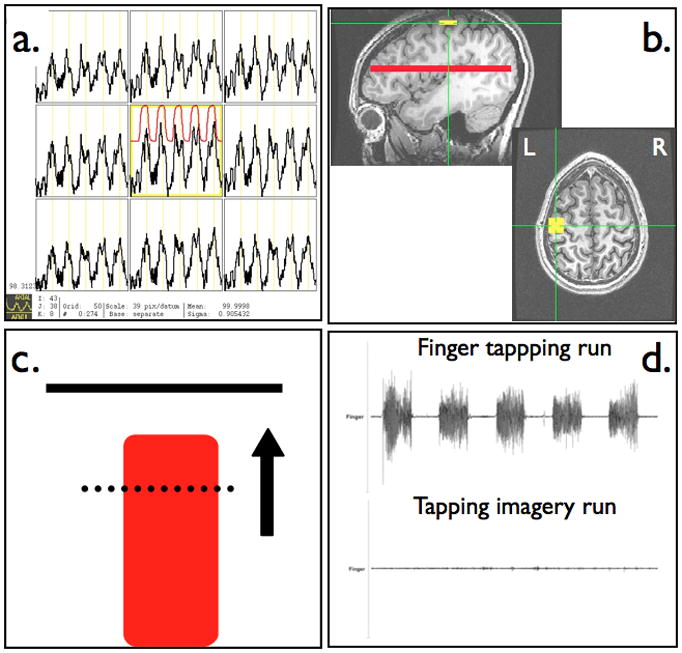
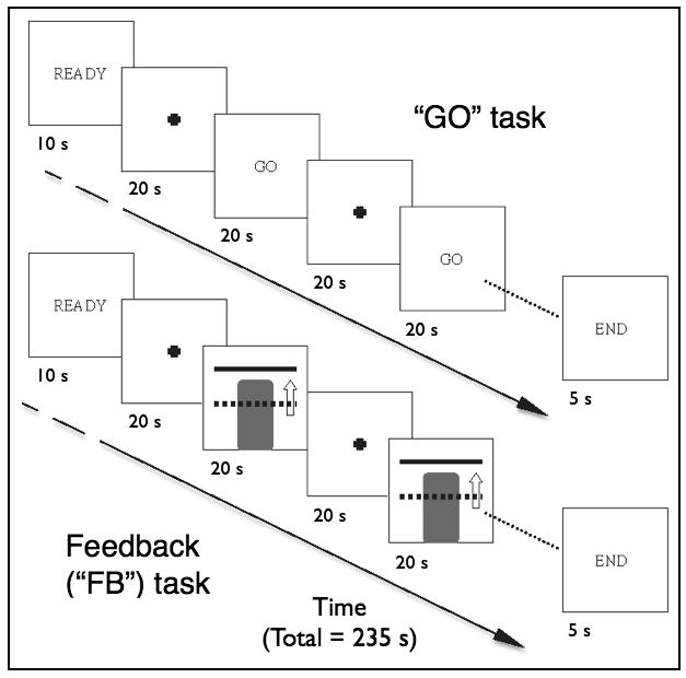
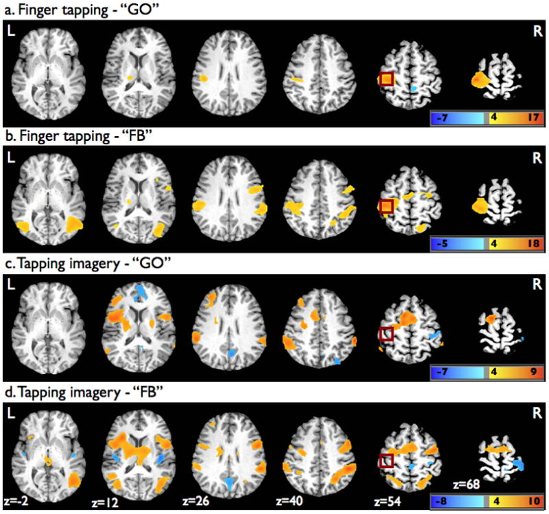
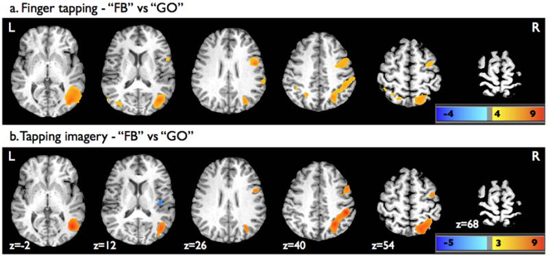
Advances in fMRI data acquisition and processing have made it possible to analyze brain activity as rapidly as the images are acquired allowing this information to be fed back to subjects in the scanner. The ability of subjects to learn to volitionally control localized brain activity within motor cortex using such real-time fMRI-based neurofeedback (NF) is actively being investigated as it may have clinical implications for motor rehabilitation after central nervous system injury and brain-computer interfaces. We investigated the ability of fifteen healthy volunteers to use NF to modulate brain activity within the primary motor cortex (M1) during a finger tapping and tapping imagery task. The M1 hand area ROI (ROI(m)) was functionally localized during finger tapping and a visual representation of BOLD signal changes within the ROI(m) fed back to the subject in the scanner. Surface EMG was used to assess motor output during tapping and ensure no motor activity was present during motor imagery task. Subjects quickly learned to modulate brain activity within their ROI(m) during the finger-tapping task, which could be dissociated from the magnitude of the tapping, but did not show a significant increase within the ROI(m) during the hand motor imagery task at the group level despite strongly activating a network consistent with the performance of motor imagery. The inability of subjects to modulate M1 proper with motor imagery may reflect an inherent difficulty in activating synapses in this area, with or without NF, since such activation may lead to M1 neuronal output and obligatory muscle activity. Future real-time fMRI-based NF investigations involving motor cortex may benefit from focusing attention on cortical regions other than M1 for feedback training or alternative feedback strategies such as measures of functional connectivity within the motor system.
Copyright © 2011 Elsevier Inc. All rights reserved.
Figures

Similar articles
-
Intermittent "real-time" fMRI feedback is superior to continuous presentation for a motor imagery task: a pilot study.J Neuroimaging. 2012 Jan;22(1):58-66. doi: 10.1111/j.1552-6569.2010.00529.x. Epub 2010 Oct 26. J Neuroimaging. 2012. PMID: 20977537 Free PMC article.
-
Investigation of fMRI neurofeedback of differential primary motor cortex activity using kinesthetic motor imagery.Neuroimage. 2012 May 15;61(1):21-31. doi: 10.1016/j.neuroimage.2012.02.053. Epub 2012 Mar 3. Neuroimage. 2012. PMID: 22401758
-
The BOLD response in primary motor cortex and supplementary motor area during kinesthetic motor imagery based graded fMRI neurofeedback.Neuroimage. 2019 Jan 1;184:36-44. doi: 10.1016/j.neuroimage.2018.09.007. Epub 2018 Sep 8. Neuroimage. 2019. PMID: 30205210 Free PMC article.
-
Optimized Motor Imagery Paradigm Based on Imagining Chinese Characters Writing Movement.IEEE Trans Neural Syst Rehabil Eng. 2017 Jul;25(7):1009-1017. doi: 10.1109/TNSRE.2017.2655542. Epub 2017 Jan 19. IEEE Trans Neural Syst Rehabil Eng. 2017. PMID: 28113345
-
EEG-Based Strategies to Detect Motor Imagery for Control and Rehabilitation.IEEE Trans Neural Syst Rehabil Eng. 2017 Apr;25(4):392-401. doi: 10.1109/TNSRE.2016.2646763. Epub 2016 Dec 30. IEEE Trans Neural Syst Rehabil Eng. 2017. PMID: 28055887 Review.
Cited by
-
Frontostriatal circuitry as a target for fMRI-based neurofeedback interventions: A systematic review.Front Hum Neurosci. 2022 Aug 24;16:933718. doi: 10.3389/fnhum.2022.933718. eCollection 2022. Front Hum Neurosci. 2022. PMID: 36092647 Free PMC article.
-
Improvement in precision grip force control with self-modulation of primary motor cortex during motor imagery.Front Behav Neurosci. 2015 Feb 13;9:18. doi: 10.3389/fnbeh.2015.00018. eCollection 2015. Front Behav Neurosci. 2015. PMID: 25762907 Free PMC article.
-
One session of fMRI-Neurofeedback training on motor imagery modulates whole-brain effective connectivity and dynamical complexity.Cereb Cortex Commun. 2022 Jul 25;3(3):tgac027. doi: 10.1093/texcom/tgac027. eCollection 2022. Cereb Cortex Commun. 2022. PMID: 36072710 Free PMC article.
-
Self-regulation of ventromedial prefrontal cortex activation using real-time fMRI neurofeedback-Influence of default mode network.Hum Brain Mapp. 2020 Feb 1;41(2):342-352. doi: 10.1002/hbm.24805. Epub 2019 Oct 21. Hum Brain Mapp. 2020. PMID: 31633257 Free PMC article.
-
The potential of real-time fMRI neurofeedback for stroke rehabilitation: A systematic review.Cortex. 2018 Oct;107:148-165. doi: 10.1016/j.cortex.2017.09.006. Epub 2017 Sep 18. Cortex. 2018. PMID: 28992948 Free PMC article.
References
-
- Astafiev SV, Stanley CM, Shulman GL, Corbetta M. Extrastriate body area in human occipital cortex responds to the performance of motor actions. Nature Neuroscience. 2004;7:542–548. - PubMed
-
- Caminiti R, Ferraina S, Johnson PB. The sources of visual information to the primate frontal lobe: A novel role for the superior parietal lobule. Cerebral Cortex. 1996;6:319–328. - PubMed
-
- Caria A, Veit R, Sitaram R, Lotze M, Weiskopf N, Grodd W, Birbaumer N. Regulation of anterior insular cortex activity using real-time fMRI. Neuroimage. 2007;35:1238–1246. - PubMed
-
- Cavanna AE, Trimble MR. The precuneus: a review of its functional anatomy and behavioural correlates. Brain. 2006;129:564–583. - PubMed
Publication types
MeSH terms
Grants and funding
LinkOut - more resources
Full Text Sources
Medical
