. 2011 Aug 3;14(2):173-83.
doi: 10.1016/j.cmet.2011.06.008.

Autophagy in hypothalamic AgRP neurons regulates food intake and energy balance

Affiliations

Autophagy in hypothalamic AgRP neurons regulates food intake and energy balance

Susmita Kaushik et al. Cell Metab. .

Abstract

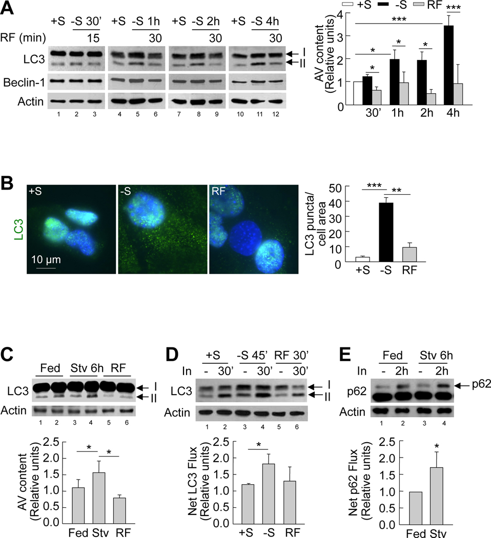
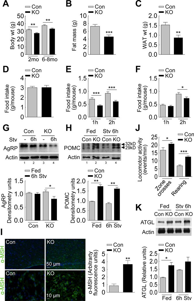
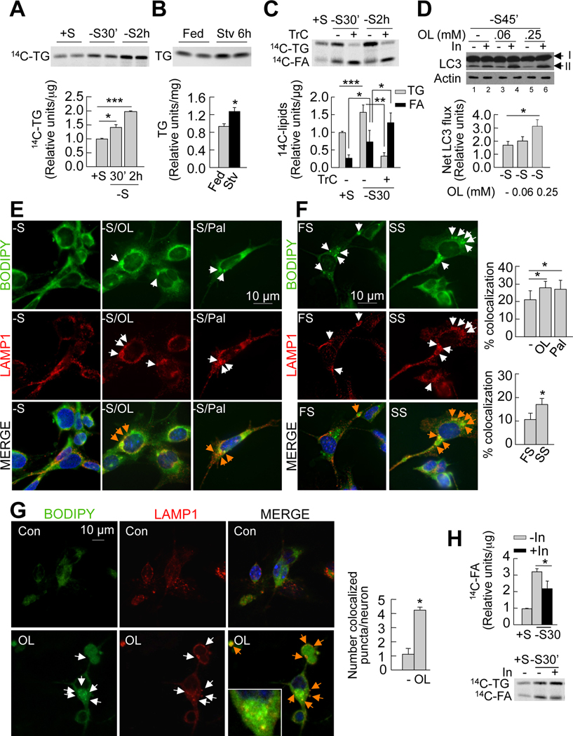
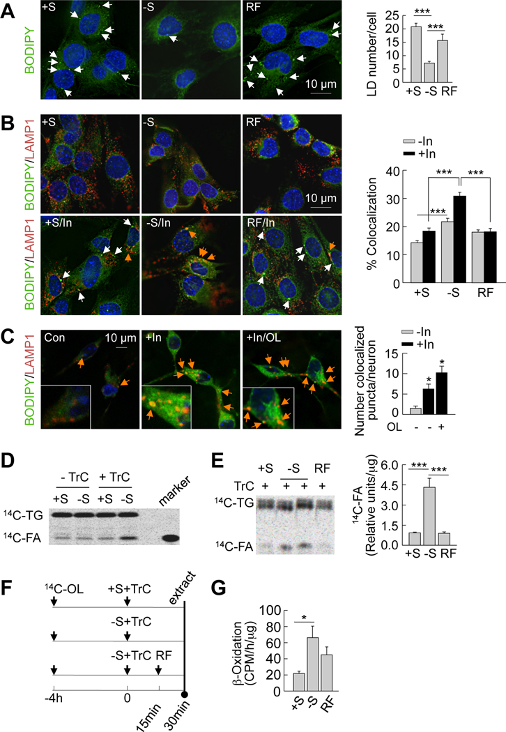
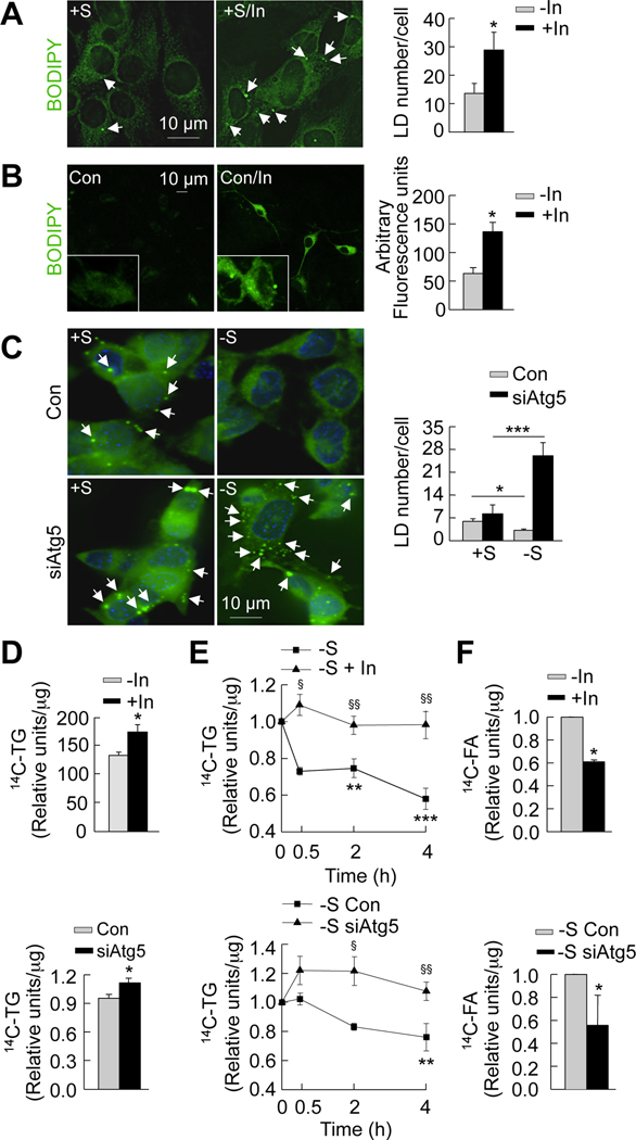
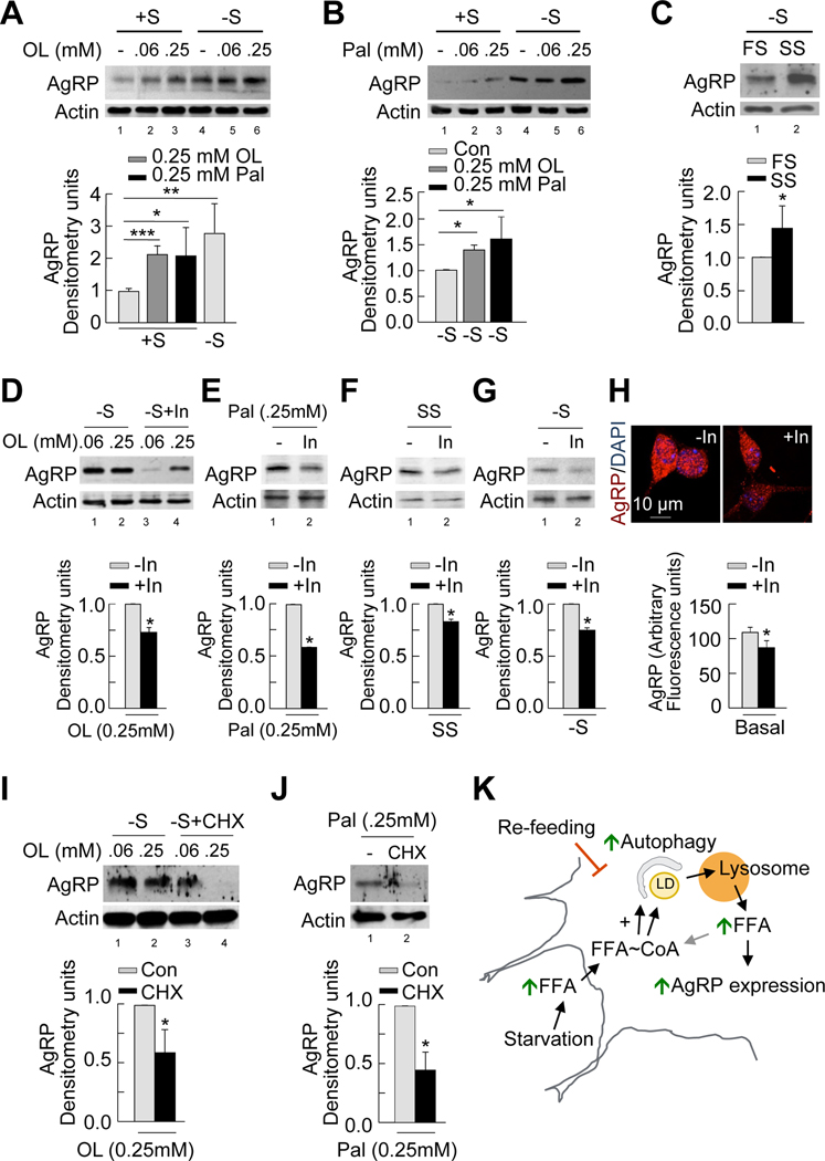
Macroautophagy is a lysosomal degradative pathway that maintains cellular homeostasis by turning over cellular components. Here we demonstrate a role for autophagy in hypothalamic agouti-related peptide (AgRP) neurons in the regulation of food intake and energy balance. We show that starvation-induced hypothalamic autophagy mobilizes neuron-intrinsic lipids to generate endogenous free fatty acids, which in turn regulate AgRP levels. The functional consequences of inhibiting autophagy are the failure to upregulate AgRP in response to starvation, and constitutive increases in hypothalamic levels of pro-opiomelanocortin and its cleavage product α-melanocyte-stimulating hormone that typically contribute to a lean phenotype. We propose a conceptual framework for considering how autophagy-regulated lipid metabolism within hypothalamic neurons may modulate neuropeptide levels to have immediate effects on food intake, as well as long-term effects on energy homeostasis. Regulation of hypothalamic autophagy could become an effective intervention in conditions such as obesity and the metabolic syndrome.


Figures

Figure 1
Figure 1. Starvation induces autophagy in the hypothalamus
(A) Immunoblots for LC3 and Beclin-1 from GT1–7 cells in presence (+S) or absence of serum (−S), or serum refed (RF) after serum removal for indicated times. Values for LC3-II represent autophagic vacuole (AV) content relative to +S (n=3). See also Figure S1. (B) Indirect immunofluorescence for LC3 (green) in GT1–7 cells in +S or −S for 1h, or RF for 30min following serum removal. DAPI-stained nuclei are in blue. Values represent LC3 puncta per cell area, and are mean+SE of 20 different cells in each experiment, n=3. (C) Immunoblots for LC3 in mediobasal hypothalamus (MBH) lysates from fed (lanes 1, 2), 6h-starved (Stv) (lanes 3, 4), or starved mice subjected to refeeding for 3h (RF) (lanes 5, 6). Values depict AV content, calculated as in A (n=5). (D) Immunoblots for LC3 from GT1–7 cells in +S or −S or RF following serum removal for times indicated. Cells were treated with (lanes 2, 4, 6) or without lysosomal inhibitors (In) (lanes 1, 3, 5) for 30min. Values represent net LC3 flux determined by subtracting LC3-II densitometric value of untreated samples from corresponding In-treated sample (n=4). (E) Immunoblot for p62 from MBH explants of fed (lanes 1, 2) and 6h-starved (Stv) (lanes 3, 4) mice cultured in presence or absence of lysosomal inhibitors (In) for 2h. Values represent net p62 flux (n=4). Values are mean+SE. *p<0.05, **p<0.01, ***p<0.001.
Figure 2
Figure 2. Food intake and energy balance in mice knock-out for atg7 in AgRP neurons
(A) Body weights of 2 month (mo) (n=10–12) and 6–8 mo-old control (Con) and Atg7F/F-AgRP-Cre (KO) mice (n=12–15). (B) Total body fat mass of 6–8 month old Con and KO mice (n=10–12). (C) Epididymal fat weights of 6–8 month old Con and KO mice (n=15–16). See also Figure S2D. (D) Food intake over 24h by Con and KO mice (n=27–31). (E) Food intake by 6h- (n=12–24), and (F) 24h-starved Con and KO mice when refed for indicated times (n=15–18). (G) Immunoblot for AgRP from mediobasal hypothalamus (MBH) of Con (lanes 1, 2) and KO (lanes 3, 4) mice fed or starved (Stv) for 6h. Values represent densitometric values for AgRP (n=10). See also Figure S2F–G. (H) Immunoblot for POMC preproprotein from MBH of fed Con and KO (lanes 1, 2) and 6h-starved Con and KO (lanes 3, 4) mice (n=6). (I) Indirect immunofluorescence (at similar exposure times) for α-MSH (green) in MBH sections from Con and KO mice (n=3). (J) Locomotor activity reflecting “zone crosses” and “rearing” by fed Con and KO rodents (n=14–19). (K) Immunoblot for ATGL from adipose tissues of fed Con and KO (lanes 1, 2) and 6h-starved Con and KO (lanes 3, 4) mice. Values represent densitometric values for ATGL (n=3–4). Values are mean+SE. *p<0.05, **p<0.01, ***p<0.001.
Figure 3
Figure 3. Fatty acid uptake during starvation induces hypothalamic autophagy
(A) Thin layer chromatogram (TLC) of 14C-triglycerides (14C-TG) from GT1–7 cells in presence (+S) or absence (−S) of serum for the indicated times. Relative densitometric values represent 14C-triglycerides per µg protein (n=3). (B) TLC of TG extracted from mediobasal hypothalamus (MBH) from fed and 6hstarved (Stv) mice. Relative densitometric values represent triglycerides per mg tissue weight (n=4–5). See also Figures S3A and S3B. (C) TLC of 14C-TG and 14C-fatty acids (14C-FA) from GT1–7 cells maintained in +S or −S treated with or without triacsin C (TrC) for indicated times. Relative densitometric values represent corresponding 14C-lipid per µg protein (n=5). (D) Immunoblot for LC3 from GT1–7 cells cultured in −S and treated with 0.06 or 0.25mM oleic acid for indicated times. Cells were treated with or without lysosomal inhibitors (In) for 30min. Values represent net LC3 flux determined by subtracting LC3-II densitometric value of untreated samples from corresponding Intreated samples (n=3). See also Figure S3D. (E) Immunofluorescence for BODIPY/LAMP1 in GT1–7 cells cultured in −S and co-treated with or without 0.25mM oleic (−S/OL) or palmitic acid (−S/Pal) for 30min, and in cells (F) cultured in 1% serum from fed (FS) or 24h-starved (SS) rats for 30min.Values represent %colocalization between BODIPY and LAMP1 (orange arrows), and are mean+SE of 20 different cells in each experiment, n=3. See also Figure S3H. (G) Confocal images for BODIPY/LAMP1 in primary hypothalamic neurons treated with or without 0.25mM oleic (OL). Values represent number of colocalized (orange arrows) puncta, and are mean+SE of more than 20 different cells in each experiment, n=3. (H) TLC of 14C-TG and 14C-FA from GT1–7 cells in +S or −S and treated with lysosomal inhibitors (In) for 30min. Relative densitometric values represent 14C-FA per µg protein (n=3). Values are mean+SE. *p<0.05, **p<0.01, ***p<0.001.
Figure 4
Figure 4. Autophagy mobilizes hypothalamic lipids to generate endogenous fatty acids
(A) BODIPY staining in GT1–7 cells cultured in presence (+S) or absence of serum (−S) for 30min or refed (RF) for 15min following serum removal. Values represents lipid droplet (LD) number per cell, and are mean+SE, for 20 different cells in each experiment, n=3. (B) Immunofluorescence for BODIPY/LAMP1 in GT1–7 cells in +S or −S for 30min or RF for 15min, and treated with or without lysosomal inhibitors (In) for 30min. Values represent % colocalization between BODIPY and LAMP1 (orange arrows), and are mean+SE for 20 different cells in each experiment, n=3. (C) Confocal images for BODIPY/LAMP1 in primary hypothalamic neurons co-treated with or without 0.25mM oleic (OL) or lysosomal inhibitors (In) for 4h. Values represent number of colocalized (orange arrows) puncta, and are mean+SE of more than 20 different cells in each experiment, n=3. See also Figure S4. (D) Thin layer chromatogram (TLC) of 14C-triglycerides (14C-TG) and 14C-fatty acids (14C-FA) from GT1–7 cells maintained in +S or −S and treated with or without triacsin C (TrC) for 30min. (E) TLC of 14C-TG and 14C-FA from TrC-treated GT1–7 cells in +S or −S for 30min or RF for 15min. Relative densitometric values represent 14C-FA per µg protein, and are mean+SE, n=3. (F) Scheme for experiment in E. (G) β-Oxidation assay in GT1–7 cells in +S or −S for 1h or RF for 30min. Values represent 14CO2 release expressed as counts per minute (CPM)/h/µg protein, and are mean+SE, n=3. *p<0.05, ***p<0.001.
Figure 5
Figure 5. Inhibition of autophagy in hypothalamic cells promotes neuronal lipid accumulation
(A) BODIPY staining (acquired at similar exposure times) in serum-fed GT1–7 cells (+S) cultured in presence or absence of lysosomal inhibitors (In) for 30min. Values represent lipid droplet (LD) number per cell, and are mean+SE for 20 different cells, n=3. (B) BODIPY staining (acquired at similar exposure times) in primary hypothalamic neurons cultured in presence or absence of lysosomal inhibitors (In) for 4h. Values represent arbitrary fluorescence intensity, and are mean+SE for 20 different cells, n=3. (C) BODIPY staining in control (Con) and autophagy-deficient (siAtg5) GT1–7 cells in +S or −S for 30min. Values represent LD number per cell, and are mean+SE for 20 different cells, n=3. (D) Relative densitometric values representing 14C-triglycerides (14C-TG) per µg protein calculated from thin layer chromatograms from GT1–7 cells cultured in +S for 4h (Con) or subjected to pharmacologic (In; Top) or genetic (siAtg5; Bottom) inhibition of autophagy. Values are mean+SE, n=3. (E) Decay assays for 14C-TG in GT1–7 cells chased in absence of serum for the indicated times (Con) or following pharmacologic (In; Top) or genetic (siAtg5; Bottom) inhibition of autophagy. Relative densitometric values represent residual 14C-TG per µg protein, and are mean+SE, n=4. (F) Relative densitometric values representing 14C-fatty acid (14C-FA) per µg protein calculated from thin layer chromatograms from GT1–7 cells cultured in −S for 30min (Con) or subjected to pharmacologic (In; Top) or genetic (siAtg5; Bottom) inhibition of autophagy. Values relative to −S control are mean+SE, n=3. *p<0.05, **p<0.01, and ***p<0.001. §p<0.05, §§p<0.005 compared to corresponding −S or −S Con.
Figure 6
Figure 6. Autophagy-derived intracellular free fatty acids promote AgRP expression
(A–C) Immunoblot for AgRP from GT1–7 cells in presence (+S) or absence of serum (−S) for 1h and treated without or with 0.06 and 0.25mM oleic acid (OL) (A) or palmitic acid (Pal) (B) or treated with 1% fed (FS) or starved rat serum (SS) (C). Densitometric values of AgRP normalized to untreated +S (lower panel: left); normalized to untreated −S (lower panel: middle); and normalized to FS (lower panel: right). Values are mean+SE, n=3. (D–G) Top: Immunoblot for AgRP from GT1–7 cells in −S in absence or presence of lysosomal inhibitors (In) for 1h and treated with 0.06 and 0.25mM OL (D), 0.25mM Pal (E), 1% SS (F), or untreated (G). Bottom: Densitometric values for AgRP normalized to In-untreated cells. Values are mean+SE, n=3. See also Figure S5. (H) Immunostaining for AgRP (acquired at similar exposure times) in primary hypothalamic neurons cultured in presence or absence of lysosomal inhibitors (In) for 4h. Values represent arbitrary fluorescence intensity for AgRP, and are mean+SE for 20 different cells, n=3. (I–J) Top: Immunoblot for AgRP from serum-deprived GT1–7 cells in absence or presence of the protein synthesis inhibitor, cycloheximide (CHX) for 1h following treatment with 0.06 and 0.25mM OL (I), or 0.25mM Pal (J). Bottom: Densitometric values for AgRP normalized to CHX-untreated cells. Values are mean+SE, n=3. (K) Proposed model depicting role of autophagy in hypothalamic AgRP expression. *p<0.05, **p<0.01, ***p<0.001.

References

    1. Andrews ZB, Liu ZW, Walllingford N, Erion DM, Borok E, Friedman JM, Tschop MH, Shanabrough M, Cline G, Shulman GI, et al. UCP2 mediates ghrelin's action on NPY/AgRP neurons by lowering free radicals. Nature. 2008;454:846–851. - PMC - PubMed
    1. Belgardt BF, Bruning JC. CNS leptin and insulin action in the control of energy homeostasis. Ann N Y Acad Sci. 2010;1212:97–113. - PubMed
    1. Boland B, Kumar A, Lee S, Platt FM, Wegiel J, Yu WH, Nixon RA. Autophagy induction and autophagosome clearance in neurons: relationship to autophagic pathology in Alzheimer's disease. J Neurosci. 2008;28:6926–6937. - PMC - PubMed
    1. Bolte S, Cordelieres FP. A guided tour into subcellular colocalization analysis in light microscopy. J Microsc. 2006;224:213–232. - PubMed
    1. Cota D, Proulx K, Smith KA, Kozma SC, Thomas G, Woods SC, Seeley RJ. Hypothalamic mTOR signaling regulates food intake. Science. 2006;312:927–930. - PubMed

Publication types

MeSH terms