. 2011 Sep 15;71(18):6010-8.
doi: 10.1158/0008-5472.CAN-11-0595. Epub 2011 Jul 29.

Activated Notch1 induces lung adenomas in mice and cooperates with Myc in the generation of lung adenocarcinoma

Affiliations

Activated Notch1 induces lung adenomas in mice and cooperates with Myc in the generation of lung adenocarcinoma

Thaddeus D Allen et al. Cancer Res. .

Abstract

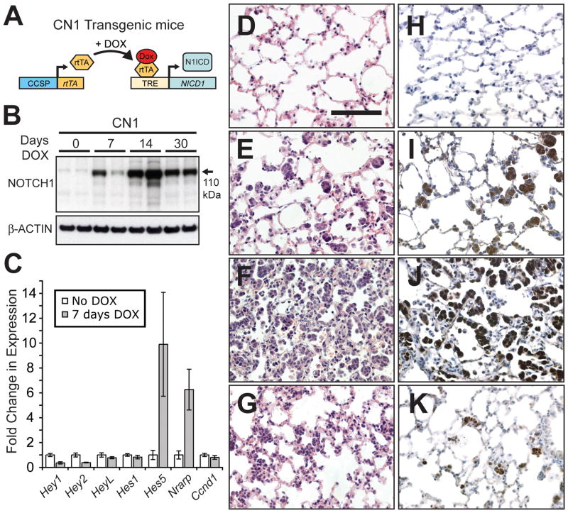
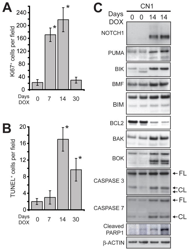
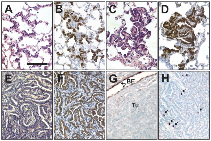
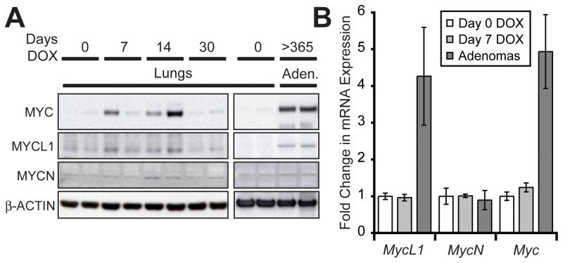
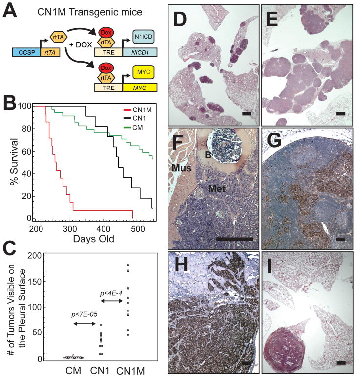
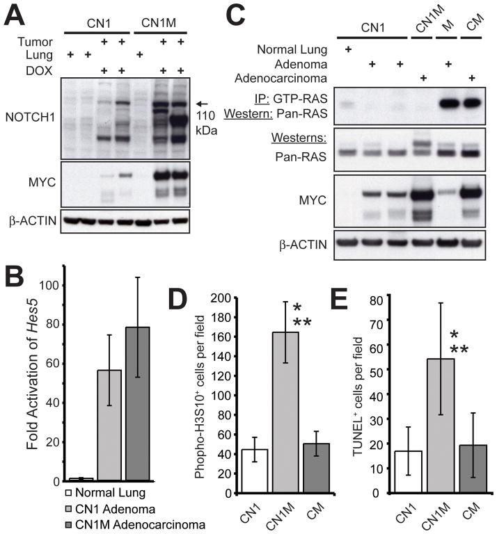
Notch1 encodes the canonical member of the mammalian Notch receptor family. Activating lesions frequently affect Notch1 in T-cell acute lymphoblastic leukemia (T-ALL) and, recently, have been found in non-small-cell lung cancer (NSCLC) as well. We explored the oncogenic potential of activated Notch1 in the lung by developing a transgenic mouse model in which activated Notch1 was overexpressed in the alveolar epithelium. The initial response to activated Notch1 was proliferation and the accumulation of alveolar hyperplasia, which was then promptly cleared by apoptosis. After an extended latency period, however, pulmonary adenomas appeared in the transgenic mice but failed to progress to become carcinomas. Interestingly, Myc and MycL1 were expressed in the adenomas, suggesting that selection for enhanced Myc activity may facilitate tumorigenesis. Using mice engineered to coexpress activated Notch1 and Myc, we found that supplementing Myc expression resulted in increased frequency of Notch1 intracellular domain (N1ICD)-induced adenoma formation and enabled progression to adenocarcinoma and metastases. Cooperation stemmed from synergistic activation of tumor cell cycling, a process that apparently countered any impedance to tumorigenesis posed by Myc and/or activated Notch1-induced apoptosis. Significantly, cooperation was independent of RAS activation. Taken together, the data suggest that activated Notch1 substitutes for RAS activation synergistically with Myc in the development of NSCLC. These tumor models should be valuable for exploring the role of activated Notch1 in the genesis of NSCLC and for testing therapies targeting either activated Notch1 or its downstream effectors.


Conflict of interest statement

Conflicts of interest: None

Figures

Figure 1
Figure 1
Overexpression of N1ICD induces transient alveolar hyperplasia. (A) The C and N1 transgenes were used to direct N1ICD expression to lung epithelial cells with DOX treatment. (B) Western analysis of N1ICD expression in lung protein extracts following 0, 7, 14 and 30 days of DOX treatment. (C) Taqman® analysis of a subset of known NOTCH target genes using reverse transcribed lung RNA from CN1 mice either not treated with DOX or treated with DOX for 7 days (n≥3 mice, error bars are ± standard deviation). (DG) H&E stained sections from CN1 lungs treated with DOX for 0 (D), 7 (E), 14 (F) and 30 (G) days. (HK) Immunohistochemical staining for NOTCH1 in lung sections from CN1 mice treated with DOX for 0 (H), 7 (I), 14 (J) and 30 (K) days (bar in D = 0.1 mm, DK same magnification).
Figure 2
Figure 2
Apoptosis contributes to the clearance of N1ICD overexpressing cells from the alveolar space. (A) Quantification of Ki67+ cells in the lungs of DOX treated CN1 mice (n≥3 mice, error bars are ± standard deviation; * p<0.01 compared to day 0 by Student’s t-test). (B) Quantification of TUNEL+ cells in the lungs of DOX treated CN1 mice (n≥3 mice, error bars are ± standard deviation; * p<0.003 compared to day 0 by Student’s t-test). (C) Western analysis of BCL2 family proteins, CASPASES 3 and 7 and PARP1 in the lungs of CN1 mice untreated and treated with DOX for 14 days (FL = full-length; CL= cleaved forms). β-ACTIN served as a loading control.
Figure 3
Figure 3
Adenomas develop in the lungs of DOX treated CN1 mice. (AD) Sections from CN1 mice maintained on DOX for 8 months. (A) H&E stained section showing organized growth along the alveolar walls. (B) Immunohistochemical staining for NOTCH1 showing that N1ICD+ cells grow along the alveolar walls instead of in cell clusters. (C) H&E stained section showing an early adenoma. (D) Immunohistochemical staining of an early adenoma for NOTCH1. (EH) Adenomas from CN1 mice maintained on DOX for > 1 year. (E) H&E stained section that shows the papillary patterning of a CN1 adenoma. Adenomas were TTF-1+ (F), but CCSP (BE = bronchiolar epithelium; Tu = tumor) (G) with rare SPC+ cells (arrows) clearly discernable in immunohistochemically stained sections (H) (bar in A = 0.1 mm, AH same magnification).
Figure 4
Figure 4
MYC and MYCL1 are upregulated in N1ICD-induced adenomas. (A) Western analysis of MYC, MYCL1 and MYCN expression in the lungs of CN1 mice treated with DOX for 0, 7, 14 and 30 days and in adenomas from CN1 mice continuously fed a DOX diet until sacrifice (>1 year). β-ACTIN served as a loading control. (B) Taqman® analysis of Myc gene mRNA in the lungs of CN1 mice not treated with DOX, treated with DOX for 7 days and in adenomas from DOX fed mice (n≥3 mice, error bars are ± standard deviation). Gene expression was normalized to β-Actin expression.
Figure 5
Figure 5
N1ICD and MYC co-expression cooperates to induce lung adenocarcinoma. (A) Schematic of transgenes used for coincident overexpression of N1ICD and MYC. (B) Kaplan-Meier plot of DOX treated CM (n=34), CN1 (n=11) and CN1M (n=14) mice monitored for 18 months (p<1E-4 by Logrank test for CN1M versus either CM or CN1). (C) Quantification of tumors visible on the pleural surface of transgenic mice (p-values calculated using Student’s t-test). The cumulative numbers of tumors up until 18 months is displayed. (DF) H&E stained sections from transgenic mice maintained on a DOX diet. (D) Lungs from a CN1 mouse with multiple adenomas. (E) Lungs from a CN1M mouse that developed multiple large adenocarcinomas. (F) Metastatic lesion growing on the wall of the thoracic cavity (B = bone; Mus = muscle; Met = metastases). (G, H) Immunohistochemical staining of metastases for the lung epithelial marker TTF-1. (G) TTF-1+ cells (brown) in an enlarged lymph node. (H) TTF-1+ cells that have invaded intercostal muscle of the ribcage. (I) H&E stained lung section from a DOX-fed CM mouse with a large adenocarcinoma (bars in D, E, I = 1 mm, FH = 0.1 mm).
Figure 6
Figure 6
Increased tumor cell cycling correlates with enhanced tumorigenesis in CN1M mice. (A) Western analysis of NOTCH1 and MYC expression in lungs and tumors from CN1 and CN1M transgenic mice. (B) Taqman® analysis comparing Hes5 expression in the lungs of CN1 mice not treated with DOX and in tumors from DOX treated CN1 and CN1M mice. (C) RAS activity assay on protein lysates from normal lung and tumors from CN1 and CN1M transgenic mice. Tumor lysates from a spontaneous adenoma arising in a mouse carrying only the MYC transgene (no rtTA transgene present) and an adenocarcinoma from a CM mouse, both known to harbor an activating mutation in Kras, served as positive controls for RAS activity and RAS and MYC expression. β-ACTIN served as a loading control. (D) Quantification of phospho-H3S10+ cells in tumors from DOX treated CN1 (n=12), CN1M (n=13) and CM (n=7) mice (* p<8.2E-10 compared to CN1 adenomas; ** p<1.7E-09 compared to CM adenocarcinomas; Student’s T-test). (E) Quantification of TUNEL+ cells in tumors from DOX treated CN1 (n=16), CN1M (n=15) and CM (n=13) mice (* p<1.2E-5 compared to CN1 adenomas; ** p<4.4E-05 compared to CM adenocarcinomas; Student’s T-test).

References

    1. Artavanis-Tsakonas S, Rand MD, Lake RJ. Notch signaling: cell fate control and signal integration in development. Science. 1999;284:770–6. - PubMed
    1. Kidd S, Lieber T, Young MW. Ligand-induced cleavage and regulation of nuclear entry of Notch in Drosophila melanogaster embryos. Genes Dev. 1998;12:3728–40. - PMC - PubMed
    1. Schroeter EH, Kisslinger JA, Kopan R. Notch-1 signalling requires ligand-induced proteolytic release of intracellular domain. Nature. 1998;393:382–6. - PubMed
    1. Jarriault S, Brou C, Logeat F, Schroeter EH, Kopan R, Israel A. Signalling downstream of activated mammalian Notch. Nature. 1995;377:355–8. - PubMed
    1. Ehebauer M, Hayward P, Martinez-Arias A. Notch signaling pathway. Sci STKE. 2006;2006:cm7. - PubMed

Publication types

MeSH terms