. 2011 Sep 23;286(38):33322-34.
doi: 10.1074/jbc.M111.263681. Epub 2011 Jul 29.

Genome-wide in silico identification of new conserved and functional retinoic acid receptor response elements (direct repeats separated by 5 bp)

Affiliations

Genome-wide in silico identification of new conserved and functional retinoic acid receptor response elements (direct repeats separated by 5 bp)

Sébastien Lalevée et al. J Biol Chem. .

Abstract

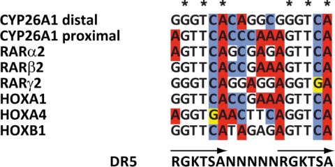
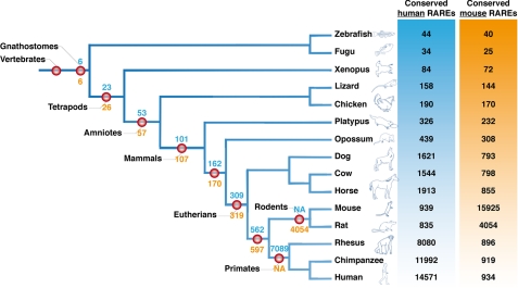
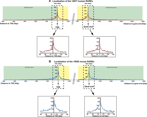
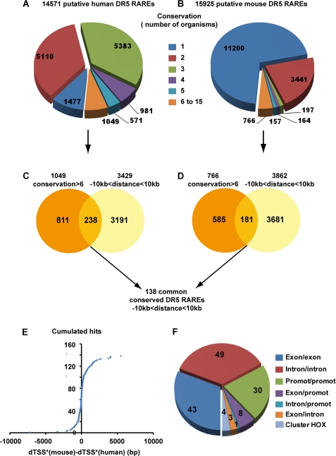
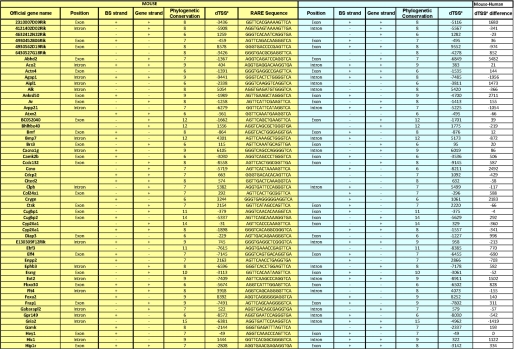
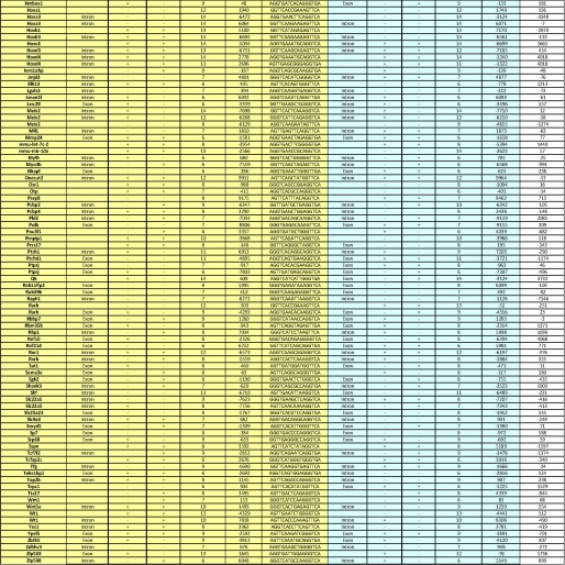
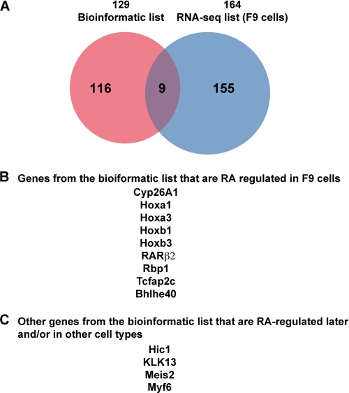
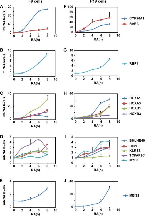
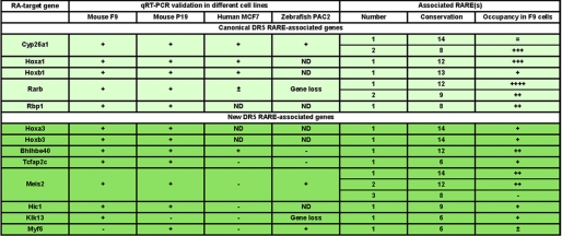
The nuclear retinoic acid receptors interact with specific retinoic acid (RA) response elements (RAREs) located in the promoters of target genes to orchestrate transcriptional networks involved in cell growth and differentiation. Here we describe a genome-wide in silico analysis of consensus DR5 RAREs based on the recurrent RGKTSA motifs. More than 15,000 DR5 RAREs were identified and analyzed for their localization and conservation in vertebrates. We selected 138 elements located ±10 kb from transcription start sites and gene ends and conserved across more than 6 species. We also validated the functionality of these RAREs by analyzing their ability to bind retinoic acid receptors (ChIP sequencing experiments) as well as the RA regulation of the corresponding genes (RNA sequencing and quantitative real time PCR experiments). Such a strategy provided a global set of high confidence RAREs expanding the known experimentally validated RAREs repertoire associated to a series of new genes involved in cell signaling, development, and tumor suppression. Finally, the present work provides a valuable knowledge base for the analysis of a wider range of RA-target genes in different species.


Figures

FIGURE 1.
FIGURE 1.
Alignments of known DR5 RARE motifs in the promoters of the Cyp26A1, RARα2, RARβ2, RARγ2, Hoxa1, Hoxa4, and Hoxb1 genes and definition of a RGKTSA motif.
FIGURE 2.
FIGURE 2.
Phylogenetic tree of jawed vertebrates showing the phylogenetic conservation of the DR5 RAREs. On the right, the number of human RAREs (blue) or mouse RAREs (orange) conserved in each species is indicated. For example, in chimpanzee, there are 11,992 RAREs conserved from the 14,571 found in human. In contrast there are 919 RAREs conserved from the 15,925 found in the mouse genome. At each relevant node of the tree, the number of RAREs conserved in the species of the relevant node is indicated in red. The blue numbers represent the human RAREs, and the orange numbers represent the mouse RAREs. For example, in the rodent primate clade we found 562 human RAREs and 597 mouse RAREs conserved in the 5 relevant species (mouse, rat, rhesus, chimpanzee, and human). NA, not applicable.
FIGURE 3.
FIGURE 3.
Genome-wide distribution of the identified human (A) and mouse (B) DR5 RAREs. The distance between the RARE and the corresponding gene was calculated by identifying its proximity to both boundaries of the genes, the TSS, and the end (end of the last exon). Following this rule for a RARE located upstream of the TSS, the distance was calculated from the TSS and was negative. For a RARE located downstream of a gene, the distance was calculated from the end of the gene and was positive. In the case of a RARE present inside the gene, both distances to the TSS and to the end of the gene were calculated, and the minimal distance in absolute value, called dTSS*, was considered (cf. Fig. 5). The genome-wide mapping of the RARE versus a canonical gene was calculated by cumulating the number of hits present in a 1-kb sliding window from each side from the TSS or from the end of the gene (the first point being attributed to 500 bp before and after TSS or gene end). These calculations were applied to a distance of 500 kb outside of the gene and 100 kb inside of the gene.
FIGURE 4.
FIGURE 4.
Selection of 138 RAREs located ±10 kb from TSSs and gene ends and conserved in more than 6 organisms. A and B, shown is conservation of the human and mouse RAREs among the 15 organisms tested. C and D, for both the human and mouse genomes, the RAREs conserved in more than 6 organisms were crossed with those located ±10 kb from gene boundaries. Crossing the resulting mouse and human RAREs led to a list of 138 RAREs with highly confident conservation and located ±10kb. E, the differences between mouse and human dTSS* were calculated and plotted into cumulative hits. F, conservation of the RARE positions (intron, exon, and promoter) between mouse and human is shown.
FIGURE 5.
FIGURE 5.
List of the 138 conserved RAREs located ±10 kb from TSSs and genes ends. For the 138 conserved RAREs located ±10 kb, gene names and orthology were analyzed manually in both the human and mouse genomes. The sequence, localization, and dTSS* of each RARE are shown as well as the name of the associated gene. BS, binding sequence.
FIGURE 5.
FIGURE 5.
List of the 138 conserved RAREs located ±10 kb from TSSs and genes ends. For the 138 conserved RAREs located ±10 kb, gene names and orthology were analyzed manually in both the human and mouse genomes. The sequence, localization, and dTSS* of each RARE are shown as well as the name of the associated gene. BS, binding sequence.
FIGURE 6.
FIGURE 6.
RA-responsiveness of the conserved DR5 RARE-associated genes identified in silico as assessed by RNA-seq in F9 cells. A, shown are Venn diagrams. B and C, shown is a summary of the RA-regulated genes.
FIGURE 7.
FIGURE 7.
Real time RT-PCR analysis of the RA regulation of the genes associated to the conserved DR5 RAREs identified in silico in F9 (A–E) and P19 (F–J) mouse embryocarcinoma cells. The results correspond to the mean ± S.D. of three independent experiments.
FIGURE 8.
FIGURE 8.
Real time RT-PCR analysis of the RA regulation of the genes associated to the conserved DR5 RAREs in human MCF7 (A) and zebrafish PAC2 (B) cells. The results correspond to the mean ± S.D. of three independent experiments.
FIGURE 9.
FIGURE 9.
Recapitulation of the conserved DR5 RAREs that are RA-activated in mouse embryocarcinoma cells (F9 and P19 cell lines), human breast cancer cells (MCF7 cells), and a zebrafish cell line (PAC2 cells).

Similar articles

Cited by

References

    1. Bour G., Taneja R., Rochette-Egly C. (2006) in Nuclear Receptors in Development (Taneja R. ed.) pp. 211–253, Elsevier Science Publishing Co., Inc., New York
    1. Duong V., Rochette-Egly C. (2011) Biochim. Biophys. Acta 1812, 1023–1031 - PubMed
    1. Mark M., Ghyselinck N. B., Chambon P. (2009) Nucl. Recept. Signal. 7, e002 - PMC - PubMed
    1. Samarut E., Rochette-Egly C. (2011) Mol. Cell. Endocrinol., in press - PubMed
    1. Rochette-Egly C., Germain P. (2009) Nucl. Recept. Signal. 7, e005 - PMC - PubMed

Publication types

MeSH terms

LinkOut - more resources