. 2011 Sep 30;286(39):34346-55.
doi: 10.1074/jbc.M111.278549. Epub 2011 Jul 29.

Acetylation of heat shock protein 20 (Hsp20) regulates human myometrial activity

Affiliations

Acetylation of heat shock protein 20 (Hsp20) regulates human myometrial activity

Magdalena Karolczak-Bayatti et al. J Biol Chem. .

Abstract

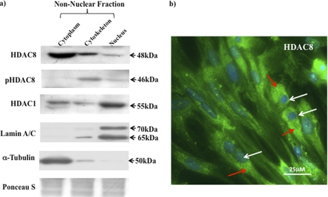
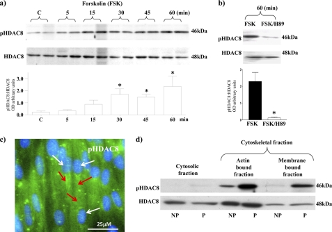
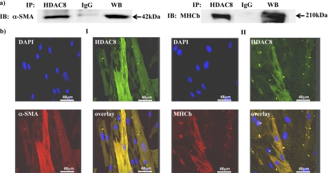
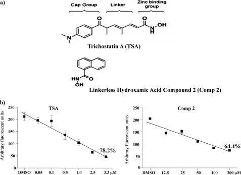
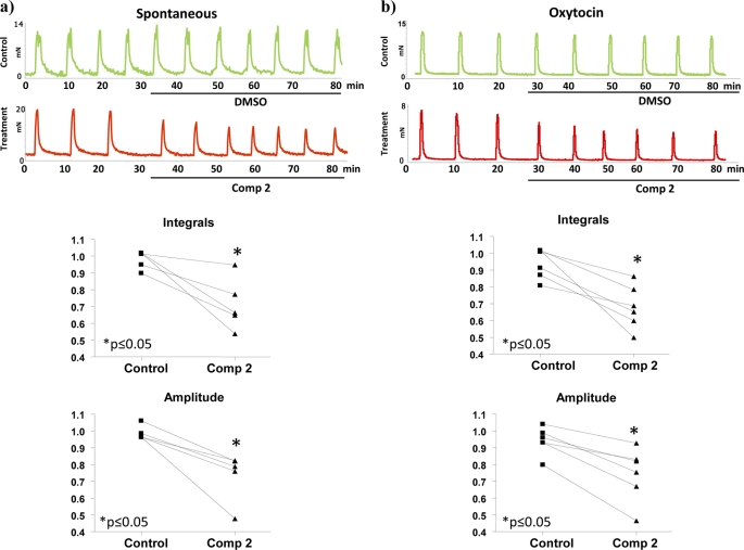
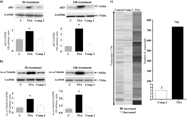
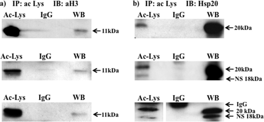
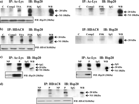
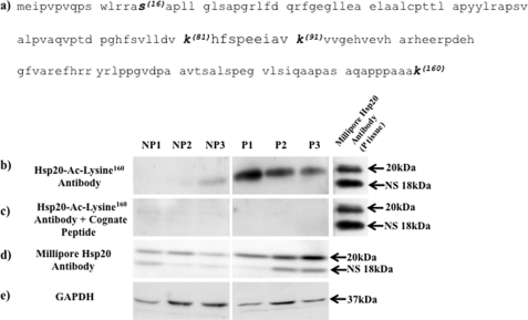
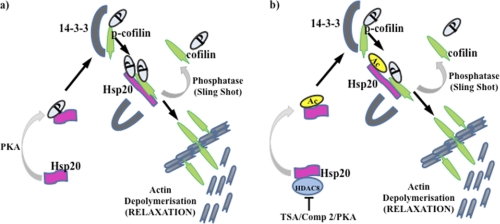
Phosphorylation of heat shock protein 20 (Hsp20) by protein kinase A (PKA) is now recognized as an important regulatory mechanism modulating contractile activity in the human myometrium. Thus agonists that stimulate cyclic AMP production may cause relaxation with resultant beneficial effects on pathologies that affect this tissue such as the onset of premature contractions prior to term. Here we describe for the first time that acetylation of Hsp20 is also a potent post-translational modification that can affect human myometrial activity. We show that histone deacetylase 8 (HDAC8) is a non-nuclear lysine deacetylase (KDAC) that can interact with Hsp20 to affect its acetylation. Importantly, use of a selective linkerless hydroxamic acid HDAC8 inhibitor increases Hsp20 acetylation with no elevation of nuclear-resident histone acetylation nor marked global gene expression changes. These effects are associated with significant inhibition of spontaneous and oxytocin-augmented contractions of ex vivo human myometrial tissue strips. A potential molecular mechanism by which Hsp20 acetylation can affect myometrial activity by liberating cofilin is described and further high-lights the use of specific effectors of KDACs as therapeutic agents in regulating contractility in this smooth muscle.


Figures

FIGURE 1.
FIGURE 1.
Intracellular localization of HDAC8 in human myometrial cells. a, localization of the HDAC1, HDAC8 and pHDAC8 in cytoplasmic/cytoskeletal (non-nuclear) and nuclear extracts from NP myometrial cells. Purity of the individual fractions was monitored with antibodies to Lamin A/C (nuclear fractions) and to α-tubulin (cytoplasmic/cytoskeletal) fractions. Ponceau S was used to stain gels for equal loading. b, immunofluorescent microscopy staining with the HDAC8 antibody (green and indicated by red arrows) and DAPI (nuclei, blue and indicated by white arrows). Results were repeated in a further two individual patient samples.
FIGURE 2.
FIGURE 2.
PKA phosphorylation of HDAC8 and cytoskeletal expression of pHDAC8 in myometrial cells. a, forskolin (FSK) (10 μm) treatment increased phosphorylation of HDAC8 (pHDAC8) from 5–60 min. HDAC8 staining was used as a control for total levels of expression. Data are expressed as mean ± S.E. *, p < 0.05, Students t test compared with control untreated cells, n = 4. b, co-treatment with FSK (10 μm) and the PKA inhibitor H89 (10 μm) for 60 min decreased pHDAC8 levels with no change in total HDAC8 levels. Data are expressed as mean ± S.E. *, n = 3. c, immunofluorescent microscopy staining of myometrial cells with the pHDAC8 antibody (green and indicated by red arrows) and DAPI (nuclei, blue and indicated by white arrows). d, subcellular localization of pHDAC8 and HDAC8 in NP and P tissues. Results were repeated in a further two individual patient samples.
FIGURE 3.
FIGURE 3.
Immunoprecipitation and co-localization of HDAC8 with α-SMA and MHCh. a, NP myometrial cell lysates were immunoprecipitated with the HDAC8 antibody and control IgG sera and precipitated proteins subjected to Western blotting with α-SMA and MHCh antibodies. WB, positive control. b, immunofluorescent microscopy staining showing co-localization of HDAC8 and cytoskeletal proteins. Panel I: HDAC8 antibody (green), α-SMA (red) DAPI (nuclei, blue), and the overlay (yellow); Panel II: HDAC8 antibody (green), ΜΗCh (red) DAPI (blue), and the overlay (yellow). n = 4 individual patient samples.
FIGURE 4.
FIGURE 4.
TSA and Compound 2 inhibit HDAC8 activity in vitro. a, structure of TSA in comparison to the linkerless hydroxamic acid derivative Compound 2 (Comp 2). b, concentration response curve of HDAC8 activity as measured by an in vitro assay. Results are derived from three assays done in duplicate. TSA and Comp 2 inhibited HDAC8 activity by 78.2 and 64.4% at 3.3 μm and at 100–200 μm, respectively.
FIGURE 5.
FIGURE 5.
Compound 2 inhibits spontaneous and oxytocin-induced myometrial contractility. Treatment with Compound 2 (100 μm) reduced both (a) spontaneous and (b) oxytocin-augmented (2 nm) contractions for up to 1 h compared with vehicle-treated controls. Integrals for contractions were calculated from three consecutive peaks (25–35 min). Data points on the graphs represent ratios of the integrals before and after treatment with vehicle or Compound 2. Significant decreases in spontaneous and oxytocin induced contractions (Integrals) were observed upon treatment with Compound 2. (p ≤ 0.05 Wilcoxon matched pair test) due to significant decreases in peak amplitudes. The duration of contractions and half peak durations were not significantly changed. n = 5–6 individual patient samples.
FIGURE 6.
FIGURE 6.
The effect TSA and Compound 2 on acetylation of histone H3/α-tubulin and myometrial gene expression. Treatment of P myometrial strips with TSA (3.3 μm) increased levels of (a) acetylated histone H3 (aH3) and (b) acetylated α-tubulin after 3 h and 24 h treatment. Compound 2 (Comp 2) (100 μm) did not affect acetylation of either of these proteins. GAPDH staining was used as a loading control. Data are expressed as mean ± S.E. *, p < 0.05, Student's t test TSA compared with control and Comp 2. n = 5 individual patient samples. c, P myometrial strips were incubated in the presence or absence of Compound 2 (Comp 2) (100 μm) or TSA (3.3 μm) for 24 h and mRNA extracted and subjected to gene microarray analysis. A heat-map of genes that change with Compound 2 (Comp 2) and TSA treatment compared with control is shown as well as the number of genes that change with Compound 2 (Comp 2) and TSA treatment normalized to the control. (n = 4 individual patient samples).
FIGURE 7.
FIGURE 7.
Acetylated lysine antibodies immunoprecipitate Hsp20 in TSA-treated tissues. NP myometrial strips were treated with TSA (3.3 μm) for 24 h, and protein lysates subjected to immunoprecipitation with a pool of 3 acetylated ϵ-lysine antibodies and then Western blotting with (a) an acetylated H3 (aH3) antibody and (b) an Hsp20 antibody. Note for the third replicates in (b) lanes were run on the same gel but were non-contiguous (line). WB, positive control. Note that the Millipore Hsp20 antibody also recognizes a nonspecific (NS) band of 18 kDa.
FIGURE 8.
FIGURE 8.
Acetylation of Hsp20 involves inhibition of HDAC8 in myometrial tissues. a, NP tissues were treated in the absence or presence of Compound 2 (Comp 2) (100 μm), TSA (3. 3 μm), and forskolin (FSK) (10 μm) for 24 h and lysates immunoprecipitated with a pool of 3 acetylated ϵ-lysine antibodies with the appropriate IgG sera control and then Western blotting with the Hsp20 antibody. Boxed insert below indicates total Hsp20 protein levels in the tissues. b, treated NP tissue lysates as in (a) were immunoprecipitated with the HDAC8 antibody and then subjected to Western blotting with the Hsp20 antibody. Lanes were run on the same gel but were non-contiguous (line). Boxed insert below indicates total HDAC8 protein levels in the tissues. Replicates of treatments with Compound 2 and forskolin in (a) and (b) are also shown. Here, lanes were run on the same gels but were non-contiguous (lines). Further data is provided in supplemental Fig. S3, a and b. c, NP and P tissue lysates were immunoprecipitated with a pool of 3 acetylated ϵ-lysine antibodies and subjected to Western blotting with the Hsp20 antibody. Boxed insert below indicates total Hsp20 protein levels in the tissues. Lanes were run on the same gel but were non-contiguous (line). Replicates of (c) are also shown. Further data is provided in supplemental Fig. S3c. d, NP and P tissue lysates were immunoprecipitated with the HDAC8 antibody and subjected to Western blotting with the Hsp20 antibody. Boxed insert below indicates total HDAC8 protein levels in the tissues. Further data are provided in supplemental Fig. S3d. Note that the Millipore Hsp20 antibody also recognizes a nonspecific (NS) band of 18 kDa.
FIGURE 9.
FIGURE 9.
Hsp20 is acetylated on the lysine 160 residue and is highly expressed in P compared with NP tissues. a, amino acid sequence of Hsp20 indicating the phosphorylation (S-Serine16) and potential acetylation (K-Lysine81,91,160) sites within the protein. The Hsp20-Ac-Lysine160 antibody was raised against a peptide sequence containing amino acids 155–160 of human Hsp20 that encompassed acetylated lysine160 and used at 1:3,000 dilution. b, Western blotting with the Hsp20-Ac-Lysine160 antibody indicates that the lysine160 residue is acetylated in the myometrium and that acetylated Hsp20 is highly expressed in P compared with NP tissues. c, no bands were observed when the Hsp20-Ac-Lysine160 antibody was pre-incubated for 1 h with the cognate peptide (40 μg/ml) to which it was raised prior to Western-blotting of NP and P tissues; thus indicating specificity of the 20 kDa band seen in b. d, control Western blotting with the Millipore Hsp20 antibody (see Table 1) confirmed increased total Hsp20 expression levels in P compared with NP tissues. e, Western blots were re-probed with the GAPDH antibody as a loading control. For b–e, samples were run on the same gels but were non-contiguous (lines). For b and c, P tissues were probed with the Millipore Hsp20 antibody as a positive control. Note that the Millipore antibody also recognizes a nonspecific (NS) band of 18 kDa. Data were obtained from three individual NP and P patient samples.
FIGURE 10.
FIGURE 10.
Inhibition of HDAC8 results in immunoprecipitation with cofilin and a decrease in p-cofilin levels. a, immunoprecipitation of lysates from NP myometrial strips treated with forskolin (FSK) (10 μm) for 24 h with the HDAC8 antibody and subjected to Western blotting with a cofilin antibody. Boxed insert below indicates total cofilin protein levels in the tissues. b, immunoprecipitation of lysates from NP and P myometrial strips with the HDAC8 antibody and subjected to Western blotting with a cofilin antibody. Boxed insert below indicates total cofilin protein levels in the tissues. Replicates of a and b are also shown. IgG, negative control sera. WB, positive control. c and d, H89 (10 μm) treated NP myometrial strips were incubated with or without TSA (3.3 μm) and Compound 2 (Comp 2) (100 μm) for 1 h and 5 h, respectively and protein lysates subjected to Western blotting with a p-cofilin antibody and a cofilin antibody to monitor total cofilin levels. Lanes in c were run on the same gel but were non-contiguous (line). Results were derived from three individual patient samples and are expressed as mean ± S.E. *, p < 0.05, Student's t test H89/TSA and H89/Comp 2 compared with H89 controls.
FIGURE 11.
FIGURE 11.
Model of Hsp20 phosphorylation and acetylation regulating myometrial actin dynamics. a, phosphorylation of Hsp20 by protein kinase A (PKA) results in displacement of p-cofilin from 14-3-3 with subsequent dephosphorylation by phosphatases such as “sling shot” resulting in liberation of cofilin, which influences the depolymerization of actin at membrane-associated dense bodies. b, acetylation of Hsp20 may act independently or in conjunction with phosphorylation to further modulate actin filamental dynamics. P, phosphorylation, Ac, acetylation.

References

    1. Goldenberg R. L., Culhane J. F., Iams J. D., Romero R. (2008) Lancet 371, 75–84 - PMC - PubMed
    1. Moynihan A. T., Hehir M. P., Sharkey A. M., Robson S. C., Europe-Finner G. N., Morrison J. J. (2008) Am. J. Obstet Gynecol 199, 167.e1–7 - PubMed
    1. Kim S. C., Sprung R., Chen Y., Xu Y., Ball H., Pei J., Cheng T., Kho Y., Xiao H., Xiao L., Grishin N. V., White M., Yang X. J., Zhao Y. (2006) Mol. Cell 23, 607–618 - PubMed
    1. Choudhary C., Kumar C., Gnad F., Nielsen M. L., Rehman M., Walther T. C., Olsen J. V., Mann M. (2009) Science 325, 834–840 - PubMed
    1. MacIntyre D. A., Tyson E. K., Read M., Smith R., Yeo G., Kwek K., Chan E. C. (2008) Endocrinology 149, 245–252 - PubMed

Publication types

MeSH terms