. 2011 Sep;77(18):6357-67.
doi: 10.1128/AEM.05203-11. Epub 2011 Jul 29.

Role of glucosyltransferase B in interactions of Candida albicans with Streptococcus mutans and with an experimental pellicle on hydroxyapatite surfaces

Affiliations

Role of glucosyltransferase B in interactions of Candida albicans with Streptococcus mutans and with an experimental pellicle on hydroxyapatite surfaces

S Gregoire et al. Appl Environ Microbiol. 2011 Sep.

Abstract

Candida albicans and mutans streptococci are frequently detected in dental plaque biofilms from toddlers afflicted with early childhood caries. Glucosyltransferases (Gtfs) secreted by Streptococcus mutans bind to saliva-coated apatite (sHA) and to bacterial surfaces, synthesizing exopolymers in situ, which promote cell clustering and adherence to tooth enamel. We investigated the potential role Gtfs may play in mediating the interactions between C. albicans SC5314 and S. mutans UA159, both with each other and with the sHA surface. GtfB adhered effectively to the C. albicans yeast cell surface in an enzymatically active form, as determined by scintillation spectroscopy and fluorescence imaging. The glucans formed on the yeast cell surface were more susceptible to dextranase than those synthesized in solution or on sHA and bacterial cell surfaces (P < 0.05), indicating an elevated α-1,6-linked glucose content. Fluorescence imaging revealed that larger numbers of S. mutans cells bound to C. albicans cells with glucans present on their surface than to yeast cells without surface glucans (uncoated). The glucans formed in situ also enhanced C. albicans interactions with sHA, as determined by a novel single-cell micromechanical method. Furthermore, the presence of glucan-coated yeast cells significantly increased the accumulation of S. mutans on the sHA surface (versus S. mutans incubated alone or mixed with uncoated C. albicans; P < 0.05). These data reveal a novel cross-kingdom interaction that is mediated by bacterial GtfB, which readily attaches to the yeast cell surface. Surface-bound GtfB promotes the formation of a glucan-rich matrix in situ and may enhance the accumulation of S. mutans on the tooth enamel surface, thereby modulating the development of virulent biofilms.


Figures
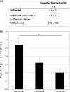
Fig. 1.
Fig. 1.
Amount of enzyme activity of GtfB adsorbed to C. albicans SC5314 cells. δ, 1 U of Gtf activity was defined as the amount of enzyme that incorporates 1 μmol of glucose into glucan over 2 h. C. albicans cells were incubated with GtfB and washed to remove unbound enzyme. Cells with adsorbed GtfB (or without GtfB bound, as a control) were then exposed to sucrose, and glucan formation was assayed after 2 h of incubation. (a) Amounts of enzyme activity of GtfB adsorbed on the yeast cell surface and unbound GtfB (data shown are means ± standard deviations; n=12). (b) Amounts of glucans formed according to the number of yeast cells available for GtfB binding. The asterisk indicates that the values (error bars shown standard deviations; n=12) are statistically significantly different from each other (P < 0.05, ANOVA, comparison for all pairs using Tukey's test).
Fig. 2.
Fig. 2.
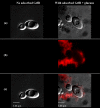
Visualization of glucans synthesized in situ by GtfB adsorbed to C. albicans SC5314 yeast cells. C. albicans cells were incubated with GtfB (or buffer) and washed to remove unbound GtfB. Cells with adsorbed GtfB (or without GtfB bound, as a control) were then exposed to sucrose, and glucan formation was assayed after 2 h of incubation. (a) DIC image of C. albicans cells after incubation with sucrose (100× oil objective, numerical aperture 1.4). (b) Image obtained with laser excitation at 633 nm for detection of glucans (Alexa Fluor 647). (c) Overlaid DIC and fluorescence images.
Fig. 3.
Fig. 3.
S. mutans UA159 cells binding to C. albicans SC5314 cells with and without surface-formed GtfB glucans. Cells of C. albicans (with or without surface-formed GtfB glucans) and S. mutans were incubated together (1 h) and then analyzed using DIC and fluorescence imaging. (a) DIC image of C. albicans and S. mutans cells after incubation (100× oil objective, numerical aperture 1.4). (b) DIC image of GtfB glucan on C. albicans and S. mutans cells after incubation. (b-1) Overlaid DIC and fluorescence images of the field of view selected in panel b. Laser excitation at 488 nm was used for detection of SYTO 9-labeled S. mutans cells (green), and laser excitation at 633 nm was used for detection of Alexa Fluor 647-labeled GtfB glucans (red).
Fig. 4.
Fig. 4.
Adherence of C. albicans (with or without in situ GtfB glucan) to experimental salivary pellicle (sHA) and to glucans synthesized in the pellicle (gsHA) formed on a hydroxyapatite surface. The data shown are mean values ± standard deviations (n=12). All of the values are statistically significantly different from each other (P < 0.05, ANOVA, comparison for all pairs using Tukey's test), but the difference between the sHA/Ca and sHA/gCa values is not statistically significant (P > 0.05).
Fig. 5.
Fig. 5.
Interaction between a sHA bead and a single yeast cell during a micropipette experiment. (a) Initial contact (when micropipette 2 moves toward micropipette 1 until the sHA bead touches the yeast cell). (b) No adhesion (after the bead is pulled back beyond the initial contact point). (c) Adhesive event (after the bead is pulled back with the attached cell beyond the initial contact point). An adhesive event is recorded when micropipette 1 (holding a C. albicans yeast cell) moves beyond the initial contact point due to binding of the cell to the sHA bead (immobilized by micropipette 2).
Fig. 6.
Fig. 6.
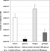
Influence of C. albicans (with or without surface-formed glucan) on S. mutans accumulation on experimental salivary pellicle (sHA) formed on a hydroxyapatite surface. The data shown are mean values ± standard deviations (n=12). The value marked with an asterisk is significantly different from the others (P < 0.05, ANOVA, comparison for all pairs using Tukey's test). Sm, S. mutans; Ca, C. albicans; Ca-GtfB, C. albicans with surface-adsorbed GtfB (no glucan); Ca-GtfB-glucan, C. albicans with surface GtfB glucan.

Similar articles

Cited by

References

    1. Ajdić D., et al. 2002. Genome sequence of Streptococcus mutans UA159, a cariogenic dental pathogen. Proc. Natl. Acad. Sci. U. S. A. 99: 14434–14439 - PMC - PubMed
    1. Ambatipudi K. S., et al. 2010. Human common salivary protein 1 (CSP-1) promotes binding of Streptococcus mutans to experimental salivary pellicle and glucans formed on hydroxyapatite surface. J. Proteome Res. 9: 6605–6614 - PMC - PubMed
    1. Bamford C. V., et al. 2009. Streptococcus gordonii modulates Candida albicans biofilm formation through intergeneric communication. Infect. Immun. 77: 3696–3704 - PMC - PubMed
    1. Banas J. A., Vickerman M. M. 2003. Glucan-binding proteins of the oral streptococci. Crit. Rev. Oral Biol. Med. 14: 89–99 - PubMed
    1. Berkowitz R. J., et al. 2009. Adjunctive chemotherapeutic suppression of mutans streptococci in the setting of severe early childhood caries. J. Public Health Dent. 69: 163–167 - PMC - PubMed

Publication types

LinkOut - more resources