. 2011 Jul;23(7):2659-79.
doi: 10.1105/tpc.111.087320. Epub 2011 Jul 29.

Arabidopsis mutants deleted in the light-harvesting protein Lhcb4 have a disrupted photosystem II macrostructure and are defective in photoprotection

Affiliations

Arabidopsis mutants deleted in the light-harvesting protein Lhcb4 have a disrupted photosystem II macrostructure and are defective in photoprotection

Silvia de Bianchi et al. Plant Cell. 2011 Jul.

Abstract

The role of the light-harvesting complex Lhcb4 (CP29) in photosynthesis was investigated in Arabidopsis thaliana by characterizing knockout lines for each of the three Lhcb4 isoforms (Lhcb4.1/4.2/4.3). Plants lacking all isoforms (koLhcb4) showed a compensatory increase of Lhcb1 and a slightly reduced photosystem II/I ratio with respect to the wild type. The absence of Lhcb4 did not result in alteration in electron transport rates. However, the kinetic of state transition was faster in the mutant, and nonphotochemical quenching activity was lower in koLhcb4 plants with respect to either wild type or mutants retaining a single Lhcb4 isoform. KoLhcb4 plants were more sensitive to photoinhibition, while this effect was not observed in knockout lines for any other photosystem II antenna subunit. Ultrastructural analysis of thylakoid grana membranes showed a lower density of photosystem II complexes in koLhcb4. Moreover, analysis of isolated supercomplexes showed a different overall shape of the C₂S₂ particles due to a different binding mode of the S-trimer to the core complex. An empty space was observed within the photosystem II supercomplex at the Lhcb4 position, implying that the missing Lhcb4 was not replaced by other Lhc subunits. This suggests that Lhcb4 is unique among photosystem II antenna proteins and determinant for photosystem II macro-organization and photoprotection.


Figures
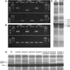
Figure 1.
Figure 1.
Genetic and Biochemical Characterization of the koLhcb4 Mutant (Triple Mutant for the Three Isoforms of Lhcb4). (A) Amplification of Lhcb4.1, Lhcb4.2, and Lhcb4.3 loci with allele-specific PCR primers. Top panel: amplification using gene-specific primers. Bands of 1378, 520, and 1046 bp were obtained for the amplification of the Lhcb4.1, Lhcb4.2, and Lhcb4.3 loci, respectively. Bottom panel: amplification using T-DNA–specific primers. Bands of 685, 661, and 773 bp were obtained for the amplification of Lhcb4.1, Lhcb4.2, and Lhcb4.3 KO loci, respectively. Details of primer sequences are reported in Methods. WT, wild type. (B) RT-PCR measurement of gene-specific transcripts. Sequences of the oligonucleotides used are reported in Methods. Top panel: for each gene, RNA extracted from the wild type and the corresponding mutant was subjected to reverse transcription, followed by 30 cycles of PCR amplification. Bottom panel: amplification of the housekeeping gene actin2 transcript from the same RNAs used as loading control. M, molecular weight marker. The expected sizes of the PCR products are as follows: Lhcb4.1, 724 bp; Lhcb4.2, 715 bp; Lhcb4.3, 730 bp; and actin, 384 bp. Each RT-PCR measurement was repeated three times. (C) SDS-PAGE analysis of wild-type and koLhcb4 mutant thylakoid proteins performed with the Tris-Tricine buffer system (Schägger and von Jagow, 1987). Selected apoprotein bands are marked. Purified thylakoid sample, corresponding to 15 μg of chlorophylls, was loaded in each lane. (D) SDS-PAGE analysis performed with the Tris-Tricine buffer system with the addition of 7 M urea to the running gel in order to separate Lhcb4 isoforms in the Lhcb4 KO mutants. Selected apoprotein bands are marked. Fifteen micrograms of chlorophylls were loaded in each lane.
Figure 2.
Figure 2.
Polypeptide Composition of Thylakoid Membranes from Wild-Type and koLhcb4 Mutant. (A) Immunoblotting used for the quantification of photosynthetic subunits in the wild type (WT) and koLhcb4 thylakoids. Immunoblot analysis was performed with antibodies directed against individual gene products: minor antenna proteins, the LHCII subunit, the PSII core subunit PsbB (CP47), the PSI core subunit (PsaA), cytochrome f, and plastocyanin. Thylakoids corresponding to 0.25, 0.5, 0.75, and 1 μg of chlorophyll were loaded for each sample. All samples were loaded on the same SDS-PAGE slab gel. (B) Results of the immunotitration of thylakoid proteins. Data of PSII antenna subunits were normalized to the core amount, PsbB content (Ballottari et al., 2007), and expressed as a percentage of the corresponding wild-type content. Significantly different values from wild-type membranes are marked with an asterisk. (C) Thylakoid pigment-protein complexes were separated by nondenaturing Deriphat-PAGE upon solubilization with α-DM. Thylakoids corresponding to 25 μg of chlorophylls were loaded in each lane. [See online article for color version of this figure.]
Figure 3.
Figure 3.
Characterization of Photosynthetic Electron Flow in Wild-Type and koLhcb4 Plants. (A) Dependence of the LEF rate on light intensity in wild-type (WT) and koLhcb4 leaves. LEF rate is calculated as ϕPSIIPSII·PAR·Aleaf·fractionPSII (see Methods for details). (B) Photosynthetic oxygen evolution in saturating CO2. Rate of oxygen evolution was measured on whole leaves during illumination with various levels of actinic red light. (C) Amplitude of qL measured at different light intensities on wild-type and koLhcb4 leaves. qL reflects the redox state of the primary electron acceptor QA, thus, the fraction of open PSII centers. (D) and (E) Measurement of state I–state II transition in the wild type (D) and koLhcb4 (E). Upon 1-h dark adaptation, plants were illuminated with blue light (40 μmol photons m−2 s−1, wavelength <500 nm) for 15 min to reach state II. A far-red light source was then superimposed on the blue light to induce a transition to state I. Values of Fm, Fm′, and Fm′′ were determined using light saturation pulses (4500 μmol photons m−2 s−1, 0.6 s). a.u., arbitrary units. (F) and (G) Dependence of the P700 oxidation ratio (ΔA/ΔAmax) on light intensity. ΔA/ΔAmax was measured on wild-type and koLhcb4 leaves at varying actinic intensities, exciting either PSII + PSI (λ = 630 nm; [F]) or PSI only (λ = 730 nm; [G]).
Figure 4.
Figure 4.
Kinetics of the Formation and Relaxation of Photoprotective Energy Dissipation. (A) Measurements of NPQ kinetics on wild-type (WT) and koLhcb4 leaves illuminated with 1200 μmol photons m−2 s−1, 24°C. (B) NPQ kinetics of wild-type and koLhcb4 plants during two consecutive periods of illumination with white light (1200 μmol photons m−2 s−1, 25 min, 24°C) with an 18-min period of darkness in between, as indicated by the white and black bars. (C) Time course of Viola deepoxidation in wild-type and koLhcb4 plants. Leaf discs from dark-adapted leaves were illuminated at 1200 μmol photons m−2 s−1, 24°C (white actinic light). At different times, discs were frozen in liquid nitrogen and total pigments extracted before HPLC analysis. (D) NPQ kinetics of the wild type, koLhcb4, and mutants retaining single Lhcb4 isoforms. The expression of the isoform Lhcb4.1 (koLhcb4.2 4.3) leads to complete compensation of the NPQ phenotype of koLhcb4; instead, the expression of the Lhcb4.2 isoform partially recovered the quenching ability in the first minutes of induction; nevertheless the mutant failed to fully match the wild type quenching capacity within 8 min of illumination. The presence of the Lhcb4.3 gene did not contribute to NPQ activity. Symbols and error bars show means ± sd (n > 3).
Figure 5.
Figure 5.
Photooxidation of Arabidopsis Wild Type and koLhcb4 Mutant Exposed to HL and Low Temperature. (A) PSII photoinhibition (Fv/Fm decay) was followed in the wild type (WT), koLhcb4, and antenna-depleted mutant (koLhcb5 Lhcb6, koLhcb3, and LHCII antisense) plants, treated at 550 μmol photons m−2 s−1, 4°C for 30 h with a 6-h period of low light (20 μmol photons m−2 s−1) between the 12 h of HL stress; low-light interval permitted the PSII efficiency recovery. (B) and (C) Detached leaves floating on water were treated at 1500 μmol photons m−2 s−1 at 4°C, and kinetics of chlorophyll bleaching (B) and MDA formation (C) were recorded. (D) Wild-type and mutant detached leaves were vacuum infiltrated with 5 μM SOSG, a 1O2-specific fluorogenic probe. SOSG increases its fluorescence emission upon reaction with singlet oxygen. The increase in the probe emission was followed with a fiber optic on the leaf surface during illumination with red actinic light (550 μmol photons m−2 s−1) at 4°C. a.u., arbitrary units.
Figure 6.
Figure 6.
Photoinhibition of the Wild Type and Mutants Retaining a Single Lhcb4 Isoform Exposed to HL and Low Temperature. Kinetics of Fv/Fm decay were measured on the wild type (WT), koLhcb4, koLhcb4.1 4.2, koLhcb4.1 4.3, and koLhcb4.2 4.3. Whole plants were treated as described for Figure 5A.
Figure 7.
Figure 7.
Kinetics of Lipid Peroxidation of Arabidopsis Detached Leaves Exposed to HL Stress. Wild-type, npq1, koLhcb4, and koLhcb4 npq1 mutant leaves floating on water were exposed to HL (800 μmol photons m−2 s−1, 4°C), and photooxidation was estimated from the extent of lipid peroxidation measured by high-temperature TL. Each experimental point corresponds to a different sample. The kinetic of oxidized lipid accumulation was described by fitting the data set with first-order exponential functions: differential kinetics npq1–wild type (WT) and koLhcb4 npq1koLhcb4 were calculated on the basis of the first-order exponential functions obtained by fitting experimental points (error bars, 95% confidence level). a.u., arbitrary units.
Figure 8.
Figure 8.
Electron Microscopy of Grana Membranes and PSII Particles. (A) and (B) Electron microscopy of negative staining grana partition membranes were obtained by partial solubilization of thylakoids from koLhcb4 (A) or wild-type (WT) plants (B) with α-DM. High-resolution micrographs show the distribution of stain-excluding tetrameric particles (arrows). Grana partitions from koLhcb4 (A) were characterized by the presence of tetrameric particles more widely spaced than wild-type membranes (B). Arrows indicate PSII core complexes (magenta) and LHCII region (violet). (C) to (E) Average projection map of a set of 1024 projections of C2S2 supercomplexes from plants lacking Lhcb4 ([C] and [D]) and the wild type (E). Contours representing PSII dimeric core (green), Lhcb4 (yellow), and whole wild-type C2S2 supercomplex (red) are superimposed. [See online article for color version of this figure.]

Similar articles

Cited by

References

    1. Ahn T.K., Avenson T.J., Ballottari M., Cheng Y.C., Niyogi K.K., Bassi R., Fleming G.R. (2008). Architecture of a charge-transfer state regulating light harvesting in a plant antenna protein. Science 320: 794–797 - PubMed
    1. Alboresi A., Ballottari M., Hienerwadel R., Giacometti G.M., Morosinotto T. (2009). Antenna complexes protect Photosystem I from photoinhibition. BMC Plant Biol. 9: 71. - PMC - PubMed
    1. Alboresi A., Caffarri S., Nogue F., Bassi R., Morosinotto T. (2008). In silico and biochemical analysis of Physcomitrella patens photosynthetic antenna: Identification of subunits which evolved upon land adaptation. PLoS ONE 3: e2033. - PMC - PubMed
    1. Alboresi A., Dall’osto L., Aprile A., Carillo P., Roncaglia E., Cattivelli L., Bassi R. (2011). Reactive oxygen species and transcript analysis upon excess light treatment in wild-type Arabidopsis thaliana vs a photosensitive mutant lacking zeaxanthin and lutein. BMC Plant Biol. 11: 62. - PMC - PubMed
    1. Allen J.F. (1992). Protein phosphorylation in regulation of photosynthesis. Biochim. Biophys. Acta 1098: 275–335 - PubMed

Publication types

MeSH terms

LinkOut - more resources