

doi: 10.1128/JB.05235-11.
Epub 2011 Jul 29.
Quantitative proteomic analysis reveals formation of an EscL-EscQ-EscN type III complex in enteropathogenic Escherichia coli
Affiliations
- PMID: 21804003
- PMCID: PMC3187409
- DOI: 10.1128/JB.05235-11
Item in Clipboard
Quantitative proteomic analysis reveals formation of an EscL-EscQ-EscN type III complex in enteropathogenic Escherichia coli
J Bacteriol.
2011 Oct.
Abstract
We characterized Orf5 and SepQ, two type III secretion (T3S) system proteins in enteropathogenic Escherichia coli, and showed that they are essential for T3S, associated with the bacterial membrane, and interact with EscN. Our findings suggest that Orf5 and SepQ are homologs of YscL and YscQ from Yersinia, respectively.
Figures
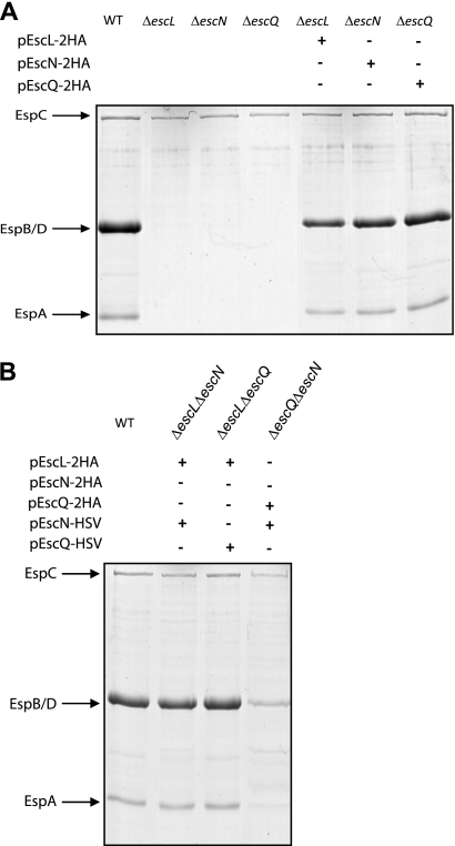
EscL, EscN, and EscQ are essential for type III secretion in EPEC. (A) Protein secretion profiles of the EPEC WT, ΔescL, ΔescN, and ΔescQ strains as well as the null mutants complemented with a double-HA-tagged version of the missing protein (pEscL-2HA, pEscN-2HA, or pEscQ-2HA). Secreted proteins were concentrated from supernatants of bacterial cultures grown in DMEM and analyzed by SDS–12.5% PAGE and Coomassie staining. The locations of the translocators EspA, EspB, and EspD are indicated at the left of the gel. The location of EPEC EspC, which is not secreted via the LEE-encoded T3SS, is also indicated. (B) Protein secretion profiles of EPEC double-deletion strains complemented with the two missing proteins. Secreted proteins were processed as described for panel A.
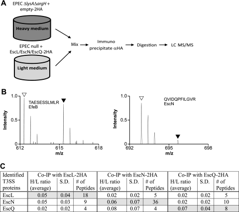
SILAC screen for interacting proteins EscL, EscN, and EscQ. (A) Schematic representation of the SILAC method. The EPECΔlysAΔargH strain expressing the double-HA tag and an EPEC null mutant complemented with the HA-tagged protein were differentially labeled, and lysates were mixed in a 1:1 ratio. Proteins were immunoprecipitated with the α-HA antibody and then trypsinized and analyzed by LC-MS/MS. (B) Representative mass spectra for peptides recovered from the SILAC experiment performed with EscL-2HA. Right panel: open and filled triangles indicate the expected mass charge (m/z) of light and heavy forms, respectively, of a peptide (QVIDQPFILGVR) from the specifically bound EscN from pulldowns with EscL-2HA and 2HA, respectively. The light peptide of EscN is present in a ratio >3.0 higher than the heavy peptide, indicating a specific interaction with EscL. Left panel: open and filled triangles indicate the expected m/z of light and heavy forms, respectively, of a peptide (TAESESSLMLR) from the nonspecifically bound EfeB protein from pulldowns with EscL-2HA and 2HA, respectively. (C) Summary of LEE-encoded proteins identified by LC-MS/MS as specifically immunoprecipitated with EscL-2HA, EscN-2HA, or EscQ-2HA in the SILAC experiments. H/L ratios represent the average values for the heavy/light isotope ratio that were determined among several peptides from either two or three independent immunoprecipitation experiments. S. D., standard deviations.
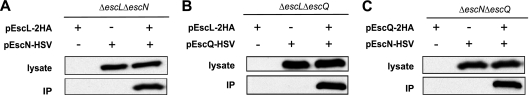
EscL, EscN, and EscQ interact with each other. (A) EscL-2HA and EscN-HSV were expressed alone or together in an EPECΔescLΔescN mutant. Lysates were immunoprecipitated with α-HA antibody. Lysates and eluted fractions (IP) were blotted against HSV. EscN-HSV was immunoprecipitated only in the presence of EscL-2HA. (B) EscL-2HA immunoprecipitated EscQ-HSV by the use of a procedure similar to that described for panel A. (C) EscQ-2HA immunoprecipitated EscN-HSV by the use of a procedure similar to that described for panel A.
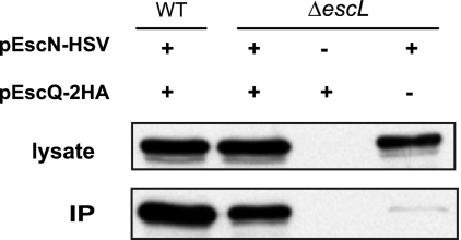
EscN-EscQ interaction is independent of EscL. EscN-HSV and EscQ-2HA were individually expressed or coexpressed in WT EPEC and in EPECΔescL strains. Lysates were immunoprecipitated with an α-HA antibody. Lysates and eluted fractions (IP) were immunoblotted against the HSV epitope.
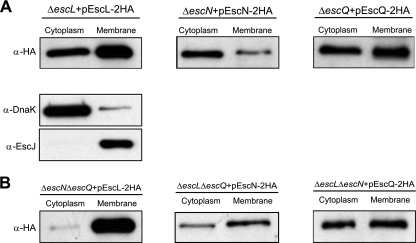
Subcellular localization of EscL, EscN, and EscQ. Bacteria expressing tagged versions of EscL, EscN, or EscQ were fractionated into soluble and membrane fractions. Samples (2 μg of each fraction) were resolved by SDS–12.5% PAGE, transferred to a polyvinylidene fluoride membrane, and probed with α-HA antisera. DnaK and EscJ were used as markers for the cytoplasmic and membrane fractions, respectively. (A) Localization was examined in the complemented strains. (B) Localization of the proteins was examined in double-mutant strains.
References
-
- Elliott S. J., et al. 1998. The complete sequence of the locus of enterocyte effacement (LEE) from enteropathogenic Escherichia coli E2348/69. Mol. Microbiol. 28:1–4 - PubMed
Publication types
MeSH terms
Substances
Grants and funding
LinkOut - more resources
Full Text Sources
Other Literature Sources
Molecular Biology Databases
