. 2011 Oct;193(19):5207-15.
doi: 10.1128/JB.05170-11. Epub 2011 Jul 29.

Peptide-regulated gene depletion system developed for use in Streptococcus pneumoniae

Affiliations

Peptide-regulated gene depletion system developed for use in Streptococcus pneumoniae

Kari Helene Berg et al. J Bacteriol. 2011 Oct.

Abstract

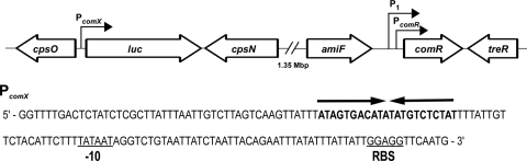
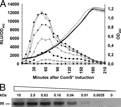
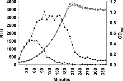
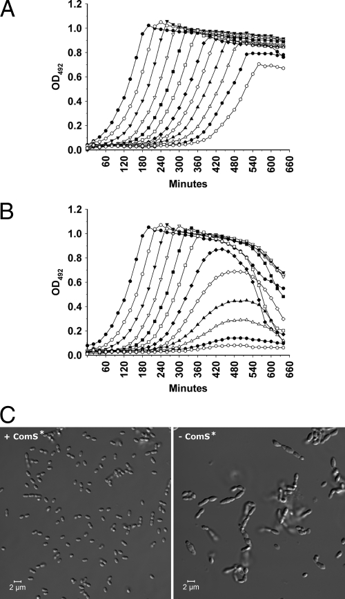
To facilitate the study of pneumococcal genes that are essential for viability or normal cell growth, we sought to develop a tightly regulated, titratable gene depletion system that interferes minimally with normal cellular functions. A possible candidate for such a system is the recently discovered signal transduction pathway regulating competence for natural transformation in Streptococcus thermophilus. This pathway, which is unrelated to the ComCDE pathway used for competence regulation in Streptococcus pneumoniae, has not been fully elucidated, but it is known to include a short unmodified signaling peptide, ComS*, an oligopeptide transport system, Ami, and a transcriptional activator, ComR. The transcriptional activator is thought to bind to an inverted repeat sequence termed the ECom box. We introduced the ComR protein and the ECom box into the genome of S. pneumoniae R6 and demonstrated that addition of synthetic ComS* peptide induced the transcription of a luciferase gene inserted downstream of the ECom box. To determine whether the ComRS system could be used for gene depletion studies, the licD1 gene was inserted behind the chromosomally located ECom box promoter by using the Janus cassette. Then, the native versions of licD1 and licD2 were deleted, and the resulting mutant was recovered in the presence of ComS*. Cultivation of the licD1 licD2 double mutant in the absence of ComS* gradually affected its ability to grow and propagate, demonstrating that the ComRS system functions as intended. In the present study, the ComRS system was developed for use in S. pneumoniae. In principle, however, it should work equally well in many other Gram-positive species.


Figures

Fig. 1.
Fig. 1.
Schematic diagram depicting two genetic regions of S. pneumoniae mutant SPH130 containing the inserted PcomX::luc and P1::PcomR::comR constructs. The promoters PcomX, PcomR, and P1 are indicated. The sequence of PcomX is shown below the diagram, with the predicted ComR-binding site (ECom box) indicated by arrows (9). PcomR represents the native ComR promoter, while P1 represents a synthetic constitutive promoter inserted upstream of PcomR. The Pribnow box and the ribosome-binding site (RBS) are underlined.
Fig. 2.
Fig. 2.
Bioluminescence (A) and Western analyses (B) showing the level of Luc expression after subjecting SPH130 cells to various concentrations of ComS*. (A) SPH130 cell cultures were induced at an OD492 of 0.1 with the following concentrations of ComS*: 0 (◆), 0.08 μM (□), 0.16 μM (■), 0.31 μM (Δ), 0.63 μM (▴), 1.25 μM (○), or 10 μM (●). Luminescence relative to cell density (in relative light units [RLU]/OD492) is indicated by lines with symbols, while lines without symbols indicate bacterial growth. Concentrations of ComS* higher than 1 to 2 μM did not increase the emitted light intensity, suggesting a saturation of the system, whereas induction with lower concentrations of ComS* displayed less production of light in a dose-dependent manner. The data shown are from a representative experiment of several replicates. (B) Detection of the luciferase enzyme by Western analysis after induction for 1 h with different concentrations of ComS*. ComS* was added to SPH130 cell cultures at an OD492 of 0.1. Luciferase was detected by using a polyclonal antiluciferase antibody produced in rabbits. The concentrations of ComS* used are indicated in μM above the respective protein bands. The bands appearing immediately below the full-size luciferase bands represent degradation products. Our results showed that the luciferase enzyme is very unstable in S. pneumoniae.
Fig. 3.
Fig. 3.
Decay of luciferase activity over time after removal of ComS* from the growth medium. The SPH130 strain was grown either in the presence of 2 μM ComS* (circles) or in the absence of ComS* (triangles). Bacterial growth is indicated by open symbols, whereas luminescence is indicated by filled symbols. After shifting the cells at time zero from a medium containing 2 μM ComS* to a ComS*-free medium, it took about 70 min before the luciferase activity started to decline (▴). In the parallel culture grown in the presence of ComS*, the luciferase activity started to decline as the culture approached stationary phase (●).
Fig. 4.
Fig. 4.
Effects of licD1 depletion on growth and morphology of SPH135 cultures. (A) A culture of SPH135 cells grown to an OD492 of 0.3 in C medium supplemented with 2 μM ComS* was pelleted, washed once in plain C medium, and resuspended to an OD492 of 0.05 in fresh C medium containing 2 μM ComS*. Then, the culture was 2-fold diluted in the same medium in a 96-well plate and incubated in a Fluostar Optima luminometer at 37°C for 11 h. (B) The same culture of SPH135 cells was washed and resuspended in ComS*-free medium but otherwise treated as describe for panel A. In cells growing in the presence of ComS* (A), ectopic expression of licD1 is driven by the ComRS system. In cells growing in ComS*-free medium (B), ectopic expression of licD1 is gradually reduced. About 5 h after the cells were shifted from a ComS*-containing to a ComS*-free medium, growth of LicD1-depleted cells started to slow down. A few hours later the growth stopped completely, and the cells started to lyse (B). The data shown are from a representative experiment of several replicates. (C) Examination of licD1-proficient (+ComS*) and licD1-deficient (−ComS*) SPH135 cells by DIC microscopy. Samples of licD1-proficient and licD1-deficient cells were collected at the transition between logarithmic and stationary phases (at 480 min) from the cultures represented by open triangles (see panels A and B). The pictures shown are representative several independent experiments. The morphology of SPH135 cells grown in the presence of ComS* was indistinguishable from that of wild-type pneumococci, while the morphology of licD1-depleted cells was clearly abnormal.
Fig. 5.
Fig. 5.
Depletion of the Kanr gene makes the SPH136 mutant sensitive to kanamycin. SPH136 cell cultures were grown in C medium containing 2 μM ComS* until they reached an OD492 of 0.3. Then, they were pelleted, washed once, and resuspended to an OD492 of 0.05 in ComS*-free C medium containing kanamycin (400 μg ml−1). The resuspended cells were 2-fold diluted in the same medium in a 96-well plate and incubated in a Fluostar Optima luminometer at 37°C for 16 h. Growth (measured as the OD492) was determined automatically by the luminometer at 10-min intervals. As Kanr was depleted over time, the growth rate of the SPH136 cells gradually slowed down. After being cultivated for about 8 h in ComS*-free medium, their growth was completely inhibited by kanamycin.
Fig. 6.
Fig. 6.
Expression levels of luc relative to the expression levels of cbpD measured by real-time RT-PCR. The late competence gene cbpD is only expressed in cells that are competent for natural genetic transformation. (A) Background transcription of luc was examined in SPH130 cells grown without ComS* to an OD550 of 0.1. (B and C) Additional samples from the same culture were collected 30 (B) and 60 min (C) later and analyzed in the same way. The results showed that the background expression of luc in uninduced cells was 10 to 30 times higher than the background expression of cbpD in noncompetent cells. Comparison of luc expression levels in cells induced with 2 μM ComS* for 30 and 60 min (D and E) with those of noninduced cells run in parallel (B and C) showed that addition of ComS* increased luc expression about 1,500-fold.

References

    1. Baur S., Marles-Wright J., Buckenmaier S., Lewis R. J., Vollmer W. 2009. Synthesis of CDP-activated ribitol for teichoic acid precursors in Streptococcus pneumoniae. J. Bacteriol. 191:1200–1210 - PMC - PubMed
    1. Blomqvist T., Steinmoen H., Håvarstein L. S. 2006. Natural genetic transformation: a novel tool for efficient genetic engineering of the dairy bacterium Streptococcus thermophilus. Appl. Environ. Microbiol. 72:6751–6756 - PMC - PubMed
    1. Chan P. F., et al. 2003. Characterization of a novel fucose-regulated promoter (PfcsK) suitable for gene essentiality and antibacterial mode-of-action studies in Streptococcus pneumoniae. J. Bacteriol. 185:2051–2058 - PMC - PubMed
    1. Chastanet A., Prudhomme M., Claverys J. P., Msadek T. 2001. Regulation of Streptococcus pneumoniae clp genes and their role in competence development and stress survival. J. Bacteriol. 183:7295–7307 - PMC - PubMed
    1. Damjanovic M., Kharat A. S., Eberhardt A., Tomasz A., Vollmer W. 2007. The essential tacF gene is responsible for the choline-dependent growth phenotype of Streptococcus pneumoniae. J. Bacteriol. 189:7105–7111 - PMC - PubMed

Publication types

MeSH terms

Substances

LinkOut - more resources