. 2011 Sep 1;187(5):2492-501.
doi: 10.4049/jimmunol.1101671. Epub 2011 Jul 29.

The multiple shark Ig H chain genes rearrange and hypermutate autonomously

Affiliations

The multiple shark Ig H chain genes rearrange and hypermutate autonomously

Catherine Zhu et al. J Immunol. .

Abstract

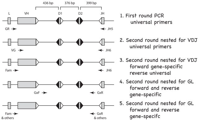
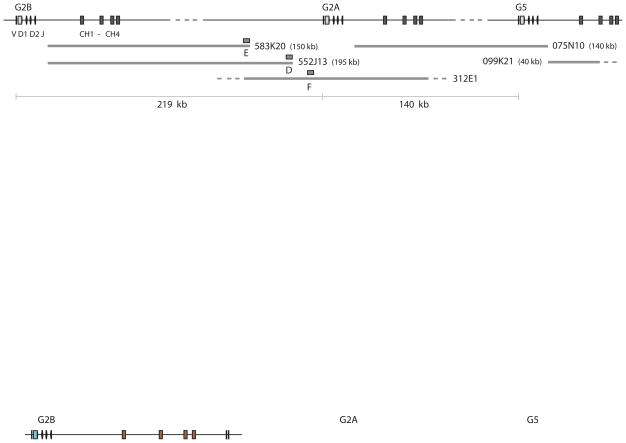
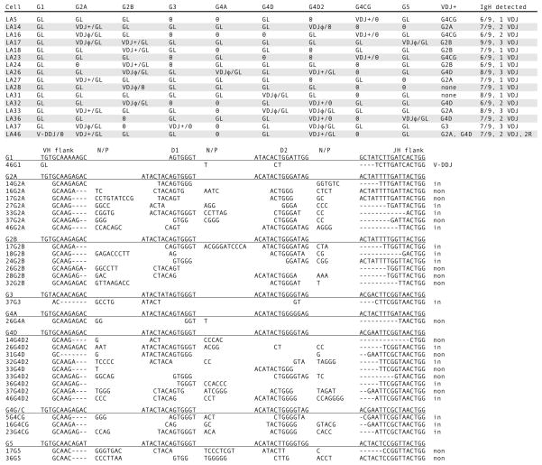
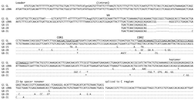
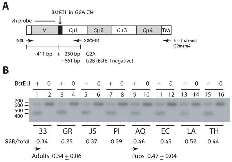
Sharks and skates are representatives of the earliest vertebrates with an immune system based on V(D)J rearrangement. They possess a unique Ig gene organization consisting of 15 to >50 individual IgM loci, each with one VH, two DH, one JH, and one set of constant region exons. The present study attempts to understand how multiple Ig genes are regulated with respect to rearrangement initiation and to targeting during somatic hypermutation. The linkage of three single-copy IgH genes was determined, and single-cell genomic PCR studies in a neonatal animal were used to examine any relationship between relative gene position and likelihood of rearrangement. Our results show that one to three IgH genes are activated independently of linkage or allelic position and the data best fit with a probability model based on the hypothesis that V(D)J rearrangement occurs as a sequence of trials within the B cell. In the neonatal cell set, two closely related IgH, G2A, and G2B, rearranged at similar frequencies, and their membrane forms were expressed at similar levels, like in other young animals. However, older animals displayed a bias in favor of the G2A isotype, which suggests that although rearrangement at G2A and G2B was randomly initiated during primary repertoire generation, the two very similar IgM sequences appear to be differentially expressed with age and exposure to Ag. We performed genomic single-cell PCR on B cells from an immunized individual to study activation-induced cytidine deaminase targeting and found that hypermutation, like V(D)J rearrangement, occurred independently among the many shark IgH.


Figures

Figure 1
Figure 1
Flow diagram for single-cell PCR in shark B cells. The rearranging gene segments (VH, D1, D2, JH) are separated by ~400 bp; the specific distances are from the gene G2A; L is leader. The flanking recombination signal sequences (RSS) with 23 bp spacers are represented as open triangles and RSS with 12 bp spacers filled triangles. Arrows indicate position and orientation of PCR primers. Line 1. First round PCR. A 40-cycle PCR was performed on B cells and on RBC, using “universal” primers detecting all functional IgH. Thereafter, the PCR products were depleted of oligonucleotides and used in the following four kinds of nested second-round reactions (25–30 cycles). Line 2. Second round nested for VDJ. Universal primers targeting FR1 and JH can amplify VDJ and/or GL fragments in B cells but only GL in the RBC samples. Line 3. Second round nested for VDJ. Specific forward primers reveal which subfamily (G1, G2, G3, G4, G5) the VDJ belongs to. VDJ is isolated and sequenced. Line 4. Second round nested for GL. Subfamily specific primers in the V-D1 and D2-J intersegmental regions amplify the non-rearranged genes. The five subfamily reactions are analyzed by restriction enzyme analyses detailed in Fig. S2, Supplementary Materials. Line 5. Second round nested for GL. The unrearranged VH is amplified for sequencing by using subfamily or gene-specific primers. Primer information is supplied in Materials and Methods.
Figure 2
Figure 2
Mapping of IgH genes G2B, G2A, G5. The orientation and order of the IgM H chain genes G2B, G2A and G5 are shown at top, with the VH gene segments and C exons, labeled at left. The distance between G2B leader and its transmembrane exons is 17.9 kb; dotted line indicates map is not drawn to scale. BAC clones, drawn as thick gray lines, include two clones carrying only the C regions of G2B (583K20, 552J13), whose insert-end sequences (box E and D) hybridized to BAC clone 312E1. The F sequence from 312E1 (box F) was located 4.4 kb upstream of the G2A leader and 16 kb downstream of the D sequence. BAC clone 075N10 bridged the C exons of G2A and the VH gene segments of G5 [15]. BAC clone 099K21 carried only the G5 C exons and extended 35 kb beyond CH1. The distances are shown at the bottom, as determined between the genes. The entire area includes: distance from G2B to G2A (219 kb), from G2A to G5 (140 kb), and the distance G5 to the end of 099K21 (43 kb), a total of 402 kb. Except for 312E1, the BAC clone insert sizes were determined by pulsed field electrophoresis. Boxes labeled D, E, F represent sequences from which probes were generated.
Figure 3
Figure 3
Single-cell PCR results from shark-LA sIgM+ cells. Top. Rearrangement configurations of the nine IgH in each cell. The IgH genes present in shark-LA were: G1, G2A, G2B, G3, G4A, G4D, G4D2, G4CG, and G5. The status of the Ig gene segments is indicated as fully rearranged (VDJ), partly rearranged as V-DDJ, or not rearranged (GL). In-frame, potentially functional VDJ are indicated by plus (+) sign; nonproductive VDJ have φ sign. Zero indicates that the gene could not be detected. The column VDJ+ shows the probable IgH gene encoding the receptor. IgH detected indicates tally of the genes characterized per B cell and the number of unique VDJ. Bottom. CDR3 of VDJ. Their junctions are shown according to assignment to VH flank, D1 gene, D2 gene, and JH flank, classified according to subfamily. N/P, nontemplated or palindromic sequence. Dashes denote nucleotide deletions at the flanks. At the right, the VDJ are indicated as in-frame or nonproductive (non). V-DDJ is sequence with only two rearrangement (2R) events, where the V-D intersegmental region was not been deleted by recombination.
Figure 4
Figure 4
Mutated pseudogene G8 sequences. The G8 cDNA sequence, consisting of leader, leader intron, germline VH gene segment directly spliced to C region, is aligned to the most closely related functional gene, G1 (top). The genomic germline G1 sequence shows leader, leader intron with splice sites (indicated in lower case) and the VH gene segment demarcated into framework (FR) and complementarity-determining region (CDR, underlined). The recombination signal sequences (heptamer, spacer, nonamer) are labeled. G8 is nonfunctional, due to a mutation disabling the leader intron splice acceptor site. G8 mutants 15, 28 and 31 are compared with the G8 cDNA, with dots representing identity and dashes gaps. The PCR primer is shown in italics.
Figure 5
Figure 5
Comparing G2A and G2B expression in individuals. A. Representation of G2 PCR products and BstE II-digested fragments. The G2 H chain is shown with labeled domains; first strand cDNA primer G2mem4 targets the transmembrane sequence (TM) of both G2A and G2B. PCR primers G2L and G2CH1R generate DNA fragments of ~661 bp (calculated average CDR3 size of 11 codons). G2A sequences can be differentiated from G2B by the BstE II site in G2A JH (black box). Bar over V region represents the vh probe. B. Relative G2A and G2B amounts in pup and adult spleen RNA. RT-PCR products from adult (shark-33 epigonal organ, GR, JS, and PI spleen) and pup RNA (shark-AQ, EC, LA, and TH spleen) were incubated with (+ lanes) or without endonuclease (0 lanes), electrophoresed on a 1.5% TBE gel, and transferred onto a nylon filter (Hybond N+, GE Healthcare). The blot was hybridized to vh probe and submitted for scanning and autoradiography (x-ray film shown). Phosphorimager values obtained: (lane 1) 9759 at 661 bp; 18,497 at 411 bp, (lane 2) 28,405 at 661 bp, (lane 3) 6930; 19,296, (lane 4) 27,624, (lane 5) 9374; 15,764, (lane 6) 24,960, (lane 7) 12,430; 19,732, (lane 8) 31,166, (lane 9) 12,997; 13,720, (lane 10) 27,911, (lane 11) 13,567; 16,951, (lane 12) 30,049, (lane 13) 14,587; 13,441, (lane 14) 27,663, (lane 15) 14,348; 18,677, (lane 16) 32,019. G2B/total is the BstE II-negative band value divided by the value of the untreated sample. For shark-33 (lanes 1 and 2) it would be 9759/18,497 or 0.34 obtained as the proportion of G2B-containing sequences. The mean value with standard deviation is given for adult and pup groups.
Figure 6
Figure 6
Single-cell PCR results from mutant shark-GR sIg+ cells. Top. Rearrangement configurations of the ten IgH in each cell. The IgH genes present in shark-GR were: G1, G2A, G2B, G3, G4A, G4D, G4E, G4C, G4G, and G5. The status of the Ig gene segments is indicated as fully rearranged (VDJ), partly rearranged as VDD-J, or not rearranged (GL). The plus or φ sign indicate respectively in-frame (highlighted) or nonproductive. The mutated VDJ are marked with asterisk. The GL VH has been sequenced in every case except where marked as NR; this means that the GL configuration was detected but a sequence with the VH could not be isolated. Bottom. CDR3 of the VDJ. Six out of nine rearrangements in 5 B cells were mutated. Substitutions indicated in lower case in the CDR3. See Fig. 3 legend. Genbank accession numbers for mutants are: 1G4CG, 15G2A, 23G2A, 25G4D, 49G4CG, and 49G5 (JN087503-JN087508).

Similar articles

Cited by

References

    1. Alt FW, Yancopoulos GD, Blackwell TK, Wood C, Thomas E, Boss M, Coffman R, Rosenberg N, Tonegawa S, Baltimore D. Ordered rearrangement of immunoglobulin heavy chain variable region segments. EMBO J. 1984;3:1209–1219. - PMC - PubMed
    1. Featherstone K, Wood AL, Bowen AJ, Corcoran AE. The mouse immunoglobulin heavy chain V-D intergenic sequence contains insulators that may regulate ordered V(D)J recombination. J Biol Chem. 2010;285:9327–9338. - PMC - PubMed
    1. Jhunjhunwala S, van Zelm MC, Peak MM, Cutchin S, Riblet R, van Dongen JJ, Grosveld FG, Knoch TA, Murre C. The 3D structure of the immunoglobulin heavy-chain locus: implications for long-range genomic interactions. Cell. 2008;133:265–279. - PMC - PubMed
    1. Chowdhury D, Sen R. Stepwise activation of the immunoglobulin μ heavy chain gene locus. EMBO J. 2001;20:6394–6403. - PMC - PubMed
    1. Fuxa M, Skok J, Souabni A, Salvagiotto G, Roldán E, Busslinger M. Pax5 induces V-to-DJ rearrangements and locus contraction of the immunoglobulin heavy-chain gene. Genes Dev. 2004;18:411–422. - PMC - PubMed

MeSH terms

Substances