. 2011 Oct;52(10):1795-809.
doi: 10.1194/jlr.M016741. Epub 2011 Jul 29.

Enhancement by LDL of transfer of L-4F and oxidized lipids to HDL in C57BL/6J mice and human plasma

Affiliations

Enhancement by LDL of transfer of L-4F and oxidized lipids to HDL in C57BL/6J mice and human plasma

David Meriwether et al. J Lipid Res. 2011 Oct.

Abstract

The apoA-I mimetic peptide L-4F [(Ac-D-W-F-K-A-F-Y-D-K-V-A-E-K-F-K-E-A-F-NH2) synthesized from all L-amino acids] has shown potential for the treatment of a variety of diseases. Here, we demonstrate that LDL promotes association between L-4F and HDL. A 2- to 3-fold greater association of L-4F with human HDL was observed in the presence of human LDL as compared with HDL by itself. This association further increased when LDL was supplemented with the oxidized lipid 15S-hydroxy-5Z, 8Z, 11Z, 13E-eicosatetraenoic acid (15HETE). Additionally, L-4F significantly (P = 0.02) promoted the transfer of 15HETE from LDL to HDL. The transfer of L-4F from LDL to HDL was demonstrated both in vitro and in C57BL/6J mice. L-4F, injected into C57BL/6J mice, associated rapidly with HDL and was then cleared quickly from the circulation. Similarly, L-4F loaded onto human HDL and injected into C57BL/6J mice was cleared quickly with T(1/2) = 23.6 min. This was accompanied by a decline in human apoA-I with little or no effect on the mouse apoA-I. Based on these results, we propose that i) LDL promotes the association of L-4F with HDL and ii) in the presence of L-4F, oxidized lipids in LDL are rapidly transferred to HDL allowing these oxidized lipids to be acted upon by HDL-associated enzymes and/or cleared from the circulation.


Figures

Fig.1.
Fig.1.
L-4F association with lipoproteins in human plasma and in C57BL/6J and apoE −/− mouse plasma. 14C-L-4F (50.0 µg with 0.25 µCi) was incubated with 200 µl human plasma for 1 h at 37°C. The mix was separated using FPLC, and radioactivity (•) and cholesterol (○) were determined for the fractions ( ). 14C-L-4F (50 µg with 1.0 µCi) was also incubated with 200 µl of either wild-type C57BL/6J or apoE −/− plasma for 1 h at 37°C. The mixes were fractionated using FPLC, and radioactivity (■, apoE−/−; ▴, BL6) and cholesterol (, apoE−/−; △,BL6) were determined for the fractions ( ). The figure depicts almost complete association with HDL in human and C57BL/6J plasma and preferential association with apoB-containing lipoproteins in plasma of the apoE −/− mouse. (The figure is a representative of four different experiments.)
Fig.2.
Fig.2.
L-4F associates with isolated human HDL and LDL. 15HETE supplementation of LDL increases the association between L-4F and LDL. hHDL and hLDL were isolated from plasma by ultracentrifugation. 1.0 mg of HDL protein ( ) was incubated for 1 h with 14C-L-4F (50 µg; 0.5 µCi), and radioactivity (■) and cholesterol (○) were determined for the isolated fractions. 15HETE enhances the association between L-4F and LDL. In a parallel experiment, samples of 1.0 mg protein of hLDL were supplemented with 0 to 5.0 µg 15HETE. The lipoprotein preparations were then incubated with 14C-L-4F (50 µg; 0.25 µCi), isolated on FPLC and the FPLC fractions were assayed for both radioactivity (◆, LDL only; ▴, LDL + 0.5 µg 15HETE; ■, LDL + 1.0 µg 15HETE; •, LDL + 5.0 µg 15HETE) and cholesterol (◇, LDL only). A dose-dependent increase in association between L-4F and LDL was observed, with calculated total counts in the various LDL fractions of 3,975, 4,653, 5,661 and 8,258 dpm accordingly. A significant 2.07-fold increase was observed between the association of 14C-L-4F with 5.0 µg 15HETE supplemented and with 15HETE unsupplemented LDL (P < 0.001, n = 3) ( ).
Fig.3.
Fig.3.
hLDL enhances the association between L-4F and hHDL, and L-4F, preloaded into hLDL, transfers to mHDL in C57BL/6J plasma. Isolated hLDL and hHDL were coincubated with 14C-L-4F, and the association pattern of L-4F in the coincubation was compared with that in individual hLDL and hHDL controls by FPLC fractionations. 2.0 mg hLDL protein was combined with 2.0 mg hHDL protein and with 14C-L-4F (100 µg; 1.0 µCi). Half was incubated at 37° for 1 h before fractionation on the FPLC, and the other half was fractionated after 5 min. In parallel, 1.0 mg hLDL protein or 1.0 mg hHDL protein was separately incubated with 14C-L-4F for 1 h before FPLC fractionation. Radioactivity (◆, LDL only; ■, HDL only; •, LDL+HDL 60 min) and cholesterol (○, LDL+HDL 60 min) were determined for the isolated fractions ( ). Radioactivity and cholesterol for the 5 min sample were very similar to those of the 60 min sample (data not shown). In a separate experiment, hLDL was first preloaded with 14C-L-4F by incubating 2.0 mg LDL protein with 100 µg (1.0 µCi) 14C-L-4F before processing by FPLC to remove unbound peptide. The LDL fractions were pooled and concentrated, and the hLDL-14C-L-4F concentrate was incubated for 1 h with isolated C57BL/6J plasma. The reaction mixture was fractionated by FPLC, and radioactivity (•) and cholesterol (○) were determined for the individual fractions ( ). This result was not plasma donor-specific and was reproducible with three different donor pairs of hHDL and hLDL preparations as well as using isolated plasma from a different mouse.
Fig.4.
Fig.4.
Loading of hLDL with 15HETE enhances the association between L-4F and hHDL compared with unsupplemented hLDL. 1.0 mg LDL protein supplemented with 0.5 µg 15HETE (see Materials and Methods) was added to 1.0 mg HDL protein and incubated with 14C-L-4F (50 µg with 0.5 µCi) for 1 h at 37°. The mixture was then analyzed by FPLC and radioactivity was determined for the isolated fractions. HDL by itself or a combination of LDL and HDL controls were treated in the same manner [◆, HDL; ■, HDL+LDL; ▴, HDL+ (LDL+15HETE)]. An increase of 1.9-fold and 2.9-fold in the uptake of L-4F by HDL was observed in the presence of LDL and the presence of 15HETE supplemented LDL, respectively (a representative of two separate experiments is depicted in ). In a separate experiment, HDL, LDL, or a mixture of both were incubated as above, isolated on FPLC; and total cholesterol was determined for the isolated fractions (○, LDL; ◇, HDL; □, HDL+LDL). No significant net transfer of total cholesterol between the lipoproteins was observed ( ). (Both are representatives of three different experiments.)
Fig.5.
Fig.5.
L-4F enhances the transfer of 15HETE from hLDL onto hHDL. LDL was supplemented with 2.5 µg deuterated 15HETE (15HETE-d8) per mg of LDL protein. 0.5 mg of HDL protein and 0.5 mg of the 15HETE-d8 supplemented LDL protein were then incubated at 37°C for 1.0 hr with or without L-4F. The mixes were fractionated on the FPLC. The isolated lipoprotein fractions were pooled and lipids were extracted from those pooled fractions. Supplemented LDL alone was treated similarly. The extracts were analyzed using LC/MS/MS. The figure shows the amount of 15HETE-d8 present in the isolated lipoproteins as a percentage of the amount originally present in the LDL. A 35-fold increase in the transferred 15HETE-d8 is observed in the presence of L-4F (P = 0.02) n = 3.
Fig.6.
Fig.6.
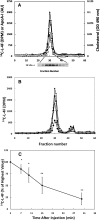
L-4F injected into the circulation of C57BL/6J mice associates rapidly and primarily with the HDL fraction of the plasma and then is cleared quickly from the circulation. 14C-L-4F (600 µg; 0.5 µCi) was injected directly into the circulation of each of five female C57BL/6J mice. Blood was collected at various time points, and the plasma was fractionated using FPLC. Radioactivity (•), cholesterol (□), and apoA-I (▴) were determined for the individual FPLC fractions of the 3 min bleed of a representative mouse ( ). Radioactivity (◆, 3 min; ■, 7 min; ▴, 15 min; •, 30 min) was determined for the FPLC fractions from all time point bleeds of that same representative mouse ( ). Radioactivity (•) in the FPLC fractions of five mice at all time points was determined. The average amount of radioactivity in the HDL fraction for each time point for the different mice is expressed as a percent of the 3 min value for each mouse. ( ). A time-dependent decline in radioactivity is observed. Significant reductions are observed between 3 min and all other time points (*: P < 0.01; **: P < 0.001). Further significant reductions were observed between 7 and 15 min (P < 0.01) and 7 and 30 min (P < 0.001); and between 10 and 30 min (P < 0.01)(n = 5).
Fig.7.
Fig.7.
Radioactivity and apoA-I from 14C-L-4F loaded hHDL injected via tail vein into C57Bl/6J mice are cleared quickly from the circulation. Increasing amounts of 14C-L-4F (0 ug to 200 µg) were loaded into samples of hHDL (1.0 mg protein). Each of the samples was then tail vein-injected into separate male C57Bl/6J mice. Blood was collected from each mouse immediately after injection (0 min) and then at 30, 60, and 120 min post injection. Radioactivity in plasma aliquots was determined for each collected sample (X, 0 µg 14C-L-4F; □, 5.0 µg 14C-L-4F; △, 50 µg 14C-L-4F; ○, 100 µg 14C-L-4F; ◇, 200 µg 14C-L-4F). A time-dependent decline of radioactivity in the plasma is shown for each of the injected L-4F concentrations with average T1/2 = 23.6 min (SD = 5.0 min) ( ). Plasma samples were also analyzed by Western blots for human-specific apoA-I using mouse albumin as a loading control. The amount of human apoA-I present in each sample is expressed as a percent of the amount at time = 0 for that mouse or, in the case of 0 µg 14C-L-4F, for those two mice (X, 0 µg 14C-L-4F; ■, 5.0 µg 14C-L-4F; ▴, 50 µg 14C-L-4F; •, 100 µg 14C-L-4F; ◆, 200 µg 14C-L-4F). Time-dependent decrease in human apoA-I is shown for the HDL samples loaded with L-4F. The most significant decrease in human apoA-I is shown for the 120 min, the longest bleed time, for all 14C-L-4F loaded samples. Western blots show human apoA-I across all four bleed times for both the mouse injected with hHDL loaded with 100 µg 14C-L-4F and for a control injection with no 14C-L-4F ( ). The same plasma bleeds were probed for mouse-specific apoA-I. The amount of mouse apoA-I present in each sample is expressed as a percent of the amount at time = 0 (X, 0 µg 14C-L-4F; ■, 5.0 µg 14C-L-4F; ▴, 50 µg 14C-L-4F; •, 100 µg 14C-L-4F; ◆, 200 µg 14C-L-4F) ( ). Unlike human apoA-I, mouse A-I showed little to no decline across time in 14C-L-4F amounts of up to 100 µg and only 12% decline is observed at 200 µg 14C-L-4F. Western blots show mouse apoA-I across all four bleed times for both the mouse injected with hHDL loaded with 100 µg 14C-L-4F and for a control mouse injected with hHDL without supplementation with 14C-L-4F. Lastly, total HDL cholesterol in these same plasma samples was determined (X, 0 µg 14C-L-4F; □, 5.0 µg 14C-L-4F; △, 50 µg 14C-L-4F; ○, 100 µg 14C-L-4F; ◇, 200 µg 14C-L-4F) ( ). HDL cholesterol showed a time-dependent decline consistent with the decline in human apoA-I. (The figure is a representative of two separate experiments.)
Fig.8.
Fig.8.
14C-L-4F transfers rapidly from preloaded hLDL to mHDL in the circulation of C57BL/6J mice and is then quickly cleared. hLDL was loaded with 14C-L-4F and the complex was purified by FPLC fractionation (see Fig 2B) and concentrated. Approximately 1.25 mg protein of the 14C-L-4F -LDL (0.10 µCi) concentrate was then injected directly into the circulation of each of three male C57BL/6J mice. Blood was drawn at 3, 10, and 60 min post injection. Plasma samples were fractionated by FPLC, and radioactivity and cholesterol were determined for all three mice. Radioactivity in the various FPLC fractions is shown for the three bleed times of a representative mouse (◆, 3 min; ■, 10 min; ▴, 60 min). Over 85% of detectable counts were already transferred to the HDL fraction of the plasma by 3 min. No counts were detected within the LDL fraction by 10 min. At 60 min, the radioactivity associated with the HDL fraction had decreased by over 85% compared with the 3 min bleed time ( ) The cholesterol values associated with this same mouse (◇, 3 min; □, 10 min; △, 60 min) indicate a minimal decline in LDL cholesterol across 60 min ( ). (The figure is a representative of three separate experiments.)
Fig.9.
Fig.9.
L-4F binds to HDL with significantly greater affinity than to LDL. A binding study was performed by surface plasmon resonance (SPR) on a BIAcore 3000 system. L-4F was immobilized on a sensor chip. Analyte solutions of isolated human and mouse lipoproteins were then passed over the chip, and binding was measured by observing the change in SPR angle. Equilibrium affinity constant (KD) values were calculated from assays performed with five different analyte concentrations. The affinities between L-4F and hHDL and mHDL were not significantly different, with KD = 3.36 × 10⁻⁸ and 1.03 × 10⁻⁸ M, respectively. By contrast, the affinity between L-4F and hLDL was significantly less than the affinity between L-4F and either hHDL or mHDL, with KD = 1.17 × 10⁻⁶ M (**, *: P = 0.001, n = 5).

Similar articles

Cited by

References

    1. Anantharamaiah G. M., Jones J. L., Brouillette C. G., Schmidt C. F., Chung B. H., Hughes T. A, Bhown A. S., Segrest J. P. 1985. Studies of synthetic peptide analogs of the amphipathic helix. Structure of complexes with dimyristoyl phosphatidylcholine. J. Biol. Chem. 260: 10248–10255. - PubMed
    1. Anantharamaiah G. M. 1986. Synthetic peptide analogs of apolipoproteins. Methods Enzymol. 128: 627–647. - PubMed
    1. Segrest J. P., Jackson R. L., Morrisett J. D., Gotto A. M., Jr 1974. A molecular theory of lipid-protein interactions in the plasma lipoproteins. FEBS Lett. 38: 247–258. - PubMed
    1. Segrest J. P., Jones M. K., De Loof H., Brouillette C. G., Venkatachalapathi Y. V., Anantharamaiah G. M. 1992. The amphipathic helix in the exchangeable apolipoproteins: a review of secondary structure and function. J. Lipid Res. 33: 141–166. - PubMed
    1. Anantharamaiah G. M., Mishra V. K., Garber D. W., Datta G., Handattu S. P., Palgunachari M. N., Chaddha M., Navab M., Reddy S. T., Segrest J. P., et al. 2007. Structural requirements for antioxidative and anti-inflammatory properties of apolipoprotein A-I mimetic peptides. J. Lipid Res. 48: 1915–1923. - PubMed

Publication types

LinkOut - more resources