. 2011 Sep;22(9):1654-64.
doi: 10.1681/ASN.2010101086. Epub 2011 Jul 29.

Pronephric tubulogenesis requires Daam1-mediated planar cell polarity signaling

Affiliations

Pronephric tubulogenesis requires Daam1-mediated planar cell polarity signaling

Rachel K Miller et al. J Am Soc Nephrol. 2011 Sep.

Abstract

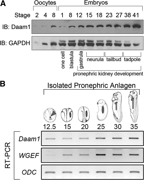
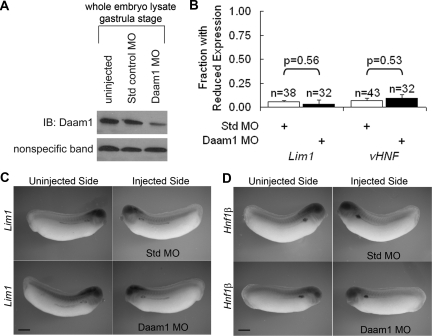
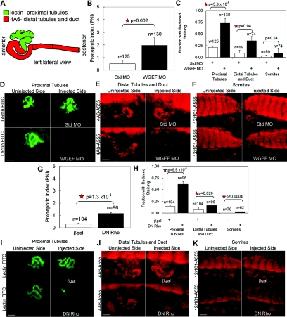
Canonical β-catenin-mediated Wnt signaling is essential for the induction of nephron development. Noncanonical Wnt/planar cell polarity (PCP) pathways contribute to processes such as cell polarization and cytoskeletal modulation in several tissues. Although PCP components likely establish the plane of polarization in kidney tubulogenesis, whether PCP effectors directly modulate the actin cytoskeleton in tubulogenesis is unknown. Here, we investigated the roles of Wnt PCP components in cytoskeletal assembly during kidney tubule morphogenesis in Xenopus laevis and zebrafish. We found that during tubulogenesis, the developing pronephric anlagen expresses Daam1 and its interacting Rho-GEF (WGEF), which compose one PCP/noncanonical Wnt pathway branch. Knockdown of Daam1 resulted in reduced expression of late pronephric epithelial markers with no apparent effect upon early markers of patterning and determination. Inhibiting various points in the Daam1 signaling pathway significantly reduced pronephric tubulogenesis. These data indicate that pronephric tubulogenesis requires the Daam1/WGEF/Rho PCP pathway.


Figures

Figure 1.
Figure 1.
Daam1 protein is detectable during Xenopus pronephric development, with Daam1 and WGEF transcripts expressed in the developing pronephros. (A) Immunoblot (IB) of lysates from stage 2 to 8 oocytes and stage 1 to 41 embryos (approximately 1/2 oocyte or embryo per lane), showing Daam1 and GAPDH (loading control) protein from oocyte through tailbud stages, a timeline encompassing pronephric development (stages 12.5 to 41). (B) RT-PCR of pronephric anlagen isolated from stage 12.5 to 35 embryos showing expression of Daam1 and WGEF, with intensification at stage 25 during pronephric differentiation and morphogenesis.
Figure 2.
Figure 2.
Daam1 knockdown in Xenopus does not affect expression of early pronephric determination and patterning markers. (A) Single-cell embryos were uninjected, injected with 40 ng of Std MO, or injected with 40 ng Daam1 MO. IB analysis from gastrula-stage embryos (approximately 1 embryo equivalent per lane) shows Daam1 protein levels were reduced in Daam1-MO-injected embryos as compared with uninjected or Std-MO-injected embryos. (B through D) The left V2 blastomere of eight-cell stage embryos was injected with 20 ng of Std MO or Daam1 MO. Embryos developed until stage 29 to 30 and were analyzed for Lim1 or Hnf1β expression levels on their injected (left) versus uninjected (right) sides. (B) The fraction of Daam1-depleted embryos with reduced Lim1 and Hnf1β marker expression on the injected side as compared with the uninjected side was similar to that of Std-MO-injected controls. (C) Injected sides of Daam1-MO and Std-MO-injected embryos do not have reduced Lim1 expression. (D) Injected sides of Daam1-MO- and Std-MO-injected animals do not have reduced Hnf1β expression. (C, D) Bars are 500 μm.
Figure 3.
Figure 3.
Daam1 knockdown reduces the expression of late pronephric markers of differentiation and morphogenesis in Xenopus. (A) Diagram of Xenopus pronephric kidney showing positions of proximal tubules, distal tubules, collecting duct, and glomus. (B through F) The left V2 blastomere of eight-cell stage embryos was injected with 20 ng of Std MO or Daam1 MO. Embryos developed until stage 38 to 40 and were analyzed for nphs1, clckb, slc5a1, and atp1a1 expression levels on their injected (left) side as compared with their uninjected (right) side. (B) The fraction with reduced nphs1 marker expression on the injected side as compared with the uninjected side for Daam1-MO-injected animals was similar to that of Std-MO-injected controls, whereas the fraction with reduced clckb, slc5a1, and atp1a1 expression on the injected side as compared with the uninjected side for Daam1-MO-injected animals is greater than that for Std-MO-injected controls. (C) Injected sides of Std-MO- and Daam1-MO-injected animals do not have reduced nphs1 expression. (D) Injected sides of Std-MO-injected animals do not have reduced clckb expression, whereas injected sides of Daam1-MO-injected animals have significantly reduced clckb expression. (E) Injected sides of Std-MO-injected animals do not have reduced slc5a1 expression, whereas injected sides of Daam1-MO-injected animals have significantly reduced slc5a1 expression. (F) Injected sides of Std-MO-injected animals do not have reduced atp1a1 expression, whereas injected sides of Daam1-MO-injected animals have significantly reduced atp1a1 expression. (C through F) Bars are 500 nm.
Figure 4.
Figure 4.
Daam1 knockdown alters morphogenesis of the pronephric tubules and duct and can be rescued by flDaam1 as assessed by markers of the mature pronephros in Xenopus. (A) Diagram of Xenopus pronephric kidney showing positions of lectin staining in the proximal tubules (green) and 4A6 antibody staining in the distal tubules and collecting duct (red). (B through F) The left V2 blastomere of eight-cell stage embryos was injected with 20 ng of Std MO or Daam1 MO. Embryos developed until stage 38 to 40 and were analyzed for lectin, 4A6 antibody, and 12/101 antibody staining within the proximal tubules, distal tubules/duct, and somites, respectively, on their injected (left) side as compared with their uninjected (right) side. (B) The PNI is greater in Daam1-MO-injected animals than in Std-MO-injected animals. (C) The fraction with reduced proximal tubule and distal tubule/duct marker staining on the injected side as compared with the uninjected side for Daam1-MO-injected animals is greater than that for Std-MO-injected controls, whereas the fraction with reduced somite marker staining on the injected side as compared with the uninjected side for Daam1-MO-injected animals was similar to that of Std-MO-injected controls. (D) Injected sides of Std-MO-injected animals do not have reduced proximal tubule marker staining, whereas injected sides of Daam1-MO-injected animals have significantly reduced proximal tubule marker staining. Bar is 100 μm. (E) Injected sides of Std-MO-injected animals do not have reduced distal tubule and duct marker staining, whereas injected sides of Daam1-MO-injected animals have significantly reduced distal tubule and duct marker staining. Bar is 100 μm. (F) Injected sides of Std-MO- or Daam1-MO-injected animals do not have reduced somite marker staining. Bar is 250 μm. (G through M) The left V2 blastomere of eight-cell stage embryos was injected with 20 ng Std MO + 1 ng β-gal, 20 ng Daam1 MO + 1 ng β-gal, or 20 ng Daam1 MO + 1 ng flDaam1. Embryos developed until stage 38 to 40 and were analyzed for lectin, 4A6 antibody, and 12/101 antibody staining within the proximal tubules, distal tubules/duct, and somites, respectively, on their injected (left) side as compared with their uninjected (right) side. (G) The PNI is greater in Daam1-MO + β-gal-injected animals than in Std-MO + β-gal embryos or Daam1-MO + flDaam1-injected rescue animals. (H) The fraction with reduced proximal tubule marker staining on the injected side as compared with the uninjected side for Daam1 MO + β-gal-injected animals was greater than that of Std-MO + β-gal-injected controls. This reduction in proximal tubule staining seen in Daam1 MO + β-gal animals was rescued in Daam1-MO + flDaam1-injected animals. (I) The fraction with reduced distal tubule and duct marker staining on the injected side as compared with the uninjected side for Daam1-MO + β-gal-injected animals was greater than that of the Std-MO + β-gal-injected controls. This reduction in distal tubule and duct staining seen in Daam1 MO + β-gal animals was rescued in Daam1 MO + flDaam1-injected animals. (J) The fraction with reduced somite marker staining on the injected side as compared with the uninjected side for was similar for Std-MO + β-gal-, Daam1-MO + β-gal-, and Daam1-MO + flDaam1-injected animals. (K) Injected sides of Std-MO + β-gal-injected animals do not have reduced proximal tubule marker staining, whereas injected sides of Daam1-MO + β-gal injected animals have significantly reduced proximal tubule marker staining. This loss of proximal tubule marker staining is rescued in Daam1-MO + flDaam1-injected animals. Bar is 100 μm. (L) Injected sides of Std-MO + β-gal-injected animals do not have reduced distal tubule and duct marker staining, whereas injected sides of Daam1-MO + β-gal-injected animals have significantly reduced distal tubule and duct marker staining. This loss of staining with the distal tubule and duct marker is rescued in Daam1-MO + flDaam1-injected animals. Bar is 100 μm. (M) Injected sides of Std-MO + β-gal-, Daam1-MO + β-gal-, and the Daam1-MO + flDaam1-injected animals do not have reduced somite marker staining. Bar is 250 μm.
Figure 5.
Figure 5.
WGEF knockdown and DN Rho alter morphogenesis of the pronephric tubules and duct as assessed by markers of the mature pronephros in Xenopus. (A) Diagram of Xenopus pronephric kidney showing positions of lectin staining in the proximal tubules (green) and 4A6 antibody staining in the distal tubules and collecting duct (red). (B through F) The left V2 blastomere of eight-cell stage embryos was injected with 20 ng of Std MO or WGEF MO. Embryos developed until stage 38 to 40 and were analyzed for lectin, 4A6 antibody, and 12/101 antibody staining within the proximal tubules, distal tubules/duct, and somites, respectively, on their injected (left) side as compared with their uninjected (right) side. (B) The PNI is greater in WGEF-MO-injected animals than in Std-MO-injected animals. (C) The fraction with reduced proximal tubule and distal tubule/duct marker staining on the injected side as compared with the uninjected side for WGEF-MO-injected animals is greater than that for Std-MO-injected controls, whereas the fraction with reduced somite marker staining on the injected side as compared with the uninjected side for WGEF-MO-injected animals was similar to that of Std-MO-injected controls. (D) Injected sides of Std-MO-injected animals do not have reduced proximal tubule marker staining, whereas injected sides of WGEF-MO-injected animals have significantly reduced proximal tubule marker staining. Bar is 100 μm. (E) Injected sides of Std-MO-injected animals do not have reduced distal tubule and duct marker staining, whereas injected sides of WGEF-MO-injected animals have significantly reduced distal tubule and duct marker staining. Bar is 100 μm. (F) Injected sides of Std-MO- or WGEF-MO-injected animals do not have reduced somite marker staining. Bar is 250 μm. (G through K) The left V2 blastomere of eight-cell stage embryos was injected with 200 pg of β-gal or DN Rho. Embryos developed until stage 38 to 40 and were analyzed for lectin and 4A6 antibody staining within the proximal tubules and distal tubules/duct, respectively, on their injected (left) side as compared with their uninjected (right) side. (G) The PNI is greater in the DN-Rho-injected animals than the β-gal-injected animals. (H) The fraction with reduced proximal tubule and distal tubule/duct marker staining on the injected side as compared with the uninjected side for DN-Rho-injected animals is greater than that for β-gal-injected controls. (I) Injected sides of β-gal-injected animals do not have reduced proximal tubule marker staining, whereas injected sides of DN-Rho-injected animals have significantly reduced proximal tubule marker staining. (J) Injected sides of β-gal-injected animals do not have reduced distal tubule and duct marker staining, whereas injected sides of DN-Rho-injected animals have significantly reduced distal tubule and duct marker staining. (I, J) Bars are 100 μm. (K) Injected sides of DN-Rho-injected animals do not have reduced somite marker staining. Bar is 250 μm.

Comment in

  • Making a tubule the noncanonical way.
    Hukriede NA, Dawid IB. Hukriede NA, et al. J Am Soc Nephrol. 2011 Sep;22(9):1575-7. doi: 10.1681/ASN.2011070710. Epub 2011 Aug 11. J Am Soc Nephrol. 2011. PMID: 21836144 No abstract available.

Similar articles

Cited by

References

    1. Park JS, Valerius MT, McMahon AP: Wnt/beta-catenin signaling regulates nephron induction during mouse kidney development. Development 134: 2533–2539, 2007 - PubMed
    1. Lyons JP, Miller RK, Zhou X, Weidinger G, Deroo T, Denayer T, Park JI, Ji H, Hong JY, Li A, Moon RT, Jones EA, Vleminckx K, Vize PD, McCrea PD: Requirement of Wnt/beta-catenin signaling in pronephric kidney development. Mech Dev 126: 142–159, 2009 - PMC - PubMed
    1. Rajagopal J, Carroll TJ, Guseh JS, Bores SA, Blank LJ, Anderson WJ, Yu J, Zhou Q, McMahon AP, Melton DA: Wnt7b stimulates embryonic lung growth by coordinately increasing the replication of epithelium and mesenchyme. Development 135: 1625–1634, 2008 - PMC - PubMed
    1. Shu W, Jiang YQ, Lu MM, Morrisey EE: Wnt7b regulates mesenchymal proliferation and vascular development in the lung. Development 129: 4831–4842, 2002 - PubMed
    1. De Langhe SP, Sala FG, Del Moral PM, Fairbanks TJ, Yamada KM, Warburton D, Burns RC, Bellusci S: Dickkopf-1 (DKK1) reveals that fibronectin is a major target of Wnt signaling in branching morphogenesis of the mouse embryonic lung. Dev Biol 277: 316–331, 2005 - PubMed

Publication types

LinkOut - more resources