. 2011 Sep;22(9):1621-30.
doi: 10.1681/ASN.2010111146. Epub 2011 Jul 29.

Activation of RhoA in podocytes induces focal segmental glomerulosclerosis

Affiliations

Activation of RhoA in podocytes induces focal segmental glomerulosclerosis

Lei Zhu et al. J Am Soc Nephrol. 2011 Sep.

Abstract

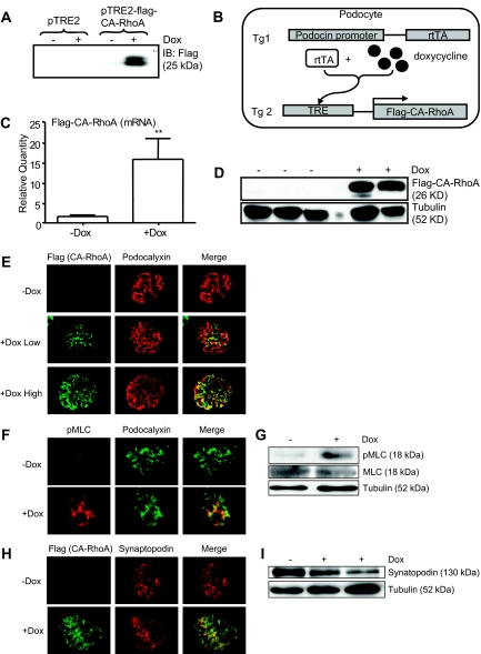
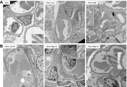
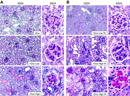
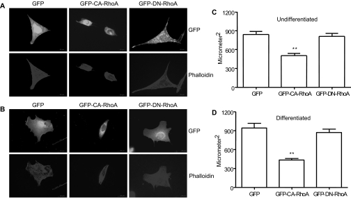
Proper organization of the actin cytoskeleton is essential for the normal structure and function of podocytes. RhoA modulates actin dynamics but its role in podocyte biology is controversial. Here, we generated transgenic mice that express a constitutively active form of RhoA in a podocyte-specific and doxycycline-inducible manner. Induction of activated RhoA with doxycycline resulted in significant albuminuria. Furthermore, both the degree of albuminuria and the histologic changes in the glomerulus positively correlated with the level of constitutively active RhoA expression: low levels of expression associated with segmental foot-process effacement without changes observable by light microscopy, whereas higher levels of expression associated with both extensive foot-process effacement and histologic features of focal segmental glomerulosclerosis (FSGS). In addition, induction of activated RhoA markedly upregulated glomerular mRNA expression of fibronectin and collagen IA1, and the degree of upregulation positively correlated with the level of albuminuria. Withdrawal of doxycycline led to a decline in albuminuria toward basal levels in most mice, but heavy albuminuria persisted in some mice. Taken together, these data suggest that activation of RhoA in podocytes leads to albuminuria accompanied by a range of histologic changes characteristic of minimal change disease and FSGS in humans. Although most changes are reversible, severe and prolonged activation of RhoA may cause irreversible glomerulosclerosis.


Figures

Figure 1.
Figure 1.
Establishment of podocyte-specific, Dox-inducible expression of CA-RhoA in mice. (A) Flag-tagged CA-RhoA was subcloned into the vector, pTRE2, and was transfected into HeLa cells stably expressing rtTA. Cells were treated with Dox (1 μg/ml) for 16 hours, and cell lysates were subjected to immunoblotting with anti-Flag antibody. Inducibility of CA-RhoA by Dox was confirmed. (B) Schematic representation of the strategy to establish the mouse line (see the Concise Methods section). (C through I) Double transgenic mice (2 to 3 months old) were treated with Dox (4 mg/ml in drinking water) or vehicle (−Dox) for 4 weeks. (C) Glomerular lysates were analyzed by real-time PCR for mRNA expression of the transgene. Flag-CA-RhoA mRNA was significantly induced by Dox (n = 8) versus −Dox (n = 9), **P < 0.001. (D) Glomerular lysates were analyzed by immunoblotting using anti-Flag antibody. Flag-CA-RhoA protein was induced by Dox. (E, F, and H) Kidney sections were immunostained for Flag (for CA-RhoA), pMLC, podocalyxin, and synaptopodin. Dox induced the expression of CA-RhoA in glomeruli, in a pattern consistent with the podocyte distribution. See Figure 3 for definition of low/high responders in (E). (G and I) Glomerular lysates were immunoblotted for pMLC, MLC, synaptopodin, and tubulin.
Figure 2.
Figure 2.
Induction of CA-RhoA by Dox causes albuminuria in mice. (A) Double transgenic mice were treated with Dox or vehicle for 4 weeks, and the urine ACR (μg/mg) was determined before and after the treatment. The ACR in Dox-treated mice (n = 15) was significantly higher than in vehicle-treated mice (n = 9) after 4 weeks of treatment. **P < 0.001 versus −Dox (4 weeks). (B) After 4 weeks of treatment with Dox, 3 μl of urine samples were separated by SDS-PAGE and stained with Coomassie Brilliant Blue. Dox-treated mice showed intense albuminuria.
Figure 3.
Figure 3.
The level of CA-RhoA expression correlates with the level of albuminuria. (A) Double transgenic mice (DT) and control littermates (Cntl) were treated with Dox as in Figure 2 for 4 weeks, and the urine ACR was determined at 2 and 4 weeks. The degree of Dox-induced albuminuria was highly variable among mice (see Figure 2A). We defined mice with urine ACR < 1000 μg/mg as low-responders (+Dox Low), and those with >1000 μg /mg as high responders (+Dox High). Both groups (n = 7 and 8) showed significantly higher ACR at 2 and 4 weeks of treatment, compared with vehicle-treated mice (−Dox, n = 9), *P < 0.05 and **<0.001. (B) Double transgenic mice were treated with Dox or vehicle for 4 weeks, and glomerular mRNA expression of the transgene was quantified by real-time PCR for the three groups. High responders (n = 4) showed significantly higher transgene expression, compared with low responders (n = 4) and vehicle-treated mice (n = 9). **P < 0.001 versus −Dox. (C) Urine ACR was plotted against transgene expression for individual mice. Positive correlation was seen (n = 17, R2 = 0.7762).
Figure 4.
Figure 4.
Albuminuria induced by CA-RhoA is, at least partially, reversible. Double transgenic mice were treated with Dox for 4 weeks, and then Dox was discontinued. Urine ACR was measured for 2 additional weeks after the withdrawal. (A) Urine ACR of low responders returned to baseline (n = 4). *P < 0.05. **P < 0.001 versus 0 weeks. (B) Time courses of four individual high responder mice are shown. High responders had variable rates of recovery; some mice showed a slow return toward the baseline (mice 1 and 2), whereas others showed sustained heavy albuminuria even at 2 weeks after Dox withdrawal (mice 3 and 4). On average, the urine ACR tended to decline after Dox withdrawal but remained high even after 2 weeks. (C) Glomerular lysates of high responders obtained 2 weeks after Dox withdrawal were analyzed by immunoblotting using anti-Flag antibody. In two of three mice (lanes 1 and 2), expression of Flag-CA-RhoA returned to minimal, whereas in one mouse (lane 3), intense expression was still detected. Lanes 1 to 3 correspond to mice 1 to 3 in Figure 4B. Lanes 4 to 6: double transgenic mice treated with vehicle for 6 weeks.
Figure 5.
Figure 5.
Foot process effacement caused by CA-RhoA is reversible. Transmission electron micrographs are shown. (A) Double transgenic mice were treated with Dox or vehicle (−Dox) for 4 weeks. Vehicle-treated mice (left) showed normal podocyte ultrastructure (foot process effacement <10%). Low responders (middle) showed a mixture of areas with normal foot processes (inset a) and foot process effacement (inset b). High responders (right) showed more extensive effacement. (B) Double transgenic mice were treated with Dox for 4 weeks and then Dox was withdrawn (W). Kidneys were harvested 2 weeks later. Low responders (left) showed normal foot processes 2 weeks after Dox withdrawal. High responders, which had a trend of declining albuminuria (middle), showed partial restoration of foot processes (inset a), whereas other areas remained fused (inset b). High responders with sustained heavy albuminuria (right) showed persistent extensive foot process effacement even at 2 weeks after Dox withdrawal. Magnification, ×6000, Scale bars = 2 μm.
Figure 6.
Figure 6.
High level of CA-RhoA expression induces FSGS. PAS staining of kidney sections is shown. (A) Double transgenic mice were treated with Dox or vehicle (−Dox) for 4 weeks. Glomeruli of low responders (middle) showed no abnormality and were not significantly different from vehicle-treated mice (top). Glomeruli of high responders (bottom) showed various degree of sclerosis. Note that cuboidal parietal glomerular epithelial cells (bottom) are often seen even in normal male mice. (B) Double transgenic mice were treated with Dox for 4 weeks, and then Dox was withdrawn (W). Kidneys were harvested 2 weeks later. Low responders (+Dox Low W, top) consistently showed normal kidney histology. Many high responders had a trend of declining albuminuria after withdrawal. PAS staining of these mice showed minimal sclerosis (+Dox High W, middle). Other high responders with sustained heavy albuminuria showed typical FSGS with interstitial leukocyte infiltration (arrow) (+Dox High W, bottom). Magnification, ×200 and ×600.
Figure 7.
Figure 7.
Activation of RhoA causes loss of processes and cell contraction in cultured mouse podocytes. Mouse podocytes, either undifferentiated or differentiated for 10 days, were transfected with pEGFP, GFP-CA-RhoA, or GFP-Dominant Negative (DN)-RhoA. After 24 hours, cells were permeabilized and stained for phalloidin. (A and B) Immunofluorescence for GFP and phalloidin are shown for undifferentiated cells (A) and differentiated cells (B). Magnification, ×600. Cells transfected with CA-RhoA showed significant cell contraction. (C and D) Cell size was quantified as in the Concise Methods section. (C) Undifferentiated cells. n = 30. (D) Differentiated cells, n = 20, **P < 0.001 versus GFP.
Figure 8.
Figure 8.
CA-RhoA upregulates fibronectin in vivo. (A) Double transgenic mice were treated with Dox or vehicle (−Dox) for 4 weeks. (A) Glomerular mRNA expression of the indicated genes was analyzed by real-time PCR. Only fibronectin and collagen IA1 were significantly upregulated. n = 8, **P < 0.001 versus vehicle-treated (n = 9). (B) Quantification of fibronectin mRNA by real-time PCR is shown according to the level of the urine ACR. Both high (n = 4) and low responders (n = 4) showed a significant upregulation of fibronectin, compared with vehicle-treated mice (n = 9). **P < 0.001 and *P < 0.05 versus −Dox. (C). Immunofluorescence staining of fibronectin is shown. Dox-treated mice (high responder, left) showed increased expression of fibronectin in glomeruli, compared with vehicle-treated mice (right). Magnification, ×100 (top) and ×600 (bottom). Scale bars = 20 μm.
Figure 9.
Figure 9.
CA-RhoA transactivates the fibronectin gene in vitro. (A) The reporter construct, p1900FN-luc, was transfected into HEK293T with CA-RhoA or empty vector, and cell lysates were analyzed for luciferase activity after 24 hours. Rho kinase inhibitor Y27632 (10 μM) was added overnight before the harvest. n = 6 in each group, **P < 0.001 versus empty vector. (B) Cell lysates were analyzed by immunoblotting using anti-fibronectin or anti-RhoA antibody. Rat visceral glomerular epithelial cells, which express CA-RhoA in an inducible manner, were stimulated with ponasterone A or vehicle (ethanol) overnight. Fibronectin protein was induced concurrently with CA-RhoA.

Similar articles

Cited by

References

    1. Faul C, Asanuma K, Yanagida-Asanuma E, Kim K, Mundel P: Actin up: Regulation of podocyte structure and function by components of the actin cytoskeleton. Trends Cell Biol 17: 428–437, 2007 - PubMed
    1. Burridge K, Wennerberg K: Rho and Rac take center stage. Cell 116: 167–179, 2004 - PubMed
    1. Ridley AJ, Hall A: The small GTP-binding protein rho regulates the assembly of focal adhesions and actin stress fibers in response to growth factors. Cell 70: 389–399, 1992 - PubMed
    1. Asanuma K, Yanagida-Asanuma E, Faul C, Tomino Y, Kim K, Mundel P: Synaptopodin orchestrates actin organization and cell motility via regulation of RhoA signalling. Nat Cell Biol 8: 485–491, 2006 - PubMed
    1. Faul C, Donnelly M, Merscher-Gomez S, Chang YH, Franz S, Delfgaauw J, Chang JM, Choi HY, Campbell KN, Kim K, Reiser J, Mundel P: The actin cytoskeleton of kidney podocytes is a direct target of the antiproteinuric effect of cyclosporine A. Nat Med 14: 931–938, 2008 - PMC - PubMed

Publication types

LinkOut - more resources