Review
. 2011 Aug;121(8):2955-68.
doi: 10.1172/JCI46315. Epub 2011 Aug 1.

Recent advances in the molecular pathophysiology of atrial fibrillation

Affiliations
Review

Recent advances in the molecular pathophysiology of atrial fibrillation

Reza Wakili et al. J Clin Invest. 2011 Aug.

Abstract

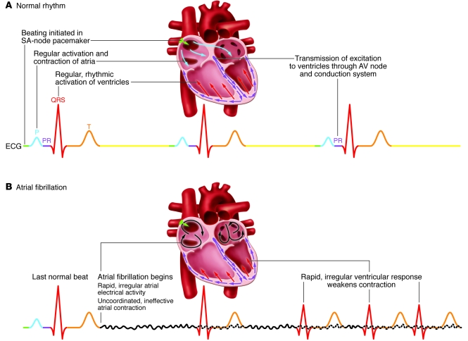
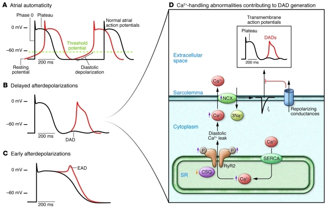
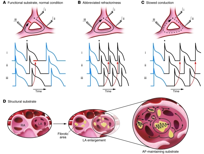
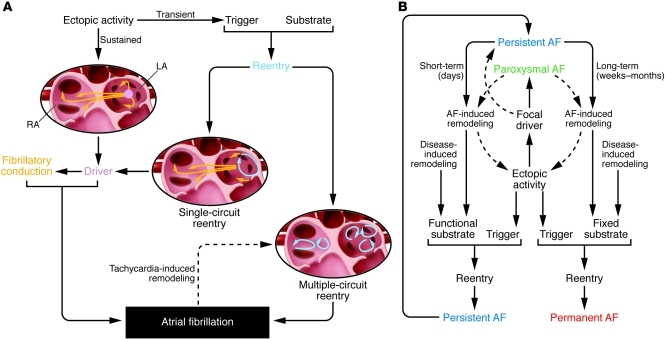
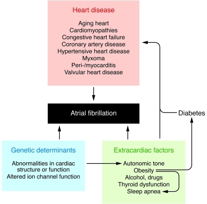
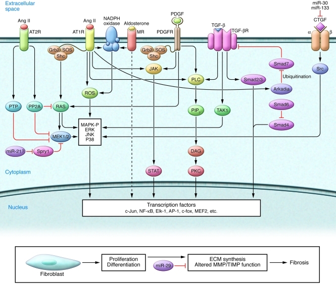
Atrial fibrillation (AF) is an extremely common cardiac rhythm disorder that causes substantial morbidity and contributes to mortality. The mechanisms underlying AF are complex, involving both increased spontaneous ectopic firing of atrial cells and impulse reentry through atrial tissue. Over the past ten years, there has been enormous progress in understanding the underlying molecular pathobiology. This article reviews the basic mechanisms and molecular processes causing AF. We discuss the ways in which cardiac disease states, extracardiac factors, and abnormal genetic control lead to the arrhythmia. We conclude with a discussion of the potential therapeutic implications that might arise from an improved mechanistic understanding.


Figures

Figure 1
Figure 1. ECG recordings of sinus rhythm and AF.
(A) Bottom: A normal ECG recording showing sinus rhythm. Top: Schematics of major events in one cardiac activation cycle: rhythm is initiated by the SA node pacemaker, resulting in atrial activation, followed by atrioventricular conduction via the AV node and His-Purkinje conducting system, leading to ventricular activation. (B) ECG showing onset of AF after one regular sinus beat. Atrial activation is now rapid and irregular, producing an undulating baseline that is visible when not obscured by larger QRS and T waves (continuous atrial activity during this phase is represented by dotted lines). During AF, rapid and uncoordinated atrial activity leads to ineffective atrial contraction. Ventricular activations (QRS complexes) now driven by the fibrillating atria occur rapidly and irregularly, weakening cardiac contraction efficiency and causing clinical symptoms.
Figure 2
Figure 2. Cellular mechanisms underlying focal ectopic activity.
(A) The normal atrial action potential (transmembrane potential as a function of time, in black) has a stable resting value close to –80 mV. Cell firing causes rapid depolarization (phase 0) to a positive value. Following initial repolarization, there is a flat (plateau) phase and then repolarization back to the resting potential. Normal atrial cells remain at the resting potential until they are fired through the SA node pacemaking system. Abnormal atrial automaticity results from spontaneous diastolic depolarization to a threshold value for activation. (B and C) Afterdepolarizations: abnormal membrane depolarizations after completion of the AP. DADs occur after full repolarization (B); EADs precede full repolarization (C). (D) Fundamental mechanisms leading to DADs, the most important source of ectopic activity in AF. DADs result from spontaneous diastolic SR Ca2+ releases through channels called RyR2s. RyR2s are sensitive to intra-SR free Ca2+ concentration. Abnormal diastolic RyR2 Ca2+ releases can result from excess SR intraluminal Ca2+ (pumped into the SR by SERCA) or reduced SR Ca2+ binding by the principal SR Ca2+ buffer, calsequestrin (CSQ). RyR2 hyperphosphorylation increases sensitivity to SR Ca2+, causing abnormal RyR2 Ca2+ release events. Diastolic RyR2 Ca2+ release increases cytosolic Ca2+, which has to be removed by the NCX. NCX moves three Na+ ions into the cell in exchange for each Ca2+ ion moved out, creating an inward movement of positive charges that produces a depolarizing Iti. Repolarizing conductances oppose Iti, protecting against excessive diastolic membrane voltage oscillations.
Figure 3
Figure 3. Factors promoting reentry.
Reentry occurs via interactions between interconnected zones of tissue, initiated by a premature beat (*). i, ii, and iii indicate microelectrode recordings in three zones of a potential reentry circuit. (A) Normal atrial tissue is unlikely to maintain reentry. Reentry maintenance can result from either a shortened refractory period (B) or slowed conduction (C). (D) A variety of cardiac conditions cause structural reentry substrates characterized by atrial enlargement and fibrosis. LA, left atrium; RA, right atrium.
Figure 4
Figure 4. Tissue mechanisms leading to AF and clinical forms.
(A) Ectopic activity can act as a driver maintaining AF or as a trigger on a vulnerable substrate resulting in reentry (single- or multiple-circuit). Local driver mechanisms (ectopic or single-circuit reentrant) produce irregular fibrillatory activity via fibrillatory conduction. Rapid atrial activity (tachycardia) causes atrial remodeling, promoting multiple-circuit reentry. (B) Clinical AF can manifest as paroxysmal AF (self-terminating), persistent AF (requires drug therapy or electrical cardioversion to terminate), and permanent AF (non-terminating). Focal ectopic drivers are principally associated with paroxysmal forms, functional reentrant substrates with persistent AF, and increasingly fixed substrates with permanent forms.
Figure 5
Figure 5. Schematic overview of factors governing AF occurrence.
Figure 6
Figure 6. Factors promoting AF by inducing SR diastolic Ca2+ leak.
RyR dysfunction may result from hyperphosphorylation or exposure to excess intraluminal Ca2+. SR Ca2+ overload can result from phospholamban hyperphosphorylation or reduced sarcolipin (SLN) expression, which disinhibit SERCA and enhance SR Ca2+ uptake. Increased cellular Ca2+ entry due to high atrial rate facilitates Ca2+/calmodulin (CaM) binding to the regulatory domain of CaMKII, resulting in disinhibition of the catalytic subunit. After initial activation of the holoenzyme by Ca2+/CaM, oxidation at Met281/282 or phosphorylation at Thr287 blocks reassociation of the catalytic domain, yielding persistent CaMKII activity. PP1 suppression by enhanced SR compartment inhibitor–1 (I-1 activity) contributes to increased phospholamban (PLN) and RyR phosphorylation. NADPHox, oxidized NADPH.
Figure 7
Figure 7. Genetic variants predisposing to AF.
Genetic variants (single-gene mutations: red; single nucleotide polymorphisms: blue) are displayed in relation to presumed AF-promoting mechanisms: (A) ectopic activity; (B) reentry with functional substrates; (C) reentry with fixed structural substrates.
Figure 8
Figure 8. Remodeling of ICa,L and inward-rectifier K+ currents by AF/tachycardia.
(A) The high atrial rate in AF increases intracellular Ca2+ load, activating calcineurin via the Ca2+/calmodulin system. Calcineurin stimulates nuclear translocation of nuclear factor of activated T lymphocytes (NFAT), reducing transcription of the principal ICa,L subunit, Cav1.2. Increased mRNA degradation/impaired protein translation of Cav1.2 and breakdown of Cav1.2 protein by calpains may also contribute. Increased expression of zinc transporter–1 (ZnT-1) impairs membrane trafficking of Cav1.2. Reduced Cav1.2 phosphorylation due to increased protein phosphatase (PP) activity and increased channel nitrosylation may also decrease ICa,L. GSH, glutathione. (B) Increased IK1 density results from upregulation of the principle Kir2.1 subunit, likely due to reduced levels of inhibitory miRNAs (miR-101, miR-26, miR-1). Increased IK,AChc is caused by altered PKC regulation: increased membrane abundance of stimulatory PKCε and reduced expression of inhibitory PKCα.
Figure 9
Figure 9. Molecular mechanisms leading to atrial fibrosis.
Major profibrotic signaling pathways involved in fibrosis generation are shown. MR, mineralocorticoid receptor.

References

    1. Miyasaka Y, et al. Secular trends in incidence of atrial fibrillation in Olmsted County, Minnesota, 1980 to 2000, and implications on the projections for future prevalence. Circulation. 2006;114(2):119–125. doi: 10.1161/CIRCULATIONAHA.105.595140. - DOI - PubMed
    1. Dobrev D, Nattel S. New antiarrhythmic drugs for treatment of atrial fibrillation. Lancet. 2010;375(9721):1212–1223. - PubMed
    1. Nattel S. From guidelines to bench: implications of unresolved clinical issues for basic investigations of atrial fibrillation mechanisms. Can J Cardiol. 2011;27(1):19–26. doi: 10.1016/j.cjca.2010.11.004. - DOI - PubMed
    1. Nattel S. New ideas about atrial fibrillation 50 years on. Nature. 2002;415(6868):219–226. doi: 10.1038/415219a. - DOI - PubMed
    1. Nattel S, Burstein B, Dobrev D. Atrial remodeling and atrial fibrillation: mechanisms and implications. Circ Arrhythm Electrophysiol. 2008;1(1):62–73. doi: 10.1161/CIRCEP.107.754564. - DOI - PubMed

Publication types