. 2011 Jul 29;30(18):3714-28.
doi: 10.1038/emboj.2011.250.

3'UTR elements inhibit Ras-induced C/EBPβ post-translational activation and senescence in tumour cells

Affiliations

3'UTR elements inhibit Ras-induced C/EBPβ post-translational activation and senescence in tumour cells

Sandip K Basu et al. EMBO J. .

Abstract

C/EBPβ is an auto-repressed protein that becomes post-translationally activated by Ras-MEK-ERK signalling. C/EBPβ is required for oncogene-induced senescence (OIS) of primary fibroblasts, but also displays pro-oncogenic functions in many tumour cells. Here, we show that C/EBPβ activation by H-Ras(V12) is suppressed in immortalized/transformed cells, but not in primary cells, by its 3' untranslated region (3'UTR). 3'UTR sequences inhibited Ras-induced cytostatic activity of C/EBPβ, DNA binding, transactivation, phosphorylation, and homodimerization, without significantly affecting protein expression. The 3'UTR suppressed induction of senescence-associated C/EBPβ target genes, while promoting expression of genes linked to cancers and TGFβ signalling. An AU-rich element (ARE) and its cognate RNA-binding protein, HuR, were required for 3'UTR inhibition. These components also excluded the Cebpb mRNA from a perinuclear cytoplasmic region that contains activated ERK1/2, indicating that the site of C/EBPβ translation controls de-repression by Ras signalling. Notably, 3'UTR inhibition and Cebpb mRNA compartmentalization were absent in primary fibroblasts, allowing Ras-induced C/EBPβ activation and OIS to proceed. Our findings reveal a novel mechanism whereby non-coding mRNA sequences selectively regulate C/EBPβ activity and suppress its anti-oncogenic functions.


Conflict of interest statement

The authors declare that they have no conflict of interest.

Figures
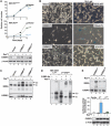
Figure 1
Figure 1
Suppression of Ras-induced C/EBPβ activation and its anti-proliferative functions by the 3′UTR. (A) Growth rates of control NIH 3T3 and 3T3Ras cells infected with retroviruses expressing the C/EBPβ coding region alone (C/EBPβCR) or coding region+3′UTR (C/EBPβUTR). Data are the mean values±s.d. from three experiments, each point assayed in triplicate. Lower panel: C/EBPβ western blot of nuclear extracts from the infected cells. (B) Morphology and senescence features of 3T3Ras cells transduced with C/EBPβCR or C/EBPβUTR. Cells expressing C/EBPβCR exhibit decreased proliferation and a flattened cellular morphology (left panels). SA-βGal staining (right) demonstrates senescent-like properties of many cells expressing C/EBPβCR but not C/EBPβUTR. The percentage of SA-βGal-positive cells is shown in the inset. (C) DNA-binding assays (EMSA) of nuclear extracts from cells described in (A). EMSA was performed using a canonical C/EBP site probe. The upper band represents a C/EBPβ homodimer; the intermediate band is composed of β:γ heterodimers and a second unidentified species. Bottom panel: C/EBPβ levels assessed by western blotting. (D) Antibody supershift assays were used to determine the composition of C/EBP EMSA complexes in C/EBPβCR-expressing NIH 3T3 cells. Lanes 1 and 2 show HEK293T cells transfected with C/EBPβCR±RasV12; the upper complex corresponds to C/EBPβ homodimers. The faster-migrating complex present in C/EBPβCR-expressing 3T3Ras cells (lanes 3–5) is a mixture of β:γ heterodimers and a second unidentified DNA-binding species. (E) DNA-binding and transactivation assays of C/EBPβ proteins transiently expressed in 293T cells. Cells were transfected with the indicated vectors and, after serum starvation, nuclear extracts were prepared, normalized for C/EBPβ levels and analysed by EMSA (upper). Transactivation (lower) was analysed using a C/EBP reporter (2xC/EBP-luc). Values represent fold activation over the reporter alone and are the mean values±s.d. of four experiments. C/EBPβ protein levels are shown below the graph.
Figure 2
Figure 2
An AU-rich element (ARE) region is necessary and sufficient for 3′UTR inhibition. (A) Analysis of Cebpb 3′UTR deletion mutants in 293T cells. The constructs depicted in the diagram were tested for C/EBPβ DNA binding (left) and transactivation (right) in transiently transfected cells. Transactivation data are the mean values±s.d. from three experiments. (B) Critical role of the ARE region for Cebpb 3′UTR repression. The constructs shown were analysed as described in (A). Transactivation data are the mean values±s.d. from three experiments. (C) Cytostatic activity of C/EBPβ proteins expressed from WT and 3′UTR mutant constructs in 3T3Ras cells. Data are the mean values±s.d. from two experiments, each point assayed in triplicate. Western blot shows C/EBPβ expression levels in the transduced cells. (D) DNA-binding assays of nuclear extracts from cells described in (C). Extracts from non-transformed NIH 3T3 cells (i.e., without RasV12) infected with the same constructs are included for comparison.
Figure 3
Figure 3
HuR is required for 3′UTR inhibition. (A) 3′UTR inhibition is disrupted in HuR knockdown (HuRKD) 293T cells. Nuclear extracts normalized for C/EBPβ levels were analysed by EMSA. Western blot shows HuR levels in control and HuRKD cells. (B) Re-expression of HuR in HuRKD cells restores 3′UTR inhibition. HuR was co-transfected with the indicated constructs in 293T-HuRKD cells. (C) Transcriptional activity of C/EBPβUTR depends on HuR. Transactivation of the 2 × C/EBP-luc reporter was compared in control and HuRKD cells. Re-expression of HuR restored inhibition by the 3′UTR without significantly affecting C/EBPβCR activity. Data are the mean values±s.d. from two experiments. (D) HuR knockdown in NIH 3T3 cells abolishes 3′UTR inhibition of C/EBPβ cytostatic activity. Control NIH 3T3 cells (upper panel) or 3T3Ras cells (lower panel) were infected with retroviruses expressing shHuR or the empty vector. The cells were subsequently infected with the indicated C/EBPβ constructs and proliferation rates were determined. Data are the mean values±s.d. from two experiments, each point assayed in triplicate. Extracts were analysed for C/EBPβ and HuR expression by western blotting (right). (E) 3′UTR inhibition of C/EBPβ transactivation is impaired in HuRKD NIH 3T3 cells. Transient transactivation assays using the 2 × C/EBP-luc reporter were performed in control and HuRKD NIH 3T3 cells. Data are the mean values±s.d. from two experiments. Ectopic C/EBPβ protein expression could not be assessed due to high levels of endogenous C/EBPβ in these cells.
Figure 4
Figure 4
The 3′UTR inhibits Ras-induced post-translational modifications and dimerization of C/EBPβ. (A) Ras-induced p-Ser64 and p-Thr188 modifications are suppressed by the 3′UTR. C/EBPβCR and C/EBPβUTR were expressed in 293T cells without or with RasV12. Nuclear extracts were analysed by western blotting using the indicated phospho-specific or conventional C/EBPβ antibodies. (B) 3′UTR inhibition of C/EBPβ phosphorylation is disrupted in HuR knockdown cells. The experiment described in (A) was performed using 293T-HuRKD and control cells. (C) 3′UTR inhibition maintains efficient C/EBPβ heterodimerization with C/EBPγ in the presence of Ras signalling. C/EBPβCR and C/EBPβUTR were expressed with RasV12 and increasing amounts of C/EBPγ in 293T cells, and nuclear extracts were analysed by EMSA. (D) C/EBPβCR and C/EBPβUTR transactivation of a luciferase reporter in the presence of RasV12 and increasing amounts of C/EBPγ. Luciferase activity for the C/EBPβ constructs in the absence of C/EBPγ (control) was set to 100%, and the other values represent percent of control. Data are the mean values±s.d. of three experiments.
Figure 5
Figure 5
The Cebpb 3′UTR alters C/EBPβ target gene specificity. (A) Heat maps depicting expression levels of genes selectively induced by C/EBPβCR (left), C/EBPβUTR (right), or both proteins (middle). Maps show microarray data from three independently derived cell lines each for 3T3Ras, C/EBPβCR-3T3Ras, and C/EBPβUTR-3T3Ras cells. Darker shading indicates higher expression; dendrograms show hierarchical clustering patterns. (B) Venn diagram showing differential activation of target genes by C/EBPβCR and C/EBPβUTR in 3T3Ras cells. Gene array data were filtered as described in the text to identify significantly upregulated genes. Genes induced significantly by both proteins are depicted in the intersection. (C) qPCR analysis of C/EBPβ-induced SASP genes and other pro-inflammatory mediators. RNA from the indicated NIH 3T3 cell lines was analysed for expression of individual transcripts using quantitative PCR. Data are the mean values±s.d. from two experiments, each assayed in triplicate. (D) Pathway and disease associations of gene signatures selectively activated by C/EBPβCR or C/EBPβUTR in 3T3Ras cells. Genes corresponding to the non-overlapping groups shown in this figure and Supplementary Table S1 were analysed using GeneGo MetaCore software to identify associations with canonical biological pathways or disease states. Pattern matches were ranked by P-value (LOD score) and the top 10 categories are shown.
Figure 6
Figure 6
The 3′UTR excludes Cebpb mRNA from a perinuclear region of the cytoplasm. (A) Localization of CebpbCR and CebpbUTR transcripts in NIH 3T3 cells using a GFP tethering assay. Cells were transiently transfected with the GFP-MS2-nls reporter alone or with tagged CebpbCR or CebpbUTR vectors. GFP-positive cells were imaged by confocal fluorescence microscopy. Nuclei were visualized by DAPI staining. The percentage of cells exhibiting the GFP fluorescence pattern depicted is indicated in parentheses. (B) The ARE is necessary and sufficient to direct Cebpb mRNA to the cytoplasmic periphery. Tagged CebpbUTRΔARE and CebpbARE transcripts were expressed with GFP-MS2-nls in NIH 3T3 cells and imaged by confocal microscopy. (C) HuR is required for perinuclear exclusion of CebpbUTR mRNA. HuRKD NIH 3T3 cells were transfected with tagged CebpbCR or CebpbUTR vectors and the GFP reporter. Control cells expressing the empty knockdown vector were analysed in parallel (Supplementary Figure S9).
Figure 7
Figure 7
Activated ERK1/2 localizes to the perinuclear region of RasV12-expressing NIH 3T3 cells. (A) Phospho-ERK1/2 is induced upon 4-OHT treatment of NIH 3T3 cells stably expressing ER:RasV12. Cells were starved for serum and lysates analysed for p-ERK and total ERK by immunoblotting following 4-OHT stimulation. p-ERK levels in non-transduced, serum-starved NIH 3T3 cells are shown for comparison. (B) Phospho-ERK1/2 is located in the perinuclear region that lacks CebpbUTR mRNA. 3T3ER:RasV12 cells were co-transfected with the GFP-MS2-nls reporter and tagged Cebpb vectors, induced with 4-OHT for 48 h, immunostained for p-ERK (Texas Red), and both markers visualized by confocal fluorescence microscopy.
Figure 8
Figure 8
C/EBPβ 3′UTR repression is lacking in primary fibroblasts. (A) DNA binding of endogenous C/EBPβ in MEFs is activated by oncogenic Ras. Nuclear extracts from MEFs infected with control or RasV12 retroviruses were analysed for C/EBPβ DNA binding. Western blotting shows similar levels of C/EBPβ in the extracts. (B) Proliferation assays of low passage Cebpb−/− MEFs infected with C/EBPβCR or C/EBPβUTR retroviruses. Data are the mean values±s.d. from two experiments assayed in triplicate. (C) C/EBPβCR and C/EBPβUTR display equivalent RasV12-induced transcriptional activity in MEFs. Cebpb−/− MEFs were transfected with 2xC/EBP-luc and C/EBPβ plasmids, with or without RasV12. Data are the mean values±s.d. from two experiments. (D) The 3′UTR does not regulate Cebpb mRNA localization in MEFs. MEFs were transfected with the GFP-MS2-nls reporter, alone or with tagged CebpbCR or CebpbUTR vectors, and imaged by confocal fluorescence microscopy.
Figure 9
Figure 9
Model depicting 3′UTR regulatory functions and the proposed mechanism of C/EBPβ 3′UTR inhibition. (A) The 3′UTR regulation of protein activity (UPA) mechanism identified in this study extends the previously known roles of 3′UTRs, which include control of mRNA decay and regulation of protein translation. (B) UPA is operative in immortalized/transformed cells but not in primary cells. The 3′UTR restricts Cebpb mRNA to the peripheral cytoplasm in tumour cells, in part due to increased levels of cytoplasmic HuR. Other factors are likely to contribute to this HuR effect, as depicted. In primary cells, Cebpb mRNA can enter the perinuclear region, where newly synthesized C/EBPβ is available for activation by p-ERK and possibly other Ras effector kinases. Increased homodimerization of activated C/EBPβ regulates inflammation, cell-cycle arrest, and senescence, whereas in tumour cells the predominantly heterodimeric form of C/EBPβ may contribute to the transformed phenotype.

Comment in

Similar articles

Cited by

References

    1. Abdelmohsen K, Kuwano Y, Kim HH, Gorospe M (2008) Posttranscriptional gene regulation by RNA-binding proteins during oxidative stress: implications for cellular senescence. Biol Chem 389: 243–255 - PMC - PubMed
    1. Acosta JC, O’Loghlen A, Banito A, Guijarro MV, Augert A, Raguz S, Fumagalli M, Da Costa M, Brown C, Popov N, Takatsu Y, Melamed J, d’Adda di Fagagna F, Bernard D, Hernando E, Gil J (2008) Chemokine signaling via the CXCR2 receptor reinforces senescence. Cell 133: 1006–1018 - PubMed
    1. Adams PD (2009) Healing and hurting: molecular mechanisms, functions, and pathologies of cellular senescence. Mol Cell 36: 2–14 - PubMed
    1. Bakheet T, Williams BR, Khabar KS (2003) ARED 2.0: an update of AU-rich element mRNA database. Nucleic Acids Res 31: 421–423 - PMC - PubMed
    1. Barreau C, Paillard L, Osborne HB (2005) AU-rich elements and associated factors: are there unifying principles? Nucleic Acids Res 33: 7138–7150 - PMC - PubMed

Publication types