. 2012 Mar 8;31(10):1264-74.
doi: 10.1038/onc.2011.324. Epub 2011 Aug 1.

PTEN, NHERF1 and PHLPP form a tumor suppressor network that is disabled in glioblastoma

Affiliations

PTEN, NHERF1 and PHLPP form a tumor suppressor network that is disabled in glioblastoma

J R Molina et al. Oncogene. .

Abstract

The phosphatidylinositol-3-OH kinase (PI3K)-Akt pathway is activated in cancer by genetic or epigenetic events and efforts are under way to develop targeted therapies. phosphatase and tensin homolog deleted on chromosome 10 (PTEN) tumor suppressor is the major brake of the pathway and a common target for inactivation in glioblastoma, one of the most aggressive and therapy-resistant cancers. To achieve potent inhibition of the PI3K-Akt pathway in glioblastoma, we need to understand its mechanism of activation by investigating the interplay between its regulators. We show here that PTEN modulates the PI3K-Akt pathway in glioblastoma within a tumor suppressor network that includes Na(+)/H(+) exchanger regulatory factor 1 (NHERF1) and pleckstrin-homology domain leucine-rich repeat protein phosphatases 1 (PHLPP1). The NHERF1 adaptor, previously characterized by our group as a PTEN ligand and regulator, shows also PTEN-independent Akt-modulating effects that led us to identify the PHLPP1/PHLPP2 Akt phosphatases as NHERF1 ligands. NHERF1 interacts via its PDZ domains with PHLPP1/PHLPP2 and scaffolds heterotrimeric complexes with PTEN. Functionally, PHLPP1 requires NHERF1 for membrane localization and growth-suppressive effects. PHLPP1 loss boosts Akt phosphorylation only in PTEN-negative cells and cooperates with PTEN loss for tumor growth. In a panel of low-grade and high-grade glioma patient samples, we show for the first time a significant disruption of all three members of the PTEN-NHERF1-PHLPP1 tumor suppressor network in high-grade tumors, correlating with Akt activation and patient's abysmal survival. We thus propose a PTEN-NHERF1-PHLPP PI3K-Akt pathway inhibitory network that relies on molecular interactions and can undergo parallel synergistic hits in glioblastoma.


Conflict of interest statement

CONFLICT OF INTEREST

The authors have no competing financial interests in relation to this work.

Figures

Figure 1
Figure 1
Synergistic Akt activation by combined PTEN and NHERF1 knockdown points to NHERF1 as a scaffolding platform for PTEN and PHLPP proteins. A. Western blot of cell lysates from LN229 glioblastoma cells with knockdown of PTEN and NHERF1 by shRNA (sh). V, vector controls. The % of actin-normalized PTEN and NHERF1 levels relatively to vector control levels are shown. The graph shows the Akt activation as phosphorylated Akt S473 levels normalized to total Akt levels (P-Akt/Akt) from two independent infections. B-C. NHERF1 associates with PHLPP proteins. Schematic organization of NHERF1 (B) and PHLPP (C) with amino acid boundaries. EB, ERM-binding region; FL, full length; ΔPDZ, PDZ-binding-motif-deleted; RA, Ras-associating domain; PH domain; PP2C, protein phosphatase 2C domain. Reciprocal overlays with either My-tagged PHLPP1-CT or PHLPP2-CT on filter-immobilized NHERF1 GST-fusion proteins (B) or with Myc-tagged NHERF1-PDZ1-2 on filter-immobilized PHLPP1 and PHLPP2 GST-fusion proteins (C) were probed with Myc-antibody to detect interactions. Re-probing of the membranes with anti-GST antibody showed equal amounts of immobilized GST-fusion proteins. D. Bridging two-step overlay assay in which the indicated filter-immobilized proteins were overlaid in a first step (1st overlay) with the NHERF1 PDZ domains and, in a second step (2nd overlay), with Myc-PHLPP1-CT or Myc-PHLPP2-CT. Probing with Myc antibody revealed the bound Myc-tagged PHLPP1/2. The filters stripped and reprobed with GST antibody show the input amounts of immobilized proteins. The graph shows the PTEN-band intensities normalized to the corresponding input and expressed as % from the normalized values of the positive control. The cartoon is a simplified view of complex assembly by NHERF1.
Figure 2
Figure 2
NHERF1 interacts with PHLPP1/2 in vivo. A. Reciprocal co-immunoprecipitation (IP) between PHLPP1α-FL and 1α–ΔPDZ forms and FLAG-tagged NHERF1 overexpressed in 293T cells. TL, total lysate; HC, heavy chain. B. Co-immunoprecipitation of FLAG-tagged NHERF1 with PHLPP1β (upper panels) and PHLPP2 (lower panels). C. Co-immunoprecipitation of endogenous PHLPP1 and NHERF1 from LN229 total cell lysate. Two rabbit NHERF1 antibodies–I (Abcam) and II (Affinity BioReagents)–and rabbit IgG control were used. D. Immunofluorescence analysis (x400 with oil immersion) with Myc antibody (red), NHERF1 antibody (green) and DAPI (blue) of LN18 cells overexpressing the proteins indicated on top. Note recruitment of PHLPP1α but not PHLPP1α-ΔPDZ to the plasma membrane by Myr-NHERF1 (arrows).
Figure 3
Figure 3
NHERF1 stabilizes PHLPP1 at the membrane and regulates its growth suppression. A-B. Subcellular fractionation of LN229 glioblastoma cells transduced with vector (V) or NHERF1 shRNA (shRF1) (A), and of NHERF1 wild-type (WT) and knockout (KO) primary MEFs (B) in cytosolic (C) and membrane (M) fractions. Erk2 and N-cadherin were used as cytosolic and membrane fractionation markers, respectively. The membrane and cytoplasmic PHLPP1β or PTEN levels were normalized to the levels of N-cadherin and Erk2, respectively. C. Stable PHLPP1α (α) expression in A172 glioblastoma cells with and without prior NHERF1 knockdown by shRNA (sh). V, corresponding vector control. D. Proliferation assay with the 4 sets of cells from (A) showing significant proliferation suppression by PHLPP1α only in the presence of NHERF1.
Figure 4
Figure 4
Synergistic Akt activation and tumor growth by concomitant PTEN and PHLPP1 silencing. A. PTEN followed by PHLPP1 shRNA (sh) knockdown in LN229 cells. V, corresponding vector control. The PTEN and PHLPP1 GAPDH-normalized levels, and % of P-Akt-S473/Akt activation are shown (from two independent infections). B. Soft agar colony formation images (100x) scoring the colonies as small, medium and large, 18 days after seeding the indicated shRNA-depleted LN229 cells. Single cells were not counted. C. Kaplan-Meier survival of mice (n=3) inoculated with the indicated shRNA-depleted LN229 cells. Significant difference and median survival are indicated. D. Immunohistochemistry (x200) of serial sections from tumors isolated from the mice in (C) with GFP antibody (recognizing the GFP moiety of the pGIPZ PTEN shRNA- cloning vector) and with P-Akt S473 antibody. Arrowheads indicate invasive cells.
Figure 5
Figure 5
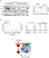
The PTEN-NHERF1-PHLPP inhibitory network is disabled in glioblastoma. A. Western blot analysis with indicated antibodies of protein extracts (8-10μg) from 9 low-grade glioma and 11 glioblastoma (GBM) samples. The average PHLPP1, NHERF1 and PTEN suppressor levels normalized to IKKβ and P-Akt/Akt levels show statistical significant differences in low-grade versus high-grade tumors. B. The correlation between PHLPP1, NHERF1 and PTEN suppressor levels and P-Akt/Akt levels for individual tumor samples, as sorted by increasing P-Akt/Akt levels (graph), is summarized in the table. Note significant inverse correlation between PHLPP1α levels and Akt phosphorylation in PTEN-negative tumors. C. Model depicting the molecular and functional interrelationships between the triple inhibitory network (gray circle) of PI3K-Akt. The white arrows indicate recruitment of PTEN and PHLPP to the membrane by NHERF1 via PDZ-motif/PDZ-domain interactions and the yellow arrow denotes an alternative lipid membrane association mechanism for PTEN. The Akt-independent suppression of tumor growth is shown with blue blocking lines.

References

    1. Alessi DR, Kozlowski MT, Weng QP, Morrice N, Avruch J. 3-Phosphoinositide-dependent protein kinase 1 (PDK1) phosphorylates and activates the p70 S6 kinase in vivo and in vitro. Curr Biol. 1998;8:69–81. - PubMed
    1. Barrett T, Troup DB, Wilhite SE, Ledoux P, Rudnev D, Evangelista C, et al. NCBI GEO: mining tens of millions of expression profiles--database and tools update. Nucleic Acids Res. 2007;35:D760–5. - PMC - PubMed
    1. Brognard J, Newton AC. PHLiPPing the switch on Akt and protein kinase C signaling. Trends Endocrinol Metab. 2008;19:223–30. - PMC - PubMed
    1. Brognard J, Sierecki E, Gao T, Newton AC. PHLPP and a second isoform, PHLPP2, differentially attenuate the amplitude of Akt signaling by regulating distinct Akt isoforms. Mol Cell. 2007;25:917–31. - PubMed
    1. Cardone RA, Bellizzi A, Busco G, Weinman EJ, Dell'Aquila ME, Casavola V, et al. The NHERF1 PDZ2 domain regulates PKA-RhoA-p38-mediated NHE1 activation and invasion in breast tumor cells. Mol Biol Cell. 2007;18:1768–1780. - PMC - PubMed

Publication types

MeSH terms