

Tissue oxygenation in brain, muscle, and fat in a rat model of sleep apnea: differential effect of obstructive apneas and intermittent hypoxia
- PMID: 21804675
- PMCID: PMC3138168
- DOI: 10.5665/SLEEP.1176
Tissue oxygenation in brain, muscle, and fat in a rat model of sleep apnea: differential effect of obstructive apneas and intermittent hypoxia
Abstract
Study objectives: To test the hypotheses that the dynamic changes in brain oxygen partial pressure (PtO(2)) in response to obstructive apneas or to intermittent hypoxia differ from those in other organs and that the changes in brain PtO(2) in response to obstructive apneas is a source of oxidative stress.
Design: Prospective controlled animal study.
Setting: University laboratory.
Participants: 98 Sprague-Dawley rats.
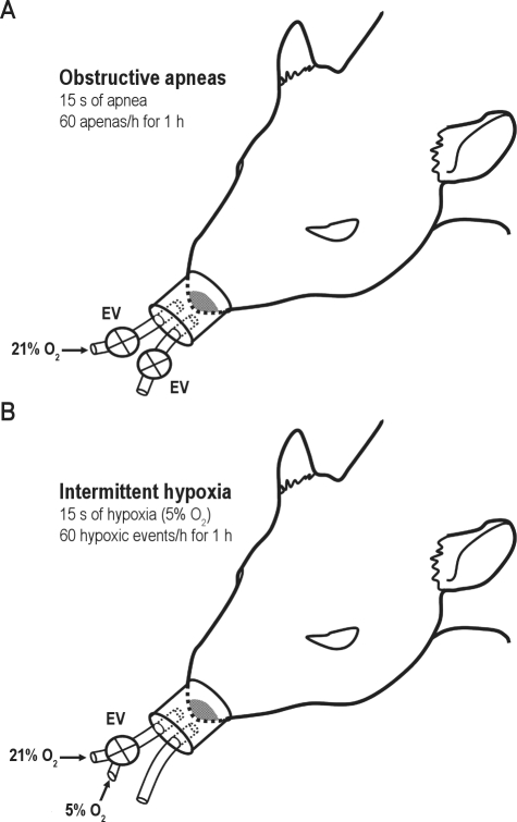
Interventions: Cerebral cortex, skeletal muscle, or visceral fat tissues were exposed in anesthetized animals subjected to either obstructive apneas or intermittent hypoxia (apneic and hypoxic events of 15 s each and 60 events/h) for 1 h.
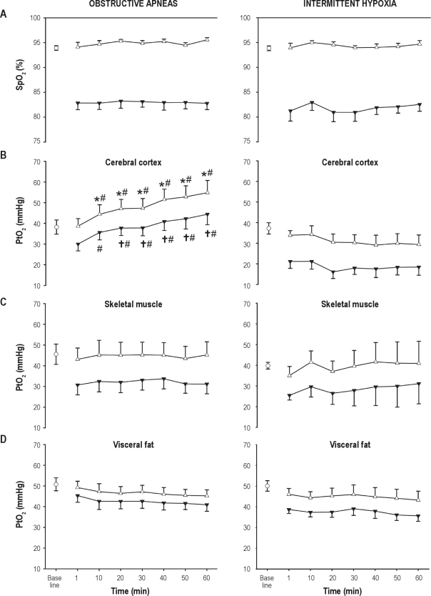
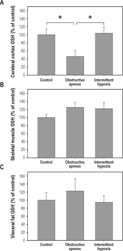
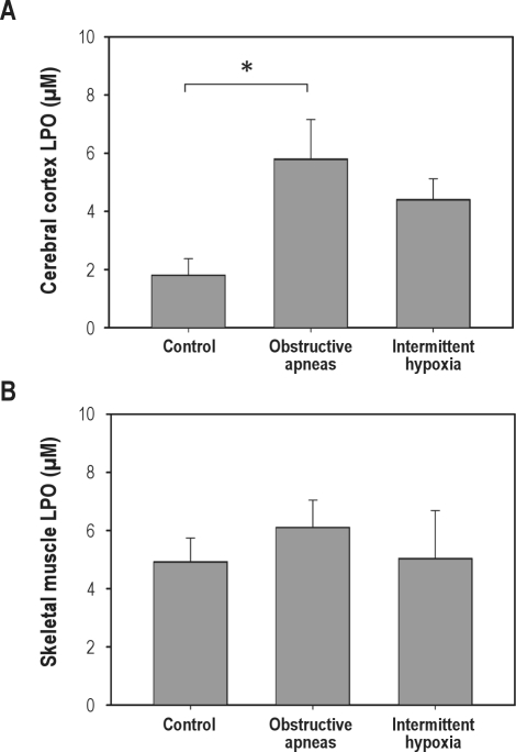
Measurements and results: Arterial oxygen saturation (SpO(2)) presented a stable pattern, with similar desaturations during both stimuli. The PtO(2) was measured by a microelectrode. During obstructive apneas, a fast increase in cerebral PtO(2) was observed (38.2 ± 3.4 vs. 54.8 ± 5.9 mm Hg) but not in the rest of tissues. This particular cerebral response was not found during intermittent hypoxia. The cerebral content of reduced glutathione was decreased after obstructive apneas (46.2% ± 15.2%) compared to controls (100.0% ± 14.7%), but not after intermittent hypoxia. This antioxidant consumption after obstructive apneas was accompanied by increased cerebral lipid peroxidation under this condition. No changes were observed for these markers in the other tissues.
Conclusions: These results suggest that cerebral cortex could be protected in some way from hypoxic periods caused by obstructive apneas. The increased cerebral PtO(2) during obstructive apneas may, however, cause harmful effects (oxidative stress). The obstructive apnea model appears to be more adequate than the intermittent hypoxia model for studying brain changes associated with OSA.
Keywords: Tissue oxygenation; animal model; intermittent hypoxia; obstructive apnea; oxidative stress.
Figures
References
-
- Duran J, Esnaola S, Rubio R, Iztueta A. Obstructive sleep apnea-hypopnea and related clinical features in a population-based sample of subjects aged 30 to 70 yr. Am J Respir Crit Care Med. 2001;163:685–9. - PubMed
-
- Young T, Palta M, Dempsey J, Skatrud J, Weber S, Badr S. The occurrence of sleep-disordered breathing among middle-aged adults. N Engl J Med. 1993;328:1230–5. - PubMed
-
- Prabhakar NR, Kumar GK. Oxidative stress in the systemic and cellular responses to intermittent hypoxia. Biol Chem. 2004;385:217–21. - PubMed
-
- Banasiak KJ, Haddad GG. Hypoxia-induced apoptosis: Effect of hypoxic severity and role of p53 in neuronal cell death. Brain Res. 1998;797:295–304. - PubMed
Publication types
MeSH terms
Substances
LinkOut - more resources
Full Text Sources
Medical
