

Expression of Pigment Cell-Specific Genes in the Ontogenesis of the Sea Urchin Strongylocentrotus intermedius
- PMID: 21804858
- PMCID: PMC3144734
- DOI: 10.1155/2011/730356
Expression of Pigment Cell-Specific Genes in the Ontogenesis of the Sea Urchin Strongylocentrotus intermedius
Abstract
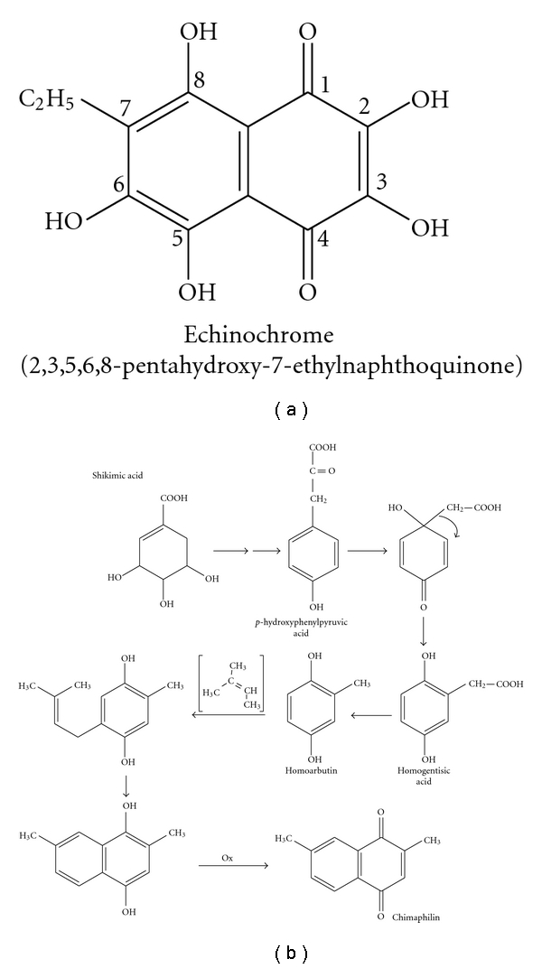
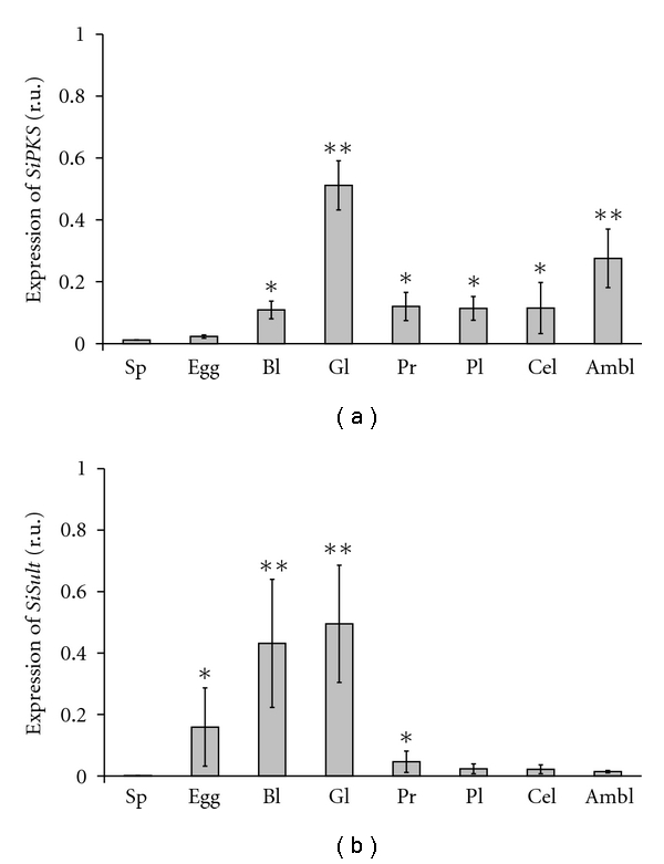
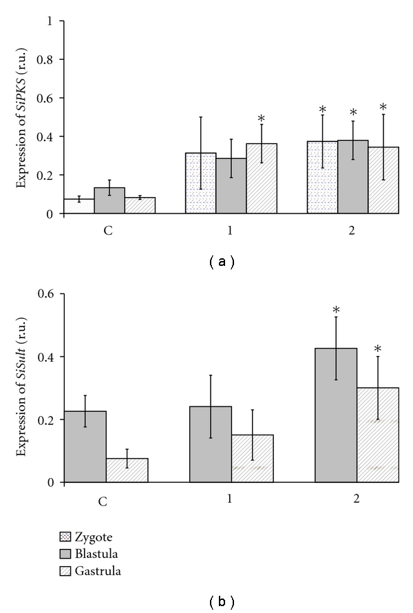
One of the polyketide compounds, the naphthoquinone pigment echinochrome, is synthesized in sea urchin pigment cells. We analyzed polyketide synthase (pks) and sulfotransferase (sult) gene expression in embryos and larvae of the sea urchin Strongylocentrotus intermedius from various stages of development and in specific tissues of the adults. We observed the highest level of expression of the pks and sult genes at the gastrula stage. In unfertilized eggs, only trace amounts of the pks and sult transcripts were detected, whereas no transcripts of these genes were observed in spermatozoids. The addition of shikimic acid, a precursor of naphthoquinone pigments, to zygotes and embryos increased the expression of the pks and sult genes. Our findings, including the development of specific conditions to promote pigment cell differentiation of embryonic sea urchin cells in culture, represent a definitive study on the molecular signaling pathways that are involved in the biosynthesis of pigments during sea urchin development.
Figures
References
-
- Fox DL, Scheer BT. Comparative studies of the pigments some Pacific Coast echinoderms. Biological Bulletin. 1941;80:441–455.
-
- Griffiths M. A study of the synthesis of naphthaquinone pigments by the larvae of two species of sea urchins and their reciprocal hybrids. Developmental Biology. 1965;11(3):433–447. - PubMed
-
- Fedoreyev SA, Mischenko NP, Kol'tsova EA, et al. Drug, Histochrome, from the sea urchin. In: Abstracts of 5th International Marine Biotechnology Conference; 2000; Townsville, Australia. p. 53.
-
- Mishchenko NP, Fedoreev SA, Bagirova VL. Histochrome: a new original domestic drug. Pharmaceutical Chemistry Journal. 2003;37(1):48–52.
-
- Koltsova EA, Boguslavskaya LV, Maximov OV. On the functions of quinonoid pigment production in sea urchin embryos. International Journal of Invertebrate Reproduction. 1981;4:17–28.
LinkOut - more resources
Full Text Sources
Miscellaneous
