. 2012:2012:415231.
doi: 10.1155/2012/415231. Epub 2011 Jul 17.

Apoptosis Induction in Primary Human Colorectal Cancer Cell Lines and Retarded Tumor Growth in SCID Mice by Sulforaphane

Affiliations

Apoptosis Induction in Primary Human Colorectal Cancer Cell Lines and Retarded Tumor Growth in SCID Mice by Sulforaphane

Ming-Jenn Chen et al. Evid Based Complement Alternat Med. 2012.

Abstract

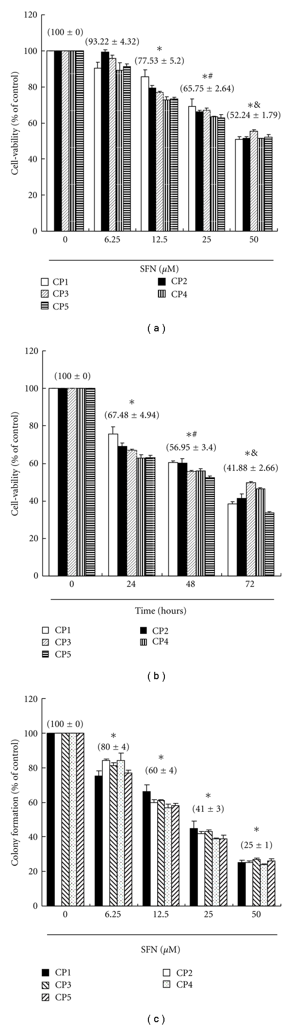
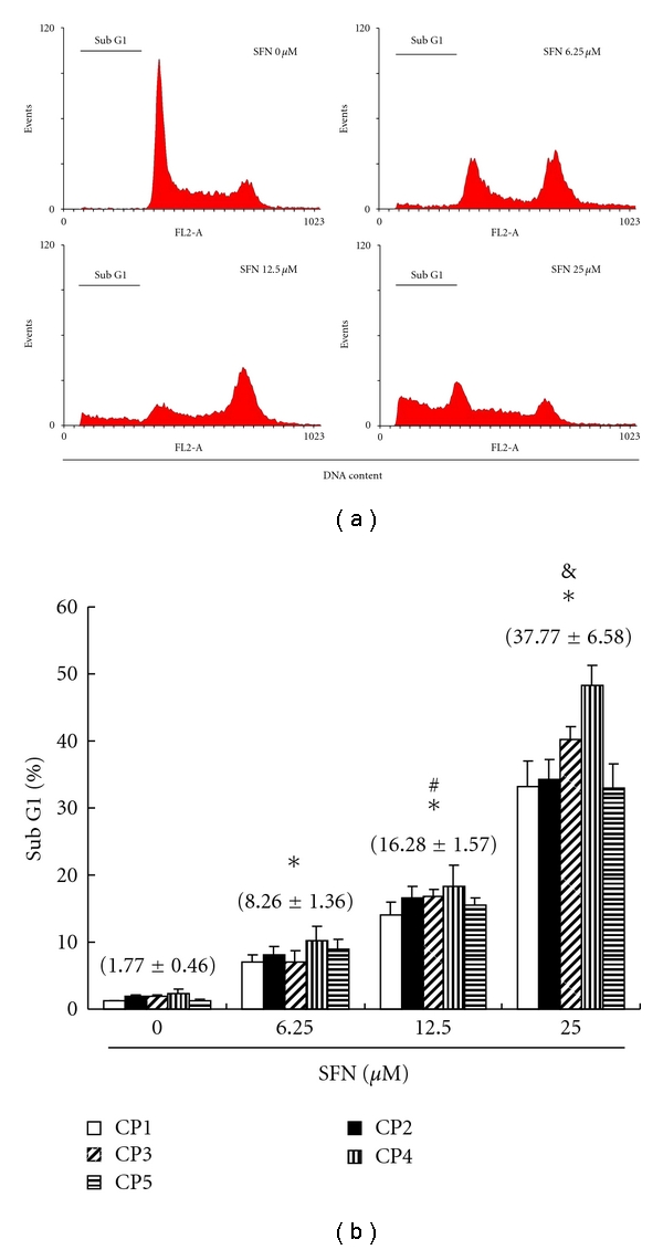
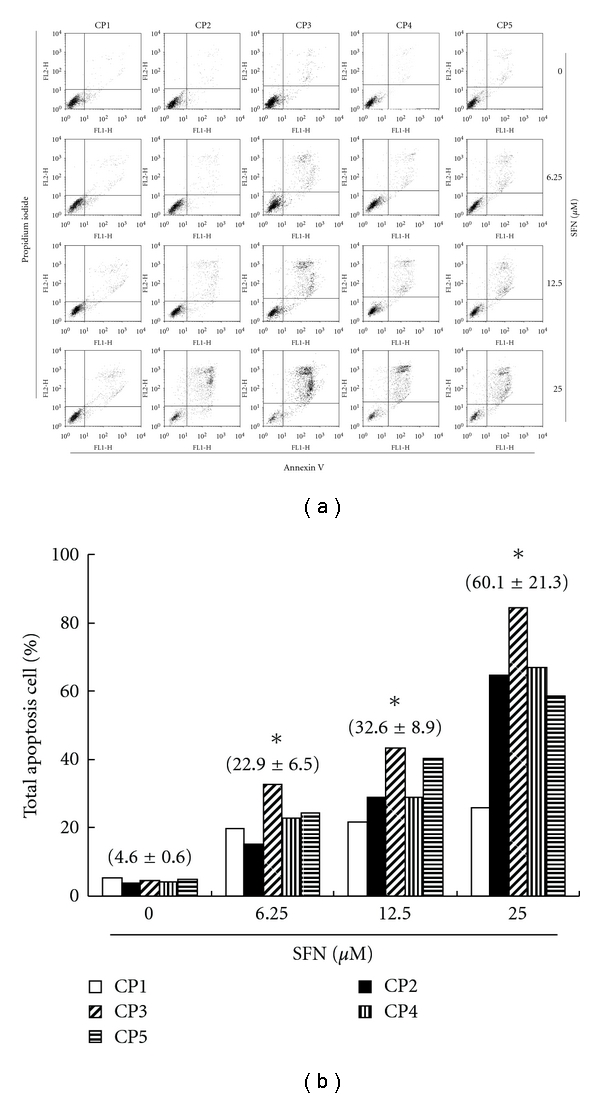
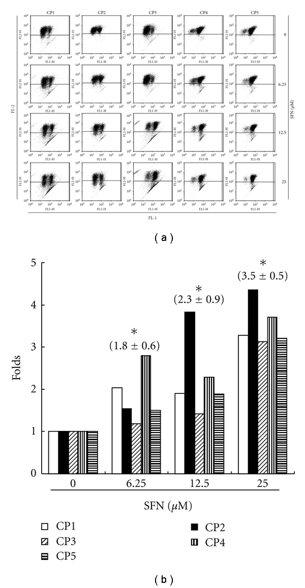
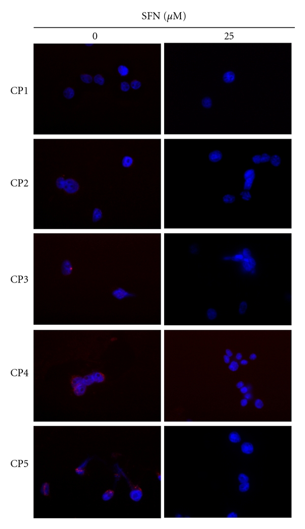
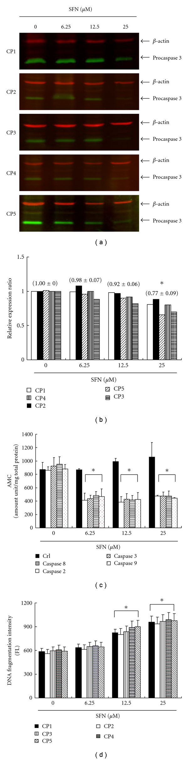
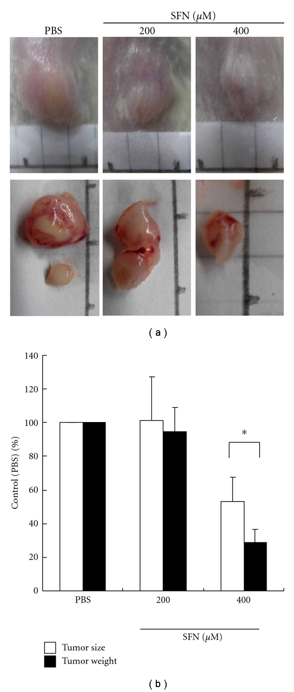
We have investigated the anticancer effects of the dietary isothiocyanate sulforaphane (SFN) on colorectal cancer (CRC), using primary cancer cells lines isolated from five Taiwanese colorectal cancer patients as the model for colorectal cancer. SFN-treated cells accumulated in metaphase (SFN 6.25 μM) and subG1 (SFN 12.5 and 25 μM) as determined by flow cytometry. In addition, treated cells showed nuclear apoptotic morphology that coincided with an activation of caspase-3, and loss of mitochondrial membrane potential (ΔΨm). Incubations at higher SFN doses (12.5 and 25 μM) resulted in cleavage of procaspase-3 and elevated caspase-2, -3, -8, and -9 activity, suggesting that the induction of apoptosis and the sulforaphane-induced mitosis delay at the lower dose are independently regulated. Daily SFN s.c. injections (400 micromol/kg/d for 3 weeks) in severe combined immunodeficient mice with primary human CRC (CP1 to CP5) s.c. tumors resulted in a decrease of mean tumor weight by 70% compared with vehicle-treated controls. Our findings suggest that, in addition to the known effects on cancer prevention, sulforaphane may have antitumor activity in established colorectal cancer.


Figures

Figure 1
Figure 1
(a) Sulforaphane mediates the survival of five human primary colorectal cancer (CRC) cell lines (five groups, n = 6 per group) and thus inhibits their proliferation. In vitro study was initiated by treating each of the cell lines to the increasing doses of SFN (0, 6.25, 12.5, 25, and 50 μM) for 24 hours. Statistical analysis used the t-test, with the significant differences determined at the level of *P < 0.05 versus control group (SFN 0 μM), while the symbol on the bar denotes the difference which is statistically significant at P < 0.05 as compared to the 12.5 (#) or 25 μM (&). (b) Reversibility of the growth inhibitory effect of SFN. The CRC cell lines were each first treated with SFN 12.5 μM for 24 hours. After the treatment was terminated by washing off SFN, the cultures were reincubated for 24–72 hours to check the extent of recovery of cancer cells. The survival of these SFN-treated CRC cells was then measured by MTT method. (c) Effect of SFN on colony formation. CRC cells were treated with SFN, and number of colonies was counted. Results were expressed as a percentage of control, which was considered as 100%. All data were reported as the means (±SEM) of at least three separate experiments. Statistical analysis used the t-test, with the significant differences determined at the level of *P < 0.05 versus time 0 group, while the symbol on the bar denotes the difference which is statistically significant at P < 0.05 as compared to the time 24 (#) or 48 (&) of the recovery study.
Figure 2
Figure 2
Arrest of cell cycle progression at G2/M and subG0/G1 in response to SFN treatment. (a) SFN-induced G2/M (SFN 6.25 μM) and G0/G1 (SFN 12.5 and 25 μM) arrest in primary CRC cell lines. The distribution of the cell cycle of CRC cell lines was assessed by flow cytometry after staining with propidium iodide (PI). (b) Results were expressed as a percentage of subG0/G1. All data were reported as the means (±SEM) of at least three separate experiments. Statistical analysis used the t-test, with the significant differences determined at the level of *P < 0.05 versus SFN 0 μM group, while the symbol on the bar denotes the difference which is statistically significant at P < 0.05 as compared to the SFN 6.25 (#) or 12.5 μM (&).
Figure 3
Figure 3
Apoptosis of CRC cell lines induced by SFN. Flow cytometric analysis of PI-Annexin-V to quantify SFN-induced apoptosis in CRC cell lines. (a) Dot plots of five CRC cell lines with SFN treatment at 0, 6.25, 12.5, and 25 μM for 6 h. (b) Results were expressed as a percentage of total apoptosis cells (early and late apoptosis).
Figure 4
Figure 4
Reduction of the mitochondrial membrane potential (ΔΨm) in the CRC cell lines by SFN, which was determined by JC-1 staining and detected by the flow cytometry. (a) Dot plots of five CRC cell lines with SFN treatment at 0, 6.25, 12.5, and 25 μM for 6 h. (b) The reduction of the ΔΨm containing polarized or depolarized mitochondria determined the ratio of the two fluorescence intensities analyzed by flow cytometry. All the data shown are the folds of five CRC cell lines. The symbol (∗) on each group of bars denotes that difference from the treatment with 0 μM SFN is statistically significant at P < 0.05.
Figure 5
Figure 5
Diminished NF-κB activity in SFN-treated CRC cell lines. Five CRC cell lines were examined for their NF-κB activity 16 hours after SFN stimulation by confocal microscopy of NF-κB subunit p50/52 localization. The CRC cell lines were stained for p50/52 (red). DAPI (blue) indicates nucleus, where active form of NF-κB subunit p50/52 is found.
Figure 6
Figure 6
SFN activates procaspase-3 degradation in five CRC cell lines. The cells were treated with SFN (0, 6.25, 12.5, and 25 μM) for 24 hours, and then Western blot analysis was performed for procaspase-3. (a) Representative blot from 3 independent experiments. (b) Quantification of band intensities by Li-COR near infrared imaging system. (c) The caspase-2, -3, -8, -9 activity was analyzed by ApoAlert Caspase assay plates. SFN induces the caspase activity of five CRC cell lines. (d) Quantification of DNA fragmentation by measuring the fluorescence intensities by flow cytometry. The data showed that DNA fragmentation levels were significantly elevated in cells incubated with SFN incubation for 24 hours. All data were reported as the means (±SEM) of five CRC cell lines. Statistical analysis used the t-test, with the significant differences determined at the level of *P < 0.05 versus 0 μM control group.
Figure 7
Figure 7
SFN inhibits in vivo tumor growth. (a) Effects of SFN on tumor growth of SCID mice subcutaneously inoculated with primary human CRC cell lines. The results showed that SFN 400 μM inhibited tumor growth significantly in both cancer xenografts. (b) Inhibition of tumor growth was compared among tested groups. The inhibitory ratio was normalized to the negative control (PBS group) (values represent percent of control, n = 3 *P < 0.05 versus PBS group).

Similar articles

Cited by

References

    1. Mukherjee S, Bhattacharya RK, Roy M. Targeting protein kinase C (PKC) and telomerase by phenethyl isothiocyanate (PEITC) sensitizes PC-3 cells towards chemotherapeutic drug-induced apoptosis. Journal of Environmental Pathology, Toxicology and Oncology. 2009;28(4):269–282. - PubMed
    1. Šmerák P, Polívková Z, Štĕtina R, Bártová J, Bárta I. Antimutagenic effect of phenethyl isothiocyanate. Central European Journal of Public Health. 2009;17(2):86–92. - PubMed
    1. Stan SD, Singh SV, Brand RE. Chemoprevention strategies for pancreatic cancer. Nature Reviews Gastroenterology and Hepatology. 2010;7(6):347–356. - PMC - PubMed
    1. Matsuda T, Maruyama T, Iizuka H, et al. Phthalate esters reveal skin-sensitizing activity of phenethyl isothiocyanate in mice. Food and Chemical Toxicology. 2010;48(6):1704–1708. - PubMed
    1. Liu T-T, Yang T-S. Stability and antimicrobial activity of allyl isothiocyanate during long-term storage in an oil-in-water emulsion. Journal of Food Science. 2010;75(5):C445–C451. - PubMed