

Ablation of breast cancer stem cells with radiation
- PMID: 21804918
- PMCID: PMC3140010
- DOI: 10.1593/tlo.10247
Ablation of breast cancer stem cells with radiation
Abstract
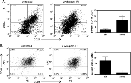
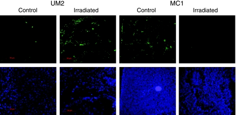
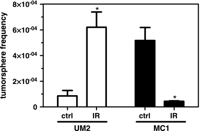
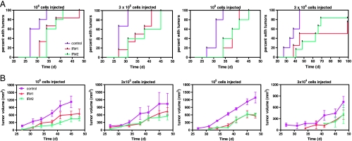
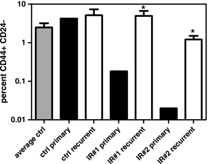
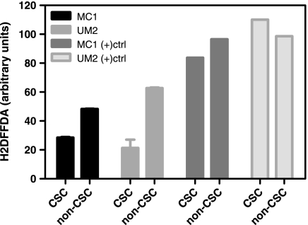
Tumor radioresistance leads to recurrence after radiation therapy. The radioresistant phenotype has been hypothesized to reside in the cancer stem cell (CSC) component of breast and other tumors and is considered to be an inherent property of CSC. In this study, we assessed the radiation resistance of breast CSCs using early passaged, patient-derived xenografts from two separate patients. We found a patient-derived tumor in which the CSC population was rapidly depleted 2 weeks after treatment with radiation, based on CD44(+) CD24(-) lin(-) phenotype and aldehyde dehydrogenase 1 immunofluorescence, suggesting sensitivity to radiotherapy. The reduction in CSCs according to phenotypic markers was accompanied by a decrease in functional CSC activity measured by tumor sphere frequency and the ability to form tumors in mice. In contrast, another patient tumor sample displayed enrichment of CSC after irradiation, signifying radioresistance, in agreement with others. CSC response to radiation did not correlate with the level of reactive oxygen species in CSC versus non-CSC. These findings demonstrate that not all breast tumor CSCs are radioresistant and suggest a mechanism for the observed variability in breast cancer local recurrence.
Figures
References
-
- Zhao L, Wang L, Ji W, Wang X, Zhu X, Hayman JA, Kalemkerian GP, Yang W, Brenner D, Lawrence TS, et al. Elevation of plasma TGF-β1 during radiation therapy predicts radiation-induced lung toxicity in patients with non-small-cell lung cancer: a combined analysis from Beijing and Michigan. Int J Radiat Oncol Biol Phys. 2009;74:1385–1390. - PubMed
-
- Jiang X, Sun Y, Chen S, Roy K, Price BD. The FATC domains of PIKK proteins are functionally equivalent and participate in the Tip60-dependent activation of DNA-PKcs and ATM. J Biol Chem. 2006;281:15741–15746. - PubMed
-
- Hamilton JP, Sato F, Greenwald BD, Suntharalingam M, Krasna MJ, Edelman MJ, Doyle A, Berki AT, Abraham JM, Mori Y, et al. Promoter methylation and response to chemotherapy and radiation in esophageal cancer. Clin Gastroenterol Hepatol. 2006;4:701–708. - PubMed
-
- Woodward WA, Chen MS, Behbod F, Rosen JM. On mammary stem cells. J Cell Sci. 2005;118:3585–3594. - PubMed
-
- Wicha MS, Liu S, Dontu G. Cancer stem cells: an old idea—a paradigm shift. Cancer Res. 2006;66:1883–1890. discussion 1895–1886. - PubMed
LinkOut - more resources
Full Text Sources
Miscellaneous