Review
. 2011 Sep;10(3):437-46.
doi: 10.1007/s10689-011-9469-3.

LKB1 as the ghostwriter of crypt history

Affiliations
Review

LKB1 as the ghostwriter of crypt history

Marnix Jansen et al. Fam Cancer. 2011 Sep.

Abstract

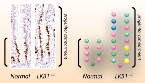
Familial cancer syndromes present rare insights into malignant tumor development. The molecular background of polyp formation and the cancer prone state in Peutz-Jeghers syndrome remain enigmatic to this day. Previously, we proposed that Peutz-Jeghers polyps are not pre-malignant lesions, but an epiphenomenon to the malignant condition. However, Peutz-Jeghers polyp formation and the cancer-prone state must both be accounted for by the same molecular mechanism. Our contribution focuses on the histopathology of the characteristic Peutz-Jeghers polyp and recent research on stem cell dynamics and how these concepts relate to Peutz-Jeghers polyposis. We discuss a protracted clonal evolution scenario in Peutz-Jeghers syndrome due to a germline LKB1 mutation. Peutz-Jeghers polyp formation and malignant transformation are separately mediated through the same molecular mechanism played out on different timescales. Thus, a single mechanism accounts for the development of benign Peutz-Jeghers polyps and for malignant transformation in Peutz-Jeghers syndrome.


Figures

Fig. 1
Fig. 1
Depiction of the original small bowel resection specimen containing the PJS polyp that led to an episode of small bowel obstruction in the index patient first described by Jan Peutz in his monograph on the pedigree
Fig. 2
Fig. 2
Overview of histological findings in a characteristic PJS polyp, in this case resected from the duodenum. A H&E staining showing an overview of the polyp. The epithelial lining demonstrates normal gobletcel-bearing epithelium without signs of dysplasia. Reactive epithelial changes can be noted at the surface of the polyp, which shows edematous villi and active inflammation, presumably due to surface erosion. Towards the center some crypts contain inspissated mucin. b Smooth muscle actin (SMA) immunohistochemical staining showing the arborizing core of smooth muscle (labeled red) that characterizes PJS polyps. c Ki67 proliferation marker immunohistochemical staining showing that proliferation is restricted to the crypt bases indicating proper compartimentalization of proliferation and differentiation. d Periodic-acid Schiff stain after diastase digestion showing normal differentiation and maturation of the secretory goblet cell lineage
Fig. 3
Fig. 3
PJS-like smooth muscle proliferation in a conventional non-PJS adenomatous polyp. a Gross macroscopy of a pedunculated colonic polyp in a 67-year old patient not affected by PJS. A right hemicolectomy was performed following the diagnosis of colon cancer in the ascending colon in this patient; the polyp shown here was found incidentally in the cecum. b Low-power photomicrograph of the polyp shown in a. The polyp has a tubulovillous architecture characterized by mucosal fronds emanating from the stalk of the polyp. Note the slender extensions of the smooth muscle core into the tips of the villous projections (indicated by asterisk in c). This arborizing core of smooth muscle resembles the histology shown in Fig. 2b for the polyp removed from the duodenum of a PJS patient. c Unlike PJS polyps, which are characterized by a non-dysplastic hyperproliferative epithelial lining, this polyp has a dysplastic epithelial covering classified as low-grade dysplasia. The boxed area in b is shown at higher power in c
Fig. 4
Fig. 4
COX-SDH enzyme histochemistry on normal colon reveals clonal evolution. The dynamics of clonal evolution can be traced by analyzing a polymorphic marker. Owing to an environment with elevated production of free radicals by the respiratory chain and limited repair mechanisms, the mitochondrial DNA (mtDNA) is prone to accumulating mutations. This accumulation is random and increases with age. Dual enzyme histochemistry can be used to simultaneously detect the mtDNA-encoded cytochrome-c oxidase (COX; complex IV of the respiratory chain) and nuclear DNA-encoded succinate dehydrogenase (SDH), a component of complex II of the respiratory chain. Sequential COX and succinate dehydrogenase histochemistry highlights deficiencies in COX enzyme activity, wherein cells lacking COX activity appear blue and normal cells retaining COX activity appear brown. a, c H&E staining showing normal mucosa. b Longitudinal section showing a mutation that has occurred in a stem cell lineage that has populated the entire crypt. All cells are COX-deficient (blue). d En face section showing a mutant clone in the process of colonizing the crypt; more than half the cells are now COX-deficient (blue); asterisk indicates clear contrast between COX-deficient (blue) and surrounding nonmutated colonocytes (ad, ×200). After extinction of all non-mutated lineages, the crypt has been clonally converted. In standard H&E preparations the dynamic process of lineage competition is obscured from our eyes
Fig. 5
Fig. 5
Increased clonal diversity in PJS crypts. This diagram illustrates the consequences of differences in clonal evolution in stem cell compartments such as the intestinal crypt. Over time stem cell lineages accumulate mutations at a set background mutation rate. By using a polymorphic marker such as COX enzyme activity we can trace the appearance of individual lineages in the crypt (see Fig. 4). We have observed an expanded crypt progenitor zone in unaffected intestinal mucosa in PJS patients [34]. By comparing CSX crypt heterogeneity patterns in unaffected PJS mucosa and age-matched control crypts, we found that PJS crypts retain a greater number of polymorphic marker mutations. This indicates that clonal evolution is protracted in Peutz-Jeghers syndrome and predicts that pre-malignant lesions will arise at an accelerated pace in comparison to the general population (see text)

References

    1. Westerman AM, Entius MM, de Baar E, et al. (1999) Peutz-Jeghers syndrome: 78-year follow-up of the original family. Lancet 353(9160): 1211–1215 - PubMed
    1. Katajisto P, Vallenius T, Vaahtomeri K, et al. The LKB1 tumor suppressor kinase in human disease. Biochim Biophys Acta. 2007;1775(1):63–75. - PubMed
    1. McGarrity TJ, Amos C. Peutz-Jeghers syndrome: clinicopathology and molecular alterations. Cell Mol Life Sci. 2006;63(18):2135–2144. doi: 10.1007/s00018-006-6080-0. - DOI - PMC - PubMed
    1. Gao H, van Lier MG, Poley JW, Kuipers EJ, van Leerdam ME, Mensink PB (2010) Endoscopic therapy of small-bowel polyps by double-balloon enteroscopy in patients with Peutz-Jeghers syndrome. Gastrointest Endosc 71(4):768–773 - PubMed
    1. Jansen M, de Leng WW, Baas AF, et al. Mucosal prolapse in the pathogenesis of Peutz-Jeghers polyposis. Gut. 2006;55(1):1–5. doi: 10.1136/gut.2005.069062. - DOI - PMC - PubMed

MeSH terms

Substances

LinkOut - more resources