. 2011 Aug 1:11:173.
doi: 10.1186/1471-2180-11-173.

The respiratory molybdo-selenoprotein formate dehydrogenases of Escherichia coli have hydrogen: benzyl viologen oxidoreductase activity

Affiliations

The respiratory molybdo-selenoprotein formate dehydrogenases of Escherichia coli have hydrogen: benzyl viologen oxidoreductase activity

Basem Soboh et al. BMC Microbiol. .

Abstract

Background: Escherichia coli synthesizes three membrane-bound molybdenum- and selenocysteine-containing formate dehydrogenases, as well as up to four membrane-bound [NiFe]-hydrogenases. Two of the formate dehydrogenases (Fdh-N and Fdh-O) and two of the hydrogenases (Hyd-1 and Hyd-2) have their respective catalytic subunits located in the periplasm and these enzymes have been shown previously to oxidize formate and hydrogen, respectively, and thus function in energy metabolism. Mutants unable to synthesize the [NiFe]-hydrogenases retain a H₂: benzyl viologen oxidoreductase activity. The aim of this study was to identify the enzyme or enzymes responsible for this activity.

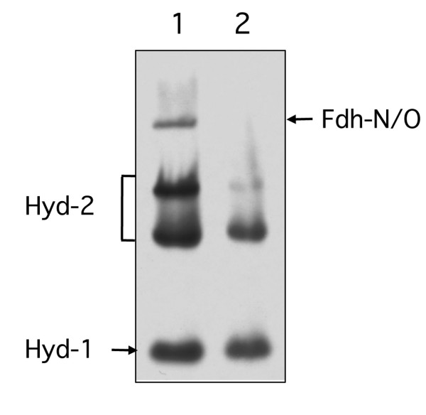
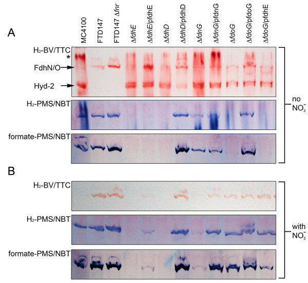
Results: Here we report the identification of a new H₂: benzyl viologen oxidoreductase enzyme activity in E. coli that is independent of the [NiFe]-hydrogenases. This enzyme activity was originally identified after non-denaturing polyacrylamide gel electrophoresis and visualization of hydrogen-oxidizing activity by specific staining. Analysis of a crude extract derived from a variety of E. coli mutants unable to synthesize any [NiFe]-hydrogenase-associated enzyme activity revealed that the mutants retained this specific hydrogen-oxidizing activity. Enrichment of this enzyme activity from solubilised membrane fractions of the hydrogenase-negative mutant FTD147 by ion-exchange, hydrophobic interaction and size-exclusion chromatographies followed by mass spectrometric analysis identified the enzymes Fdh-N and Fdh-O. Analysis of defined mutants devoid of selenocysteine biosynthetic capacity or carrying deletions in the genes encoding the catalytic subunits of Fdh-N and Fdh-O demonstrated that both enzymes catalyze hydrogen activation. Fdh-N and Fdh-O can also transfer the electrons derived from oxidation of hydrogen to other redox dyes.

Conclusions: The related respiratory molybdo-selenoproteins Fdh-N and Fdh-O of Escherichia coli have hydrogen-oxidizing activity. These findings demonstrate that the energy-conserving selenium- and molybdenum-dependent formate dehydrogenases Fdh-N and Fdh-O exhibit a degree of promiscuity with respect to the electron donor they use and identify a new class of dihydrogen-oxidizing enzyme.


Figures

Figure 1
Figure 1
A hypF mutant retains hydrogenase-independent H2: BV oxidoreductase activity. Extracts derived from MC4100 (lane 1) and the isogenic ΔhypF mutant DHP-F2 (lane 2) were separated by non-denaturing PAGE and subsequently stained for hydrogenase enzyme activity as described in the Methods section. Strains were grown in TYEP medium with 0.8% (w/v) glucose, pH 6.5. Equivalent amounts of Triton X-100-treated crude extract (50 μg of protein) were applied to each lane. The activity bands corresponding to Hyd-1 and Hyd-2 are indicated, as is the slowly migrating activity band (designated by an arrow) that corresponds to a hydrogenase-independent H2:BV oxidoreductase enzyme activity.
Figure 2
Figure 2
Chromatographic separation of the H2: BV oxidoreductase activity on a Superdex-S200 column. A. A representative elution profile of the enriched H2: BV oxidoreductase enzyme activity after size exclusion chromatography on Superdex-S200 is shown. The absorbance at 280 nm was monitored and the two main elution peaks were labelled P1 and P2. B. Samples of the fractions across the elution peaks P1 and P2 were separated by non-denaturing PAGE and subsequently stained for hydrogenase enzyme activity. Lane 1, crude cell extract (50 μg protein); lane 2, membrane fraction (50 μg protein); lane 3, solubilised membrane fraction (50 μg protein); lane 4, aliquot of the 400 mM fraction from the Q-sepharose column. The arrow identifies the H2: BV oxidoreductase enzyme activity.
Figure 3
Figure 3
A selC mutant is devoid of the hydrogenase-independent H2: BV oxidoreductase activity. Extracts derived from MC4100 (lane 1) and the isogenic ΔselC mutant FM460 (lane 2) were separated by non-denaturing PAGE and subsequently stained for hydrogenase enzyme activity. Equivalent amounts of Triton X-100-treated crude extract (50 μg of protein) were applied to each lane. The activity bands corresponding to Hyd-1 and Hyd-2 are indicated, as is the activity band due to Fdh-N/Fdh-O (designated by an arrow).
Figure 4
Figure 4
Analysis of H2- and formate-oxidizing activities of Fdh-N/O in different mutant backgrounds. Small-scale cultures of each strain were grown in TGYEP medium in the absence (A) or presence of nitrate (B). Extracts derived from the strains indicated were separated by non-denaturing PAGE and subsequently stained for H2: BV oxidoreductase (top panel), H2: PMS/NBT oxidoreductase (middle panel) or formate: PMS/NBT oxidoreductase (bottom panel) enzyme activity as described in the Methods section. Equivalent amounts of Triton X-100-treated crude extract (25 μg of protein) were applied to each lane. The activity band due to Fdh-N/Fdh-O is labelled by an arrow. The activity band due to hydrogenase 2 (Hyd-2) is also labelled in the top panel of part A and was used as a loading control for the experiment. Note the Hyd-2 activity can only be identified as a H2: BV oxidoreductase activity. The asterisk indicates hydrogenase activity associated with incompletely solubilised membrane material. The gel stained for H2: BV oxidoreductase activity was incubated for 8 h, while the gels stained with PMS/NBT were incubated for 1 h. In the interests of clarity, lanes were labelled based on the key genotype of the strain used. Lanes: MC4100 (wild type); FTD147 (ΔhyaB ΔhybC ΔhycE); FTD147 Δfnr signifies CP1104; ΔfdhE signifies JW3862 (ΔfdhE); ΔfdhE/pfdhE signifies JW3862 complemented with plasmid pCA24N-fdhE+; ΔfdhD signifies JW3866 (ΔfdhD); ΔfdhD/pfdhD signifies JW3866 complemented with plasmid pCA24N-fdhD+; ΔfdnG signifies JW1470 (ΔfdnG); ΔfdnG/pfdnG signifies JW1470 complemented with plasmid pCA24N-fdnG+; ΔfdoG signifies JW3865 (ΔfdoG); ΔfdoG/pfdoG signifies JW3865 complemented with plasmid pCA24N-fdoG+; ΔfdoG/pfdhE signifies JW3865 complemented with plasmid pCA24N-fdhE+. Note that BW25113 had an identical phenotype in these experiments to MC4100.

Similar articles

Cited by

References

    1. Sawers G. The hydrogenases and formate dehydrogenases of Escherichia coli. Antonie van Leeuvenhoek. 1994;66:57–88. doi: 10.1007/BF00871633. - DOI - PubMed
    1. Sawers G, Blokesch M, Böck A. In: EcoSal- Escherichia coli and Salmonella: Cellular and Molecular Biology. Curtiss III R.(Editor in Chief), editor. ASM Press, Washington, D.C; Anaerobic formate and hydrogen metabolism.http://www.ecosal.org September 2004, posting date.
    1. Sawers RG. Formate and its role in hydrogen production in Escherichia coli. Biochem Soc Trans. 2005;33:42–46. - PubMed
    1. Jormakka M, Törnroth S, Byrne B, Iwata S. Molecular basis of proton motive force generation: structure of formate dehydrogenase-N. Science. 2002;295:1863–1868. doi: 10.1126/science.1068186. - DOI - PubMed
    1. Berg BL, Li J, Heider J, Stewart V. Nitrate-inducible formate dehydrogenase in Escherichia coli K-12. I. Nucleotide sequence of the fdnGHI operon and evidence that opal (UGA) encodes selenocysteine. J Biol Chem. 1991;266:22380–22385. - PubMed

Publication types

MeSH terms

LinkOut - more resources