Comparative Study
. 2011 Aug 1:12:385.
doi: 10.1186/1471-2164-12-385.

Comparative analyses imply that the enigmatic Sigma factor 54 is a central controller of the bacterial exterior

Affiliations
Comparative Study

Comparative analyses imply that the enigmatic Sigma factor 54 is a central controller of the bacterial exterior

Christof Francke et al. BMC Genomics. .

Abstract

Background: Sigma-54 is a central regulator in many pathogenic bacteria and has been linked to a multitude of cellular processes like nitrogen assimilation and important functional traits such as motility, virulence, and biofilm formation. Until now it has remained obscure whether these phenomena and the control by Sigma-54 share an underlying theme.

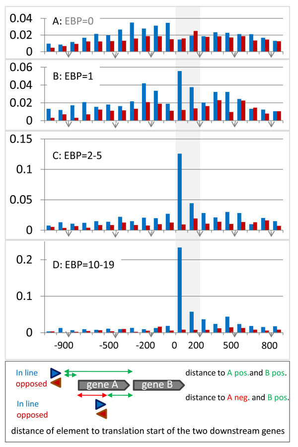
Results: We have uncovered the commonality by performing a range of comparative genome analyses. A) The presence of Sigma-54 and its associated activators was determined for all sequenced prokaryotes. We observed a phylum-dependent distribution that is suggestive of an evolutionary relationship between Sigma-54 and lipopolysaccharide and flagellar biosynthesis. B) All Sigma-54 activators were identified and annotated. The relation with phosphotransfer-mediated signaling (TCS and PTS) and the transport and assimilation of carboxylates and nitrogen containing metabolites was substantiated. C) The function annotations, that were represented within the genomic context of all genes encoding Sigma-54, its activators and its promoters, were analyzed for intra-phylum representation and inter-phylum conservation. Promoters were localized using a straightforward scoring strategy that was formulated to identify similar motifs. We found clear highly-represented and conserved genetic associations with genes that concern the transport and biosynthesis of the metabolic intermediates of exopolysaccharides, flagella, lipids, lipopolysaccharides, lipoproteins and peptidoglycan.

Conclusion: Our analyses directly implicate Sigma-54 as a central player in the control over the processes that involve the physical interaction of an organism with its environment like in the colonization of a host (virulence) or the formation of biofilm.


Figures

Figure 1
Figure 1
Taxonomic distribution of Sigma 54 and the associated Enhancer Binding Proteins. The phyla Proteobacteria and Firmicutes have been divided into the constituent classes. Between brackets, the number of genomes with Sigma-54 over the total number of sequenced genomes is listed followed by the average number of EBP54s with a discrete standard deviation. The corresponding data can be found in additional file 1. The ordering of the phyla is based on conserved gene arrangement comparisons [162], a concatenated alignment of 22 single-copy conserved genes [163] and the analysis of conserved indels [164]. Gemmatimonas was placed according to [165], Nitrospira according to [166] and Thermatogae according to [167]. On the right, the cell morphology in terms of number of membranes (monoderm vs. diderm), presence of LPS (from [64]) and nature of the cell wall peptidoglycan (Gram-, Gram+ or other) is given. The majority of phyla represent diderms, except for Tenericutes, Firmicutes and Actinobacteria. Chloroflexi are probably also monoderm [168] and some have been shown to have a thick cell wall and stain Gram positive [169]. Deinococcus radiodurans has a complex Gram + like cell wall that includes outer membrane-like structure and the cell wall and outer membrane can be shared by multiple cells [170]. Dictyoglomus thermophilum is diderm but can grow in bundles or spherical bodies which are surrounded by a common outer membrane [171]. Finally, the Thermotoga have an outer sheath-like envelope ('toga') and an atypical thin cell wall [172].
Figure 2
Figure 2
Distribution of genome size (A), the number of EBP54-activators (B), and motility (C), for species with (blue) and without (red-brown) Sigma-54. A) the analyzed species were binned according to genome size in bins of one Mbase, and divided in two groups that related to the presence or absence of Sigma-54. B) for every size-bin the fraction of genomes with a particular number of EBP-activators was determined and a height-plot was created. The grey dots indicate the data points. The contour was generated with Microsoft Office Excel 2007. C) Within every bin the fraction of motile species was determined for the genomes with Sigma-54 and without. The corresponding data can be found in additional file 1.
Figure 3
Figure 3
Sequence composition of the Sigma-54 Enhancer Binding Proteins. A) The 'GAFTGA' sequence logo of the 4970 putative functional Sigma-54 related EBPs. Data from literature and similarity in chemical structure were used to categorize the substitutions into those that relate to functional EBP54s, those that will probably relate to functional EBP54s, and those that will abolish the interaction with Sigma-54. The effect of amino acid substitutions on the EBPs capacity to activate Sigma-54 mediated transcription has been studied by [75,76]. Furthermore, some experimentally validated activators carry specific substitutions: G1 is replaced by N in the only EBP54 of Paracoccus denitrificans and Ruegeria pomeroyi (putative: ADEHS); A2 is replaced by S in LevR of the Bacilli (putative: TGIVMC; inactive: DN); F3 is replaced by Y in TouR of Pseudomonas stutzeri (other replacements inactive); T4 is replaced by S in BkdR of B. subtilis and by E in PhhR of Pseudomonas aeruginosa (putative: D; other replacements inactive); G5 is replaced by D in FlgR, the only EBP54 of Campylobacter and other Epsilon-proteobacteria (putative; EAHNS); and A6 is replaced by S in PrpR of E. coli (putative: TGIVMC; inactive DN). B) Schematic representation of the four basic architectures of functional EBP54s. The types were distinguished on basis of their domain organization: Ia) N-terminal signal recognition domain of the response regulator (RR) type, followed by the central activator domain and a C-terminal DNA-binding domain of the HTH_8 PFAM family; Ib) different N-terminal signal recognition domain(s), followed by the central activator domain and a C-terminal DNA-binding domain of the HTH_8 PFAM family; Ic) an activator domain, but lacking the signal recognition domain (e.g. PspF, HrpRS, LafK) or the DNA-binding domain (e.g. CtcC, FlgR) or both (FleT); and II) N-terminal DNA-binding domain of the NtrC family, the central domain, and four phosphorylatable domains related to the PTS.
Figure 4
Figure 4
Sequence features of Sigma-54 and its promoter. A) Functional architecture of the Sigma-54 sequence (adapted from [10]). The first HTH is responsible for recognition of the -12 element as was demonstrated by [61]. The solution structure of the C-terminal domain of Aquifex aeolicus Sigma-54 bound to the promoter implied that the RpoN box [173] and two flanking stretches interact directly with the -24 element of the promoter [174], confirming an earlier assertion of [175]. B) Sequence logo of the two HTH elements as present in all analyzed Sigma-54 proteins. The residue pairs whose substitution abolished binding activity in the elaborate Ala-Cys scanning mutagenesis study by [93], are marked by purple dots in-between. C) Reduced promoter sequence motif. The motif is based on the 85 promoters with validated transcription start site as collected by [14]. The position relative to the transcription start is given on the x-axis.
Figure 5
Figure 5
Distribution of the genomic distance between the downstream genes and the sequence elements that are most similar to the Sigma-54 promoter motif. The distance distribution (in bins of 100 nucleotides) was summed for A) all genomes that lack Sigma-54 and its activator (A; EBP = 0), and for those genomes that have Sigma-54 and one (B; EBP = 1) or multiple EBP54s (C; EBP = 2-5, D; EBP = 10-19). The distance distribution for genomes with EBP = 6-9 and EBP ≥ 20 are similar to the latter and therefore not shown. For every identified element two distances were included as indicated in the figure inset. As a result the distribution actually represents the sum of two distributions. The distance was taken from the -11 position of the promoter to the predicted translation start of the gene (situation i). In case the element was located within a gene (situation ii) the distance to the first gene was taken as negative. In blue the distance distribution is given for the cases that the gene downstream is oriented in line with the predicted promoter and in red for the cases that it opposes the promoter. The sum of the distributions was normalized.
Figure 6
Figure 6
Conserved function tendencies within the gene-associations of Sigma-54, its EBP54s and the Sigma-54 promoter. The highly represented and cross-phylum conserved metabolic reactions were mapped using iPATH [108]. The reactions that relate to only Firmicutes are colored green, those that relate to diderm organisms only are colored yellow and those reactions represented in both monoderm and diderm species are given in orange. The routes associated with phospholipid, peptidoglycan and lipopolysaccharide biosynthesis are indicated and the related precursors are given in blue boxes. The metabolites that are associated to the recovered reactions fall in 3 distinct categories. i) CoA-related: A1, acetyl-CoA; A2, propanoyl-CoA; A3, propenoyl-CoA; A4, 3-hydroxypropanoyl-CoA; A5, 2-methylpropanoyl-CoA; A6, 3-methylbutanoyl-CoA; A7, 2-methylbutanoyl-CoA; A8, (R)-2-methyl-3-oxopropanoyl-CoA; A9, 2-butenoyl-CoA; A10, (S)-3-hydroxybutanoyl-CoA; A11, succinyl-CoA; A12, glutaryl-CoA; A13, 3alpha,7alpha-dihydroxy-5beta-cholestanoyl-CoA; A14, 3-oxoadipyl-CoA; A15, hexadecanoyl-CoA; A16a, acetoacetyl-CoA; A16b, acetoacetyl-[acp]; A17, butanoyl-CoA. ii) carboxylates: C1, acetate; C2, 3-oxopropanoate; C3 glycolate; C4, malate; C5, 3-methyl-2-oxobutanoate; C6, 4-methyl-2-oxopentanoate; C7, 3-methyl-2-oxopentanoate; C8, (S)-methylmalonate semialdehyde; C9, L-aspartate; C10, butanoate; C11, 4-aminobutanoate; C12, L-glutamate; C13, succinate semialdehyde; C14, succinate; C15, hexadecanoate; C16, isocitrate; C17, citrate; C18, oxaloacetate; C19, 3-phospho-D-glycerate; C20, acetoacetate; C21, salicylate; C22, 3-oxoadipate; C23, 3,4-dihydroxymandelaldehyde; C24, chorismate; C25, 6-oxohexanoate; C26, 2-oxoglutarate. iii) amino-group containing: N1, histamine; N2, anthranilate; N3, 5-hydroxytryptamine; N4, 2-amino-4-hydroxy-6-(erythro-1,2,3-trihydroxypropyl)dihydropteridinetriphosphate; N5, Nicotinate; N6, 1,4-butanediamine; N7, 2-hydroxyethyl-ThPP.

References

    1. Gruber TM, Gross CA. Multiple sigma subunits and the partitioning of bacterial transcription space. Annu Rev Microbiol. 2003;57:441–466. - PubMed
    1. Kolter R, Siegele DA, Tormo A. The stationary phase of the bacterial life cycle. Annu Rev Microbiol. 1993;47:855–874. - PubMed
    1. Hengge R. Proteolysis of sigmaS (RpoS) and the general stress response in Escherichia coli. Res Microbiol. 2009;160(9):667–676. - PubMed
    1. Zhao K, Liu M, Burgess RR. The global transcriptional response of Escherichia coli to induced sigma 32 protein involves sigma 32 regulon activation followed by inactivation and degradation of sigma 32 in vivo. J Biol Chem. 2005;280(18):17758–17768. - PubMed
    1. Wagner MA, Zahrl D, Rieser G, Koraimann G. Growth phase- and cell division-dependent activation and inactivation of the {sigma}32 regulon in Escherichia coli. J Bacteriol. 2009;191(5):1695–1702. - PMC - PubMed

Publication types

MeSH terms

LinkOut - more resources