. 2011 Aug 1:11:48.
doi: 10.1186/1471-213X-11-48.

Amnion formation in the mouse embryo: the single amniochorionic fold model

Affiliations

Amnion formation in the mouse embryo: the single amniochorionic fold model

Paulo N G Pereira et al. BMC Dev Biol. .

Abstract

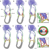
Background: Despite the detailed knowledge obtained over the last decade on the molecular regulation of gastrulation in amniotes, the process of amnion development has been poorly described and illustrated in mice, and conflicting descriptions exist. Understanding the morphogenesis and development not only of the early mouse embryo, but also of its extraembryonic tissues, is crucial for correctly interpreting fate-mapping data and mouse mutants with gastrulation defects. Moreover, the recent isolation from amnion of cells with stem cell features further argues for a better understanding of the process of amnion formation. Here, we revisit the highly dynamic process of amnion formation in the mouse. Amnion development starts early during gastrulation and is intimately related to the formation of the exocoelom and the expansion of the amniotic fold. The authoritative description involves the fusion of two amniotic folds, a big posterior and a smaller anterior fold. We challenged this 'two amniotic folds' model by performing detailed histomorphological analyses of dissected, staged embryos and 3D reconstructions using historical sections.

Results: A posterior fold of extraembryonic ectoderm and associated epiblast is formed early during gastrulation by accumulation of extraembryonic mesoderm posterior to the primitive streak. Previously called the "posterior amniotic fold", we rename it the "amniochorionic fold" (ACF) because it forms both amnion and chorion. Exocoelom formation within the ACF seems not to involve apoptosis within the mesoderm. The ACF and exocoelom expand without disrupting the anterior junction of epiblast, extraembryonic ectoderm and visceral endoderm. No separate anterior fold is formed; its absence was confirmed in 3D reconstructions. Amnion and chorion closure is eccentric, close to the anterior margin of the egg cylinder: we name it the "anterior separation point".

Conclusions: Here, we reconcile previous descriptions of amnion formation and provide new nomenclature, as well as an animation, that clarify and emphasize the arrangement of the tissues that contribute to amnion development and the dynamics of the process. According to our data, the amnion and the chorion are formed by a single amniochorionic fold initiated posteriorly. Finally, we give an overview on mutant mouse models with impaired amnion development.


Figures

Figure 1
Figure 1
Extraembryonic tissues and organs in a mouse embryo and foetus. (A-E) Progressive dissection of an E8.5 mouse conceptus revealing its extraembryonic tissues. (A) Deciduum (De) as isolated from the uterus. (B) Isolated conceptus: only the parietal yolk sac (PYS), including Reichert's membrane is visible. (C) Upon removal of the PYS the ectoplacental cone (EPCn), visceral yolk sac (VYS) and embryo proper become visible. (D-E) When the VYS is, subsequently, removed the amnion (Am), the allantois (Al) and the embryo proper become better visible. Scale bar: 500 μm (F-H) E12.5 mouse conceptus. (F) Foetus within its PYS and VYS, with intact placenta (Pl). (G) Removal of the PYS reveals the vascularized VYS. (H) Avascular amnion and the umbilical cord (UC) are visible when the foetus is dissected free from VYS. Scale bar: 1 mm (I) Schematic representation of the extraembryonic tissues at the level of the dashed line in (F), and their composition. Additional abbreviations in the scheme: AmEc: amniotic ectoderm; AmM: amniotic mesoderm; AC: amniotic cavity; EC: exocoelomic cavity; Em: embryo; ExM: extraembryonic mesoderm; PEnd: parietal endoderm; RM: Reichert's membrane; TE: trophectoderm; VEnd: visceral endoderm; YSC: yolk sac cavity.
Figure 2
Figure 2
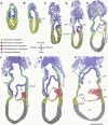
Amnion formation in mouse embryos, illustrated by longitudinal sections. Sections at the midline in the extraembryonic-embryonic region of embryos between E6.0 and E7.5 illustrate the different stages of amnion development. The tissue layers in the extraembryonic and extraembryonic-embryonic junction region were artificially painted based on histomorphological differences and the analysis of layer-specific markers (Figure 4). (A) Prestreak stage: no mesoderm, no amniochorionic fold (ACF). (B) Early streak stage: extraembryonic and embryonic mesoderm emerges at the primitive streak (PS). (C) Midstreak stage: extracellular spaces accumulate within the extraembryonic mesoderm. (D) Late streak/no bud stage: the ACF protrudes into the proamniotic cavity (PAC) and coalescence of spaces in the extraembryonic mesoderm generates the exocoelomic cavity (EC). The endodermal furrow (En-frw) marks the anterior midline at the extraembryonic-embryonic junction. (E) Between late streak/no bud and late streak/early bud stage: expansion of the EC. (F) Late streak/early bud stage: a large ACF extends from the posterior. The allantoic bud (Al-bud) has become visible. (G) Between late streak/early bud and neural plate stage: the lateral extensions of the EC meet at the focal anterior separation point (ASP). Closure and separation of the ectodermal lineages occurs. (H) Neural plate stage: the segregated amniotic (Am) and chorionic (Ch) membranes divide the PAC into the amniotic cavity (AC), EC and ectoplacental cavity (EPC). The allantois (Al) and visceral yolk sac (VYS) become clear. The amniotic ectoderm (AmEc) and mesoderm (AmM) acquire their squamous architecture. Sections do not go through the midline at the posterior (E) or anterior (H) side at the extraembryonic-embryonic junction of the conceptus. Scale bar: 200 μm.
Figure 3
Figure 3
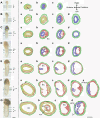
Amnion Formation In Mouse Embryos, Illustrated By Transverse Sections. Transverse sections of embryos between E6.5 and E7.5 illustrate the different stages of amnion development. Each section plane is represented by a corresponding line in the picture of a matching whole-mount embryo. Artificial colors as in Figure 2. No attempt has been made to distinguish the head process and any definitive endoderm from the embryonic mesoderm and visceral endoderm, respectively. (A) Early streak stage: extraembryonic mesoderm has inserted between extraembryonic ectoderm and visceral endoderm at the posterior side of the embryo. (B) Midstreak stage: extracellular spaces are present within the extraembryonic mesoderm. (C) Late streak/no bud stage: lacunae in extraembryonic mesoderm coalesce to form the exocoelomic cavity (EC). (D) Between late streak/no bud and late streak/early bud stage: the EC has enlarged. (E) Late streak/early bud stage: the EC extends laterally around the egg cylinder, converging on the anterior midline at the embryonic-extraembryonic junction. (F) Between late streak/early bud and neural plate stage: the proamniotic cavity (PAC) is constricted by the embryonic ectoderm and extraembryonic ectoderm of the amniochorionic fold, preceding the closure and separation of amnion and chorion. The allantoic bud (Al-bud) is prominent. (G) Neural plate stage: the amnion (Am) and chorion (Ch) are completely segregated dividing the proamniotic cavity into the amniotic cavity (AC), exocoelomic cavity and ectoplacental cavity (EPC). Scale bar: 200 μm.
Figure 4
Figure 4
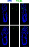
Absence of apoptosis in the amniochorionic fold of embryos undergoing exocoelom formation. (A-B) TUNEL assay in sections of embryos showing absence of programmed cell death in extraembryonic mesoderm during the process of exocoelom formation. Apoptotic cells were readily detected in the ectoplacental cone. Scale bar: 200 μm.
Figure 5
Figure 5
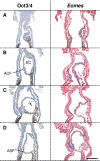
Embryonic and extraembryonic ectoderm demarcation in the amniochorionic fold. Embryonic ectoderm was marked by IHC for Oct3/4 (brown), while extraembryonic ectoderm was marked by the expression of Eomes (blue). The latter is also detected in nascent embryonic and extraembryonic mesoderm. (A) Prior to the formation of the exocoelomic cavity (EC), the amniochorionic fold (ACF) is recognized by the extraembryonic mesoderm accumulation between extraembryonic ectoderm and visceral endoderm. (B) Soon after formation of the EC, the extraembryonic ectoderm is the major contributor for the ectoderm of the ACF. (C) The contribution of embryonic ectoderm to the ACF increases progressively as the embryo continues to grow, and the EC enlarges. The black arrow indicates the transient non-alignment that often appears between the mesoderm and extraembryonic ectoderm of the ACF. (D) At the anterior separation point (ASP), embryonic ectoderm is aligned with extraembryonic mesoderm in the lower half of the ACF to form the amnion, while the extraembryonic ectoderm apposed to extraembryonic mesoderm in the upper half of the fold forms the chorion. Scale bar: 200 μm.
Figure 6
Figure 6
Series of longitudinal sections of an embryo with large exocoelomic cavity (ec). Artificial colors as in Figure 2. (A) Midline section characterized by the presence of the allantoic bud (Al-bud), and endodermal furrow (En-frw) close to the prospective anterior separation point. (B) Section adjacent to the midline section A. (C-D) Neighboring sections cut twice through the amniochorionic fold (ACF), which result seemingly in an anterior fold and posterior fold with their respective cavities. (E) A more lateral section reveals that the exocoelomic cavity extends around the egg cylinder. (F) Most lateral section of the series. Scale bar: 100 μm (G) Position of the longitudinal sections (A to F) on a schematic transverse section of the embryo at the level of the dashed line in A. Grey reflects the EC; no fill represents the proamniotic canal (PAC). (C'-D') Magnifications of the boxed areas shown in figure panel C and D, respectively. Scale bar: 50 μm.
Figure 7
Figure 7
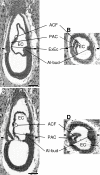
Reconstruction of embryos prepared for kaufman's the atlas of mouse development. (A) Midline longitudinal section from the embryo displayed in plate 5 a-e of The Atlas of Mouse Development. Scale bar: 100 μm (B) Transverse slice at the level of the dashes in A after 3D computer reconstruction of the consecutive sections of the embryo used for plate 5 a-e. The pixelation is mainly due to the thickness of the sections (7 μm), compared with the 0.34 μm resolution in A. (C) Longitudinal section of another embryo from Kaufman's collection. This embryo was not included in The Atlas of Mouse Development. (D) Transverse slice at the level of the dashes in C after 3D computer reconstruction of the consecutive sections of the embryo. Dashes in D indicates the level of the section shown in C. Abbreviations: ACF: amniochorionic fold; Albud: allantoic bud; EC: exocoelomic cavity; ExEc: extraembryonic ectoderm; PAC: proamniotic cavity.

References

    1. Schmidt W. The amniotic fluid compartment: the fetal habitat. Adv Anat Embryol Cell Biol. 1992;127:1–100. - PubMed
    1. Gardner RL. The relationship between cell lineage and differentiation in the early mouse embryo. Results Probl Cell Differ. 1978;9:205–41. - PubMed
    1. Kaufman MH. The Atlas of Mouse Development. London: Academic Press; 1992.
    1. George EL, Georges-Labouesse EN, Patel-King RS, Rayburn H, Hynes RO. Defects in mesoderm, neural tube and vascular development in mouse embryos lacking fibronectin. Development. 1993;119:1079–91. - PubMed
    1. Gersdorff N, Muller M, Otto S, Poschadel R, Hubner S, Miosge N. Basement membrane composition in the early mouse embryo day 7. Dev Dyn. 2005;233:1140–8. doi: 10.1002/dvdy.20425. - DOI - PubMed

Publication types

LinkOut - more resources