. 2011 Aug 2:9:106.
doi: 10.1186/1477-7827-9-106.

Spatiotemporal expression of SERPINE2 in the human placenta and its role in extravillous trophoblast migration and invasion

Affiliations

Spatiotemporal expression of SERPINE2 in the human placenta and its role in extravillous trophoblast migration and invasion

Schu-Rern Chern et al. Reprod Biol Endocrinol. .

Abstract

Background: SERPINE2, one of the potent serpins belonging to the plasminogen activator (PA) system, is involved in the tissue remodeling. We previously demonstrated the expression patterns of Serpine2 in the mouse placenta and uterus, indicating that Serpine2 is a major PA inhibitor in the placenta and uterus during the estrous cycle, pregnancy, and lactation. In this study, we further investigated the expression pattern of SERPINE2 in the human placenta and explored possible functional roles of SERPINE2 in regulating trophoblast activity.

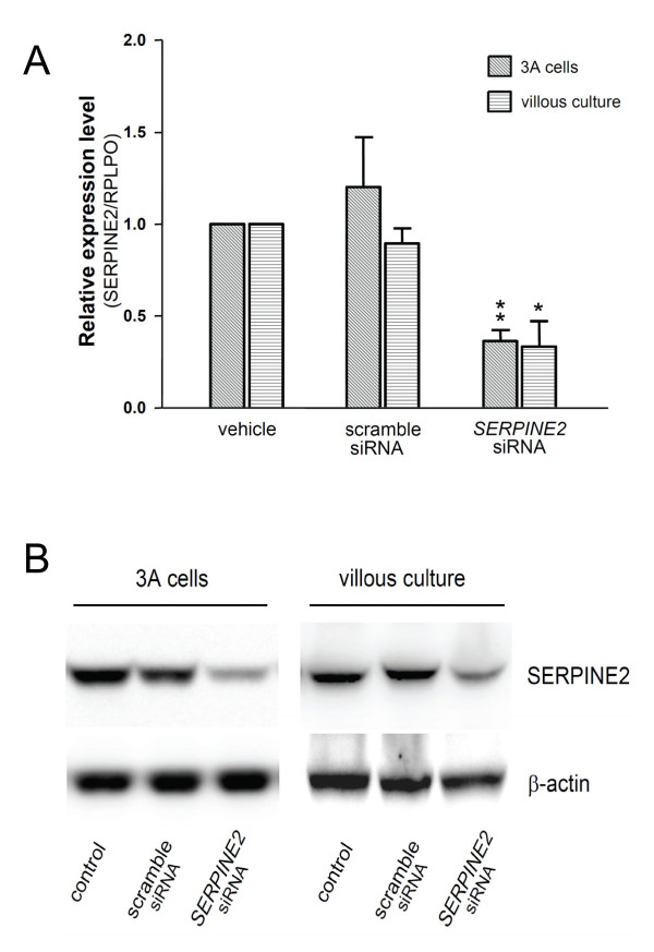
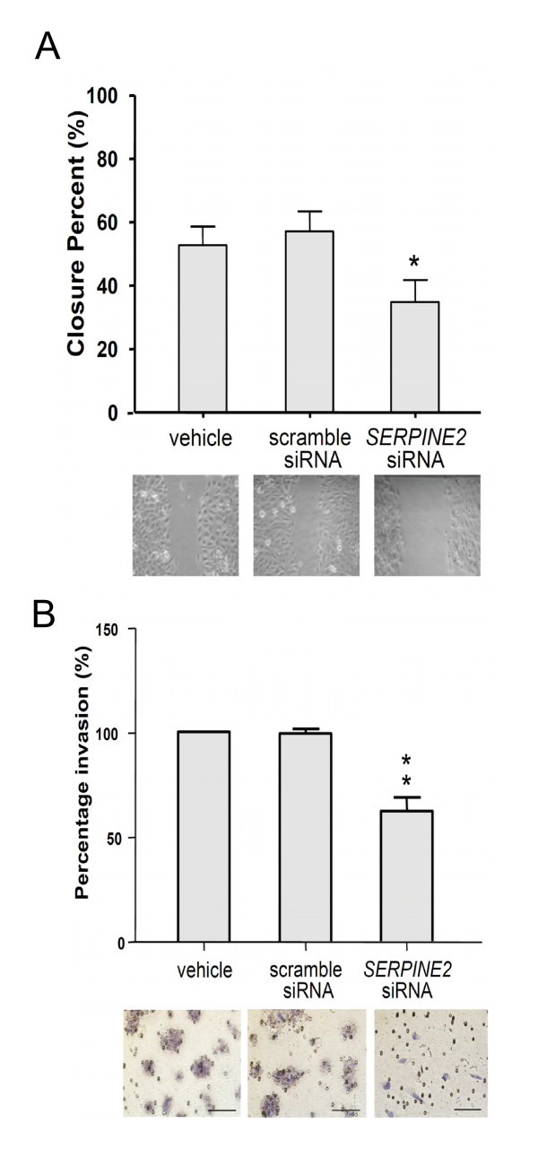
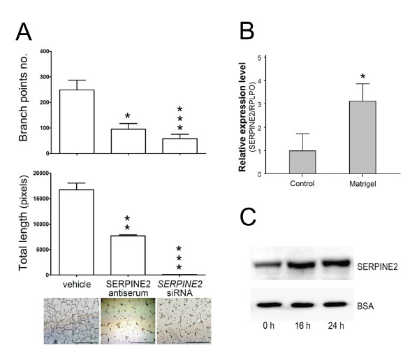
Methods: Placental tissues from various trimesters were collected for real-time reverse-transcription polymerase chain reaction quantification. Immunohistochemical staining was performed in placental tissues to assure localization of SERPINE2. SERPINE2 small interfering (si) RNA was applied to suppress its expression in villous explants and extravillous trophoblast-like 3A cells. Subsequent experiments to evaluate SERPINE2 levels, villous outgrowth, trophoblast invasion, and tube formation were performed.

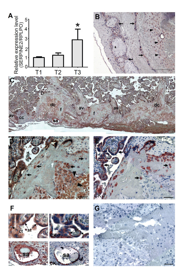
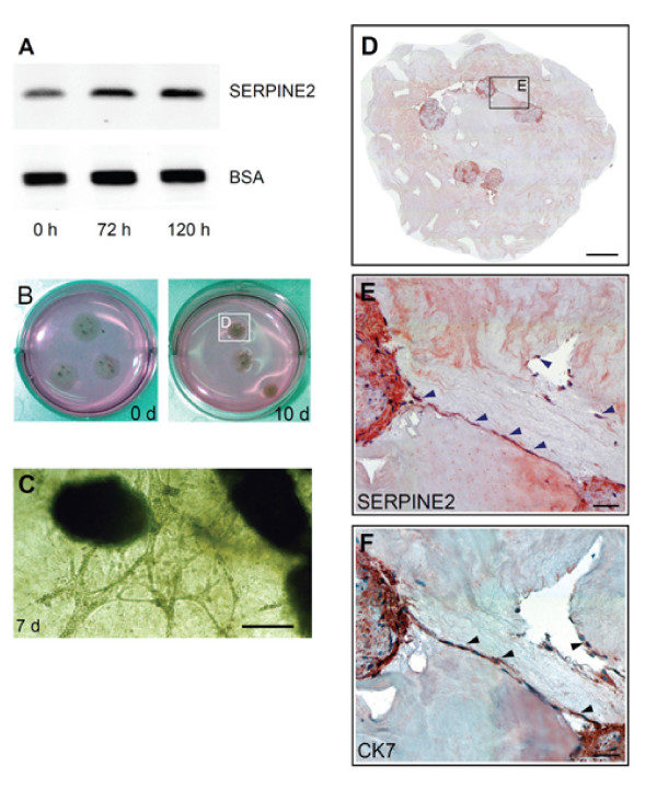
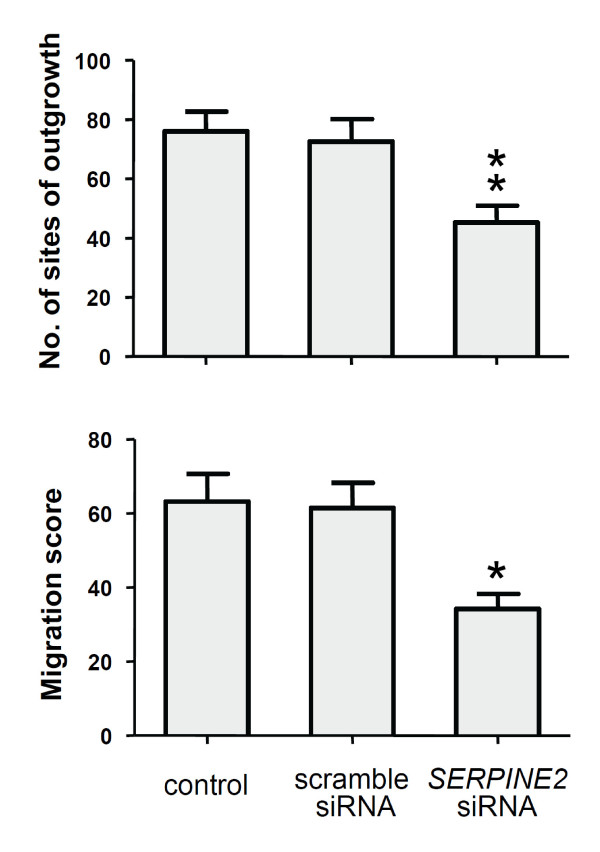
Results: SERPINE2 messenger RNA was detected in the human placenta during pregnancy with the highest levels in the third trimester. The SERPINE2 protein was present in villous syncytiotrophoblasts and trophoblasts of chorionic villi for anti-SERPINE2 immunostaining. Extravillous trophoblasts in the chorionic plate and basal plate confronting the invasive face of anchoring villi were also positive. In most decidual cells, SERPINE2 was observed in the cytoplasm. In addition, fibrinoid deposit was weakly immunoreactive. Introduction of SERPINE2 siRNA into villous explants and trophoblast cells led to significantly reduced villous outgrowth, and trophoblastic migration and invasion. Moreover, capillary-like network formation of 3A cells in Matrigel was greatly attenuated by SERPINE2 siRNA and SERPINE2 antiserum.

Conclusions: These data identify the temporal and spatial SERPINE2 distribution in the human placenta and suggest its possible role in modulating tissue remodeling of extravillous trophoblasts in the placenta during pregnancy.


Figures

Figure 1
Figure 1
Spatiotemporal expression of SERPINE2 in the human placenta. (A) Expression of SERPINE2 mRNA in human placentas in different trimesters. Levels of SERPINE2 mRNA were determined by real-time quantitative PCR in 19 placental tissues collected in three gestational trimesters (T1, T2, and T3; n = 5, 4, and 10, respectively). For each sample, the SERPINE2 expression level was normalized to the expression level of the RPLPO gene in the same sample. Data are presented as the mean ± SEM. * p < 0.05. Immunohistochemical staining showed the distribution of the SERPINE2 protein in the human placenta at gestational week 15. (B) Moderate staining of extravillous trophoblasts (arrow) and decidual cells (arrowhead) in the chorionic plate, and very low staining in the chorionic mesoderm and fibrinoid deposits. (C) Positive immunostaining was extensively detected in decidual cells (dc), cytotrophoblasts, extravillous trophoblasts at the junction zone of the cell column (cc) and anchoring villi (av), and the endothelia of the spiral artery (sa); and weak staining was found in fibrinoids (f) and the villous mesenchyme. (D) The dashed-lined region in 1C was magnified to show intense staining in syncytiotrophoblasts and cytotrophoblasts in floating villi. The invaded extravillous trophoblasts (arrow) and decidual cells (arrowhead) at the basal plate were strongly stained. (E) Positive staining of cytokeratin (CK)-7 was confirmed in syncytiotrophoblasts and cytotrophoblasts in floating villi, and invading trophoblasts (arrow). (F) Upper panels, dashed-lined rectangle regions in D and E were magnified to show strong staining of SERPINE2 (left) and CK-7 (right) in syncytiotrophoblasts (st) and cytotrophoblasts (ct) in floating villi. Lower panels, most of the endothelia of spiral arteries were positively stained with anti-SERPINE2 (left), and anti-CK-7 (right) antibodies. (G) Negative staining of control antiserum. Scale bars represent 200 μm (B, C), and 50 μm (D, E, G).
Figure 2
Figure 2
Trophoblast outgrowth and invasion of villous explants on the extracellular matrix (ECM) gel accompanying elevated SERPINE2 expression. (A) Western blot analysis was used to detect the secreted SERPINE2 protein in the supernatant of explant cultures. Significant increases of the explants during culture were evidenced compared to the loading controls (BSA from FBS in the culture medium, partially removed by affinity chromatography). (B) After long-term (10-day) explant culture, the gels had shrunk and formed condensed 'balls' and probably detached from the plastic surface of the culture dish to float in the medium. (C) Before condensation of the ECM gel, a 3D mesh could be observed under an inverted microscope. An illustrative photo of a 7-day explant is shown. (D) Explants in shrunken gel were harvested and immunochemically stained with SERPINE2 antiserum. (E) The invading, lined trophoblasts were immunoreactive with SERPINE2 (arrowhead). (F) The invading cells were mostly CK-7-positive trophoblasts. Scale bars represent 500 μm (C, D), and 100 μm (E, F).
Figure 3
Figure 3
SERPINE2 siRNA decreases expressions of SERPINE2 mRNA and protein in 3A cells and villous culture. (A) SERPINE2 mRNA levels in 3A cells and villous culture were specifically blocked by siRNA against SERPINE2. Scrambled or SERPINE2 siRNAs were delivered into cells by transfection (3A cells) or by active transport (villous culture). SERPINE2 mRNA was determined by real-time quantitative PCR after 48 h of treatment. Data are presented as the mean ± SEM. * p < 0.05, ** p < 0.01 were considered significant. (B) Western blot analysis of cell lysates isolated from siRNA-treated (48 h) villi and 3A cells. For the loading control, membranes were stripped and re-incubated with a β-actin antibody.
Figure 4
Figure 4
Semiquantitative analysis of villous explant outgrowth and migration. Explants from four placentas (at the gestational ages of 9, 12, 16, and 20 wk) were cultured in the presence or absence of SERPINE2 siRNA for 120 h, and the migration score based on a scale (see Methods and Additional file 3, Figure S2) was evaluated by 2 observers. Data are presented as the mean ± SEM. * p < 0.05 ** p < 0.01.
Figure 5
Figure 5
Suppression of SERPINE2 led to inhibition of the migration and invasion of trophoblast-derived 3A cells. (A) A motility assay demonstrated deduced cell migration of 3A cells transfected with siRNA which specifically silenced the expression of SERPINE2. (B) A Matrigel invasion assay showed a reduced invasive capacity of 3A cells treated with SERPINE2 siRNA. Percentage invasion of vehicle is set as 100%. * p < 0.5, ** p < 0.01. Data are presented as the mean ± SEM. Scale bars represent 100 μm.
Figure 6
Figure 6
Matrigel-induced network formation of trophoblast 3A cells was inhibited by SERPINE2 antiserum or siRNA. (A) Treatment of 3A cells with SERPINE2 antiserum or siRNA significantly blocked the formation of the capillary-like network, as scored by the number of branch points and total length formed following 24 h of treatment. (B) SERPINE2 mRNA levels in 3A cells in Matrigel compared to controls. (C) Western blot analysis was used to detect the secreted SERPINE2 protein in the supernatant of 3A cells in the tube formation assay. Each experiment was carried out in triplicate, and each bar represents the mean ± SEM. * p < 0.05, ** p < 0.01, *** p < 0.001 compared to the scrambled siRNA-treated controls. Representative images of capillary-like network formation by 3A cells are shown. Scale bars represent 100 μm.

Similar articles

Cited by

References

    1. Gloor S, Odink K, Guenther J, Nick H, Monard D. A glia-derived neurite promoting factor with protease inhibitory activity belongs to the protease nexins. Cell. 1986;47(5):687–93. doi: 10.1016/0092-8674(86)90511-8. - DOI - PubMed
    1. Baker JB, Low DA, Simmer RL, Cunningham DD. Protease-nexin: a cellular component that links thrombin and plasminogen activator and mediates their binding to cells. Cell. 1980;21(1):37–45. doi: 10.1016/0092-8674(80)90112-9. - DOI - PubMed
    1. Stone SR, Nick H, Hofsteenge J, Monard D. Glial-derived neurite-promoting factor is a slow-binding inhibitor of trypsin, thrombin, and urokinase. Arch Biochem Biophys. 1987;252(1):237–44. doi: 10.1016/0003-9861(87)90028-2. - DOI - PubMed
    1. Knauer DJ, Majumdar D, Fong PC, Knauer MF. SERPIN regulation of factor XIa. The novel observation that protease nexin 1 in the presence of heparin is a more potent inhibitor of factor XIa than C1 inhibitor. J Biol Chem. 2000;275(48):37340–6. doi: 10.1074/jbc.M003909200. - DOI - PubMed
    1. Chen LM, Skinner ML, Kauffman SW, Chao J, Chao L, Thaler CD, Chai KX. Prostasin is a glycosylphosphatidylinositol-anchored active serine protease. J Biol Chem. 2001;276(24):21434–42. doi: 10.1074/jbc.M011423200. - DOI - PubMed

Publication types

LinkOut - more resources