. 2011 Aug 3;101(3):651-61.
doi: 10.1016/j.bpj.2011.06.025.

Molecular determinants of U-type inactivation in Kv2.1 channels

Affiliations

Molecular determinants of U-type inactivation in Kv2.1 channels

Y M Cheng et al. Biophys J. .

Abstract

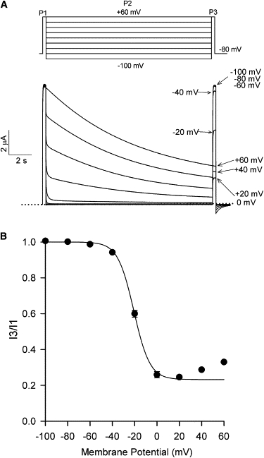
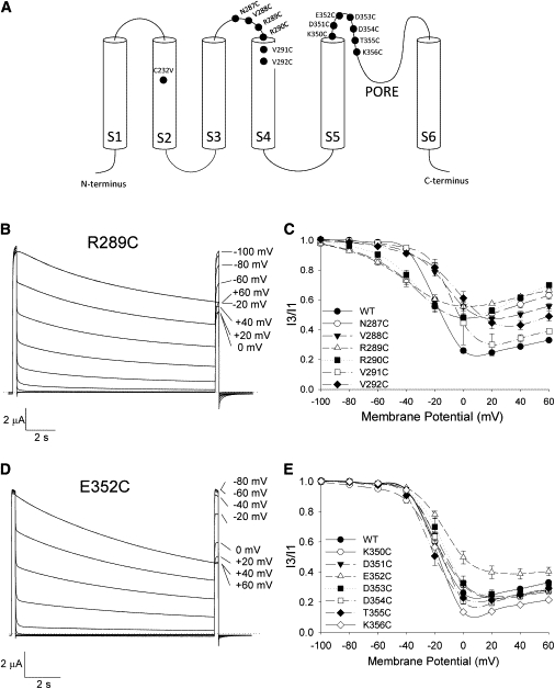
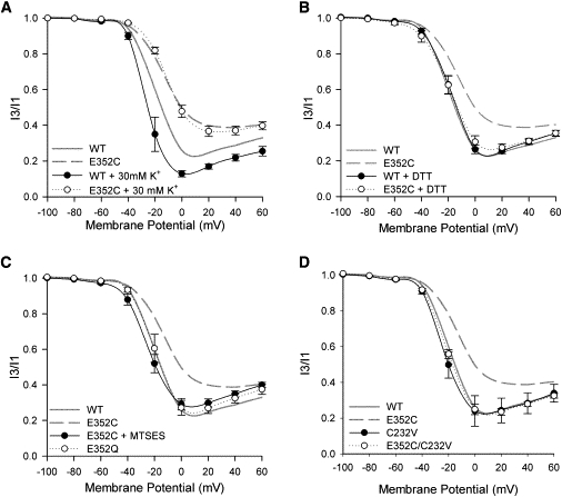
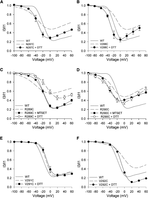
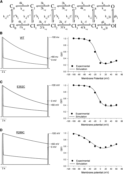
Kv2.1 channels exhibit a U-shaped voltage-dependence of inactivation that is thought to represent preferential inactivation from preopen closed states. However, the molecular mechanisms underlying so-called U-type inactivation are unknown. We have performed a cysteine scan of the S3-S4 and S5-P-loop linkers and found sites that are important for U-type inactivation. In the S5-P-loop linker, U-type inactivation was preserved in all mutant channels except E352C. This mutation, but not E352Q, abolished closed-state inactivation while preserving open-state inactivation, resulting in a loss of the U-shaped voltage profile. The reducing agent DTT, as well as the C232V mutation in S2, restored U-type inactivation to the E352C mutant, which suggests that residues 352C and C232 may interact to prevent U-type inactivation. The R289C mutation, in the S3-S4 linker, also reduced U-type inactivation. In this case, DTT had little effect but application of MTSET restored wild-type-like U-type inactivation behavior, suggestive of the importance of charge at this site. Kinetic modeling suggests that the E352C and R289C inactivation phenotypes largely resulted from reductions in the rate constants for transitions from closed to inactivated states. The data indicate that specific residues within the S3-S4 and S5-P-loop linkers may play important roles in Kv2.1 U-type inactivation.


Figures

Figure 1
Figure 1
U-type inactivation of wt Kv2.1. (A, Top) Schematic of the three-pulse (3P) protocol used to study Kv2.1 inactivation. The voltage for the 15 s P2 was increased in 20-mV increments, whereas P1 and P3 were at +60 mV for 300 ms. (Bottom) Macroscopic currents recorded using the 3P protocol from an oocyte expressing wt Kv2.1 channels. Shown are nine superimposed traces obtained in ND96 solution. The sweep interval was 60 s. (B) The inactivation-voltage relationship for wt Kv2.1 was determined by taking the ratio of current (I3/I1) measured during P3 and P1 and plotting it against the P2 voltage. (Data points) Mean ± se; where not visible, error bars are hidden within symbols. n = 7. (Solid line) Fit of the descending portion of the inactivation-voltage relationship (i.e., between −100 and +20 mV) to a Boltzmann function. See Table 1 for a summary and the fit parameters.
Figure 2
Figure 2
Cysteine substitutions in the S3-S4 and S5-P-loop linkers alter Kv2.1 inactivation. (A) Schematic of a Kv2.1 α-subunit showing the relative locations of the mutations studied. (B and D) Current recordings from oocytes expressing the R289C and E352C mutant channels, made using the 3P protocol. (C and E) The inactivation-voltage relationships for the S3-S4 and S5-P-loop linker mutants, respectively. The data for wt Kv2.1 is also shown for comparison. (Data points) Mean ± se. n > 4 for each mutant. (Lines) Spline connectors to guide the eye. For clarity, Boltzmann fits to the data are not shown; see Table 1 for a summary of the data and the fit parameters.
Figure 3
Figure 3
U-type inactivation can be restored to the E352C mutant by removing disulfide bonds. Each panel shows the inactivation-voltage relationship of wt and E352C channels under different test conditions. Unless otherwise stated, recordings were performed in ND96. Data are shown as mean ± SE, connected by simple spline curves; n > 3 for each data set. The inactivation-voltage relationships of wt and E352C channels under control conditions in ND96 are also shown for comparison (shaded and dashed spline curves, respectively). (A) Inactivation of the E352C mutant is not enhanced by 30 mM [K+]o. (B) Thirty-minute DTT pretreatment restores U-type inactivation to E352C channels. (C) Thirty-minute MTSES pretreatment restores U-type inactivation in E352C channels. Note that before treatment with MTSES, the oocyte was exposed to DTT for 30 min. U-type inactivation is unaffected by the E352Q mutation. (D) U-type inactivation is restored in the E352C/C232V double mutant. The inactivation-voltage relationship of the C232V mutation alone is not different from that of wt Kv2.1.
Figure 4
Figure 4
Modulation of U-type inactivation in the S3-S4 linker mutants. Each panel shows the inactivation-voltage relationship of a given mutant channel recorded after pretreatment with DTT and/or MTSET. Each panel also shows the inactivation-voltage relationship of wt Kv2.1 and the mutant channel (shaded and dashed lines, respectively) recorded under control conditions for comparison. (Data points) Mean ± SE, connected by spline curves; n ≥ 3 for each test condition.
Figure 5
Figure 5
Kinetic modeling of wt and mutant Kv2.1 inactivation. (A) Allosteric model used to simulate channel gating. State transitions are governed by 11 free parameters, as described in the text and Table 3. (BD, left side) Simulated macroscopic currents for wt, E352C, and R289C channels, respectively. These simulations were performed using the parameters shown in Table 3. Three superimposed traces are shown for each construct. (Right side) Comparison of the experimental and simulated inactivation-voltage relationships. (Data points) Mean ± SE experimental i3/i1 values. (Solid lines) Simulated I3/I1 values, plotted against voltage in 1-mV increments.

References

    1. Kurata H.T., Fedida D. A structural interpretation of voltage-gated potassium channel inactivation. Prog. Biophys. Mol. Biol. 2006;92:185–208. - PubMed
    1. Bähring R., Covarrubias M. Mechanisms of closed-state inactivation in voltage-gated ion channels. J. Physiol. 2011;589:461–479. - PMC - PubMed
    1. Grissmer S., Cahalan M. TEA prevents inactivation while blocking open K+ channels in human T lymphocytes. Biophys. J. 1989;55:203–206. - PMC - PubMed
    1. Choi K.L., Aldrich R.W., Yellen G. Tetraethylammonium blockade distinguishes two inactivation mechanisms in voltage-activated K+ channels. Proc. Natl. Acad. Sci. USA. 1991;88:5092–5095. - PMC - PubMed
    1. Molina A., Castellano A.G., López-Barneo J. Pore mutations in Shaker K+ channels distinguish between the sites of tetraethylammonium blockade and C-type inactivation. J. Physiol. 1997;499:361–367. - PMC - PubMed

Publication types

LinkOut - more resources