Review
. 2011 Oct;22(8):842-9.
doi: 10.1016/j.semcdb.2011.07.011. Epub 2011 Jul 23.

How signaling between cells can orient a mitotic spindle

Affiliations
Review

How signaling between cells can orient a mitotic spindle

Adam D Werts et al. Semin Cell Dev Biol. 2011 Oct.

Abstract

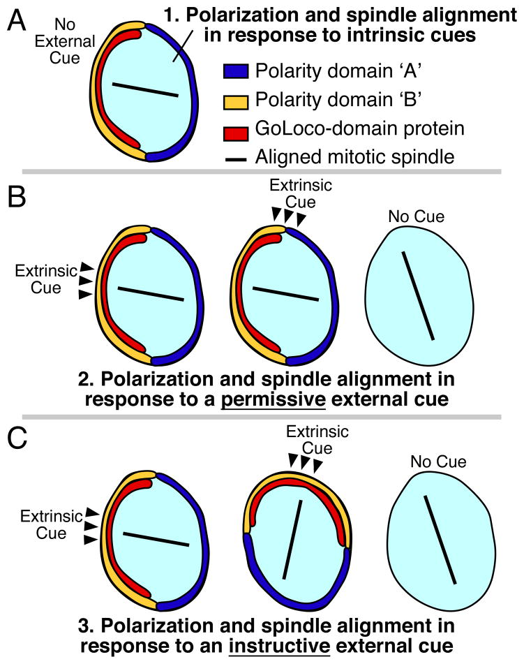
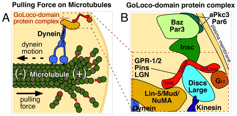
In multicellular animals, cell communication sometimes serves to orient the direction in which cells divide. Control of division orientation has been proposed to be critical for partitioning developmental determinants and for maintaining epithelial architecture. Surprisingly, there are few cases where we understand the mechanisms by which external cues, transmitted by intercellular signaling, specify the division orientation of animal cells. One would predict that cytosolic molecules or complexes exist that are capable of interpreting extrinsic cues, translating the positions of these cues into forces on microtubules of the mitotic spindle. In recent years, a key intracellular complex has been identified that is required for pulling forces on mitotic spindles in Drosophila, Caenorhabditis elegans and vertebrate systems. One member of this complex, a protein with tetratricopeptide repeat (TPR) and GoLoco (Gα-binding) domains, has been found localized in positions that coincide with the positions of spindle-orienting extracellular cues. Do TPR-GoLoco proteins function as conserved, spatially regulated mediators of spindle orientation by intercellular signaling? Here, we review the relevant evidence among cases from diverse animal systems where this protein complex has been found to localize to specific cell-cell contacts and to be involved in orienting mitotic spindles.


Figures

Figure 1
Figure 1. Polarity establishment by intrinsic cues, permissive external cues, and instructive external cues
(A) Some cells align their mitotic spindles independent of external signaling cues. Polarity domains ‘A’ and ‘B’ are nonspecific and could represent ‘Anterior’ and ‘Posterior’ polarity, ‘Apical’ and ‘Basal’ polarity, ‘Dorsal’ and ‘Ventral’ polarity, etc, depending on the specific cell type. (B) Permissive external cues: Some cells require an external cue (black arrowheads) for polarization and spindle alignment, but the position of that cue does not convey positional information to cell polarity: moving the cue has no effect on cell polarity or spindle orientation (middle). Absence of these cues leads to polarity defects and defects in spindle orientation (right). (C) Instructive external cues: Some cells are polarized by instructive external cues (black arrowheads), where changing the position of the cue changes the orientation of polarity and division. Experimentally moving the position of an extrinsic cue differentiates between permissive (B) and instructive (C) functions in spindle orientation.
Figure 2
Figure 2. Model of the TPR-GoLoco protein complex
(A) Schematic showing how the TPR-GoLoco protein complex forms a link between microtubules and the plasma membrane, based upon [62]. Red arrows show tubulin depolymerization from the plus end of the microtubule. Proteins are depicted roughly proportional to their relative sizes. (B) Enlargement of part of (A), with additional associated proteins included. See text for details. Gα is myristoylated (pink line), associating it with the plasma membrane. Inscuteable links Par3/Baz to TPR-GoLoco proteins in some cells such as Drosophila neuroblasts [53], but this interaction is absent in cells such as MDCK cyst cells and Drosophila SOPs, where Par3/Baz localizes in a reciprocal cortical pattern to TPR-GoLoco protein [37,75].
Figure 3
Figure 3. Intercellular signaling functions as an instructive cue for spindle orientation mediated by the TPR-GoLoco protein Pins in Drosophila SOP cells
(A) Extracellular signals from Frizzled, Strabismus, and Flamingo control Pins cortical localization and cell polarity of pI, the primary progenitor of the sensory organ lineage. See text for details. Two functional isoforms of Flamingo have been proposed: F-Flamingo and V-Flamingo [48]. F-Flamingo is proposed to interact with and be induced by Frizzled in the same cell, while V-Flamingo is proposed to interact with Vang in the same cell [74]. (B–C) Schematics after [35, 76]. (B) A view from the embryo’s surface of an SOP cell. Colored crescents represent polarized localization of proteins shown in (A), using the same colors. (C) Schematic of localization of the relevant cortical proteins controlling division along the axis of polarity and within the plane of the epithelium. Black lines represent the orientation of the mitotic spindle. (D) Schematic of SOP clone border analysis from experiments described in [15]. Loss of Fz or Vang on one side of a SOP cell results in altered polarization of Pins and another anterior cortical protein called Partner of Numb (Pon), and alignment of the mitotic spindle. Black arrows represent spindle alignment, and crossed black lines on the far right panels represent randomization of the division axis in the absence of external cues. SOPs in the far right two panels still polarize and divide asymmetrically in the absence of PCP signaling, but in a random orientation.

Similar articles

Cited by

References

    1. Baena-Lopez LA, Baonza A, Garcia-Bellido A. The orientation of cell divisions determines the shape of Drosophila organs. Curr Biol. 2005;15:1640–4. - PubMed
    1. Fischer E, Legue E, Doyen A, Nato F, Nicolas JF, Torres V, Yaniv M, Pontoglio M. Defective planar cell polarity in polycystic kidney disease. Nat Genet. 2006;38:21–3. - PubMed
    1. Saburi S, Hester I, Fischer E, Pontoglio M, Eremina V, Gessler M, Quaggin SE, Harrison R, Mount R, McNeill H. Loss of Fat4 disrupts PCP signaling and oriented cell division and leads to cystic kidney disease. Nat Genet. 2008;40:1010–5. - PubMed
    1. Segalen M, Bellaiche Y. Cell division orientation and planar cell polarity pathways. Semin Cell Dev Biol. 2009;20:972–7. - PubMed
    1. Karner CM, Chirumamilla R, Aoki S, Igarashi P, Wallingford JB, Carroll TJ. Wnt9b signaling regulates planar cell polarity and kidney tubule morphogenesis. Nat Genet. 2009;41:793–9. - PMC - PubMed

Publication types

LinkOut - more resources