. 2011 Oct;69(10):2564-78.
doi: 10.1016/j.joms.2011.03.005. Epub 2011 Jul 31.

Novel therapy to reverse the cellular effects of bisphosphonates on primary human oral fibroblasts

Affiliations

Novel therapy to reverse the cellular effects of bisphosphonates on primary human oral fibroblasts

Matthew Cozin et al. J Oral Maxillofac Surg. 2011 Oct.

Abstract

Purpose: Osteonecrosis of the jaws (ONJ) is a clinical condition that is characterized by a nonhealing breach in the oral mucosa resulting in exposure of bone and has been increasingly reported in patients receiving bisphosphonate (BP) therapy. Although the pathogenesis and natural history of ONJ remain ill-defined, it appears that the oral soft tissues play a critical role in the development of this condition. We examined the effects of the nitrogen-containing BPs pamidronate and zoledronate on primary human gingival fibroblasts.

Materials and methods: Primary gingival fibroblasts were exposed to clinically relevant doses of pamidronate and zoledronate. Cellular proliferation was measured with an MTS/PMS reagent-based kit (Promega, Madison, WI), scratch wound assays were performed to measure cellular migration, and apoptosis was measured by use of terminal deoxynucleotidyl transferase-mediated dUTP-FITC end labeling and caspase assays. The BP-exposed cells were treated with 10-ng/mL recombinant human platelet-derived growth factor BB (rhPDGF-BB) and 50-μmol/L geranylgeraniol (GGOH).

Results: Gingival fibroblasts are significantly more sensitive to inhibition of proliferation by zoledronate compared with pamidronate. Exposure of these cells to pamidronate but not zoledronate resulted in an increase in cellular apoptosis. Furthermore, exposure of gingival fibroblasts to pamidronate or zoledronate resulted in a decrease in cellular migration. We show that these defects are due to a loss of cell-substratum adhesion and a reduction of F-actin bundles. Finally, we show that the addition of rhPDGF-BB and GGOH in vitro is able to partially rescue the cell proliferation, migration, and adhesion defects.

Conclusion: The cytotoxic effects of BPs on oral fibroblasts and their significant reversal by the addition of GGOH and rhPDGF-BB provide both the potential mechanism and treatment options for ONJ.


Figures

Figure 1
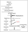
Figure 1. Nitrogen Containing BPs: Mechanism of Action
Both pamidronate and zoledronate act by inhibiting the farnesyl pyrophosphatase enzyme in the HMG-CoA reductase pathway. Inhibition of this enzyme prevents the prenylation of proteins which results in cell death of osteoclasts. The addition of exogenous GGOH is able to overcome the block of the geranylgeranylation of Rab and Rho GTPases.
Figure 2
Figure 2. Cell Proliferation of fibroblasts exposed to BPs
Cellular proliferation of oral fibroblasts was strongly inhibited after exposure to 0.06mM pamidronate for greater than 48 hours (A). A dose of 0.03mM pamidronate was sufficient to have an adverse effect on proliferation after 168 hours of exposure. Cellular proliferation of oral fibroblasts was inhibited after exposure to 0.008mM zoledronate for greater than 72 hours (B). Concentrations of 0.002mM zoledronate and higher strongly inhibited cell growth after 168 hours of exposure. * - p<0.05 when compared to controls
Figure 3
Figure 3. The effects of the BPs on cell survival
The percentage of cells that were TUNEL positive upon exposure to 0.03mM, 0.06mM or 0.1mM pamidronate were 5%, 25% and 35% respectively after 96 hours (A). Cells exposed to 0.06mM zoledronate had approximately 15% TUNEL positive cells, after 144 hours of exposure (B). Cells exposed to 0.1mM pamidronate for 96 hours displayed more number of TUNEL positive cells (D) compared to cells exposed to 0.06mM zoledronate for 144 hours (F) as judged by the number of green cells per field. Non treated cells did not have any TUNEL positive cells (C, E). Cells that were exposed to increasing levels of pamidronate displayed increased caspase activity (G).
Figure 4
Figure 4. Proliferation of primary human fibroblasts treated with bisphosphonates and rescued with PDGF-BB and GGOH
Cells were untreated or treated to 0.03mM or 0.06mM pamidronate and 0.003mM or 0.008mM zoledronate. Platelet-derived growth factor (10 ng/ml) or 50μM GGOH were added to cultures to assess the ability of these agents to overcome the inhibition of the BPs. A 20% increase in proliferation could be seen by the addition of PDGF to the cell cultures (A and B). When added to the pamidronate treated cells at 168 hours, PDGF was able to restore approximately 40% of the proliferation when compared cells treated with pamidronate alone (A). The addition of PDGF was unable to rescue the zoledronate treated cells (B). In cells treated with 0.03mM pamidronate, the addition of 50μM GGOH was able to restore the proliferation to 75% of control after 168 hours (C). The cells treated with 0.008mM and 0.03mM zoledronate the addition of 50μM GGOH restored the proliferation to 75% and 100% of control respectively (D). * - p<0.05 when compared to controls ^ - p<0.05 when compared to inhibited samples
Figure 5
Figure 5. Wound healing of primary human oral fibroblasts treated with BPs and rescued with GGOH
Cells in a confluent 6 well plate were either untreated (A, I) or treated with 50μM GGOH (C, K). Cells were exposed to 0.06 mM pamidronate alone (E) or 0.06 mM pamidronate plus 50μM GGOH (G) and 0.03mM zoledronate alone (M) or 0.03mM zoledronate plus 50μM GGOH (O) and scratch wound assays were performed. After 72 hours, untreated cells closed the wound (B, J). Cells exposed to 0.06mM pamidronate or 0.03mM zoledronate were inhibited from migrating into the wounded area (F, N). When 50μM GGOH was added to the wounded cultures, minimal rescue of pamidronate treated cultures was seen (H), while the zoledronate cultures showed a complete rescue of wound healing (P). Addition of GGOH to non-wounded cultures had no effect on the cell migration (D, L).
Figure 6
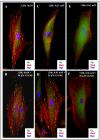
Figure 6. Effect of zoledronate on integrin mediated cell-substratum adhesions of oral fibroblasts
Cells were plated on fibronectin-coated coverslips and were exposed to 0.03mM or 0.06mM zoledronate with or without the addition of 50μM GGOH. The cells were fixed after 120 hours and then stained with vinculin and phalloidin to visualize the focal adhesions and actin cytoskeleton. In cells that were untreated or treated with 50μM GGOH alone, there was robust actin cytoskeletal network associated with numerous focal adhesions (A, B). In cells that were exposed to 0.03mM or 0.06mM zoledronate, there were very few F-actin bundles and a corresponding loss of focal adhesions (C, E). The addition of 50μM GGOH to cells treated with 0.03mM zoledronate was able to completely rescue both the focal adhesions and the actin stress fibres (D) In cells treated with 0.06mM zoledronate and 50μM GGOH, the focal adhesions and actin cytoskeleton network were also restored (F), but not to the same extent as seen in (D).

Comment in

Similar articles

Cited by

References

    1. Colella G, Campisi G, Fusco V. American Association of Oral and Maxillofacial Surgeons position paper: Bisphosphonate-Related Osteonecrosis of the Jaws-2009 update: the need to refine the BRONJ definition. J Oral Maxillofac Surg. 2009;12:2698. - PubMed
    1. Khosla S, Burr D, Cauley J, et al. Bisphosphonate-associated osteonecrosis of the jaw: report of a task force of the American Society for Bone and Mineral Research. J Bone Miner Res. 2007;10:1479. - PubMed
    1. Kyle RA, Yee GC, Somerfield MR, et al. American Society of Clinical Oncology 2007 clinical practice guideline update on the role of bisphosphonates in multiple myeloma. J Clin Oncol. 2007;17:2464. - PubMed
    1. Patel V, McLeod NM, Rogers SN, et al. Bisphosphonate osteonecrosis of the jaw-a literature review of UK policies versus international policies on bisphosphonates, risk factors and prevention. Br J Oral Maxillofac Surg. 2010 - PubMed
    1. Ruggiero SL, Dodson TB, Assael LA, et al. American Association of Oral and Maxillofacial Surgeons position paper on bisphosphonate-related osteonecrosis of the jaws--2009 update. J Oral Maxillofac Surg. 2009;5(Suppl):2. - PubMed

Publication types

MeSH terms