

Standard-based method for proton-electron double resonance imaging of oxygen
- PMID: 21807539
- PMCID: PMC3235921
- DOI: 10.1016/j.jmr.2011.06.030
Standard-based method for proton-electron double resonance imaging of oxygen
Abstract
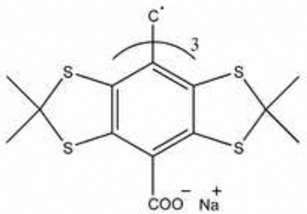
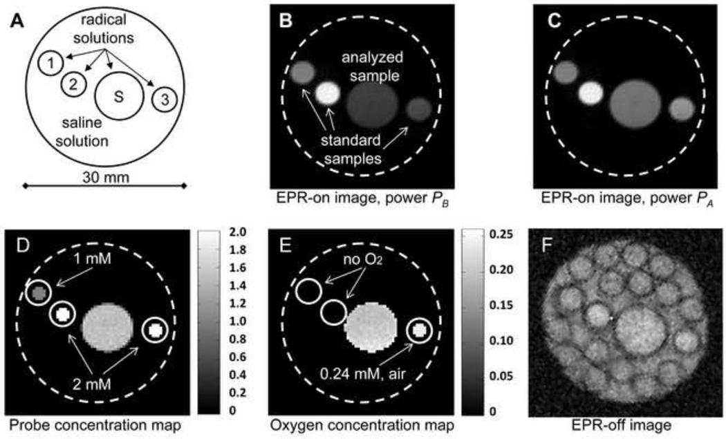
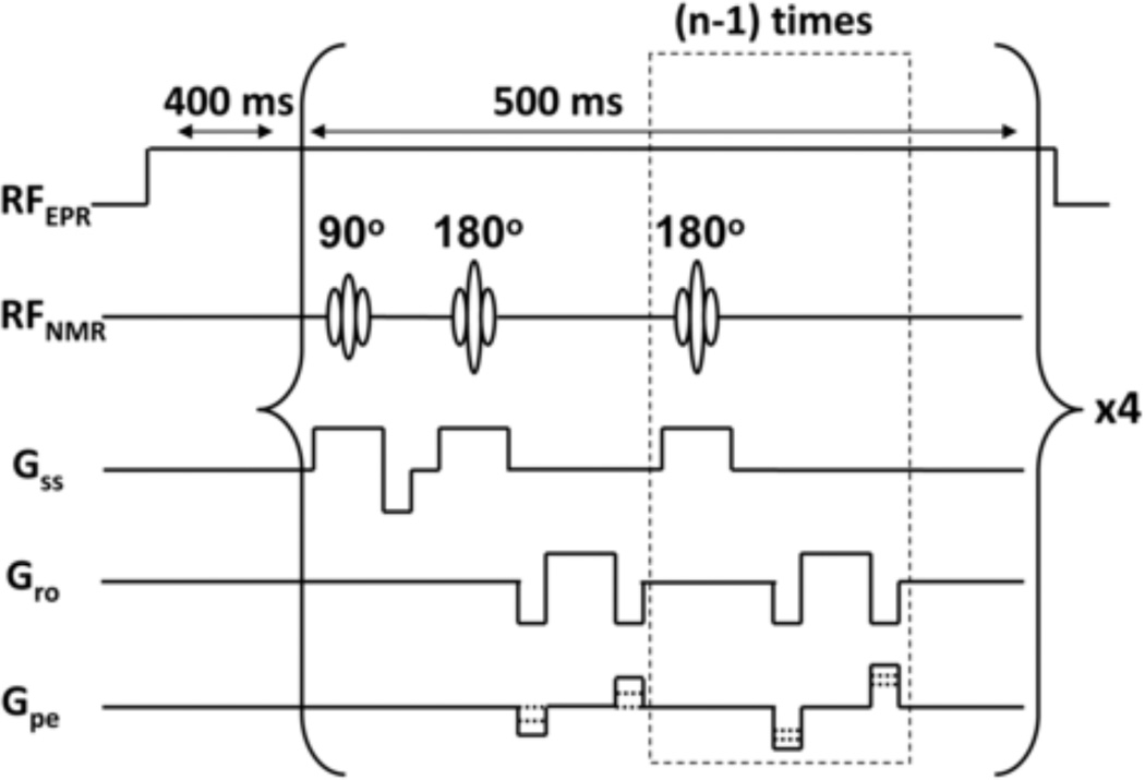
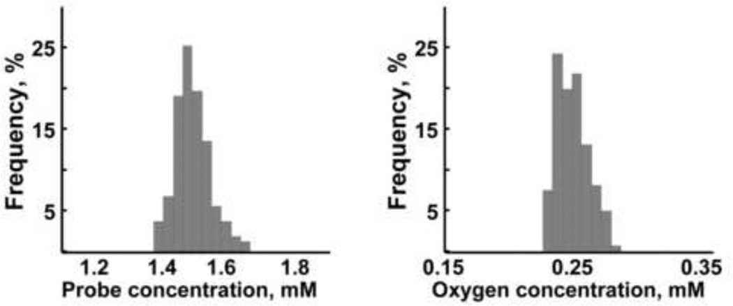
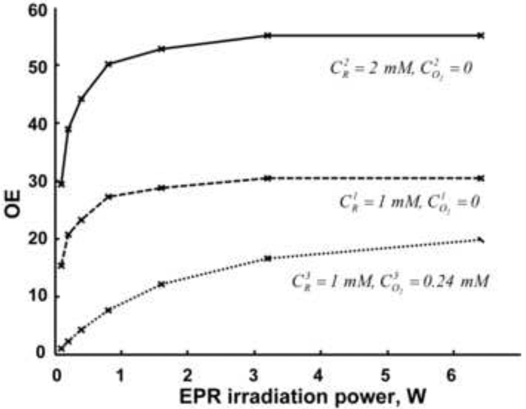
Proton-electron double resonance imaging (PEDRI) has been utilized for indirect determination of oxygen concentrations in aqueous samples and living systems. Due to the complexity of the problem, there are seven oxygen related parameters that need to be measured to determine the distribution of oxygen. We present an improved approach in which image intensities from only two PEDRI acquisitions with different EPR irradiation powers are required to determine the distribution of a paramagnetic probe and oxygen in an analyzed sample. This is achieved using three reference samples with known concentrations of a paramagnetic probe and oxygen placed inside the resonator together with the measurement sample. An EPR-off image, which has low signal intensity at low magnetic field (0.02 T) is not required for the calculations, significantly reducing the total time of the experiments and the noise while enhancing the accuracy of these oxygen measurements. The Finland trityl radical was used as the paramagnetic probe and oxygen concentrations could be accurately measured and imaged over the physiological range from 0 to 240 μM.
Published by Elsevier Inc.
Figures
Similar articles
-
In Vivo Application of Proton-Electron Double-Resonance Imaging.Antioxid Redox Signal. 2018 May 20;28(15):1345-1364. doi: 10.1089/ars.2017.7341. Epub 2017 Nov 13. Antioxid Redox Signal. 2018. PMID: 28990406 Free PMC article. Review.
-
Oxygen-induced leakage of spin polarization in Overhauser-enhanced magnetic resonance imaging: Application for oximetry in tumors.J Magn Reson. 2018 Dec;297:42-50. doi: 10.1016/j.jmr.2018.10.005. Epub 2018 Oct 10. J Magn Reson. 2018. PMID: 30359906 Free PMC article.
-
Variable radio frequency proton-electron double-resonance imaging: application to pH mapping of aqueous samples.J Magn Reson. 2011 Apr;209(2):227-32. doi: 10.1016/j.jmr.2011.01.011. Epub 2011 Jan 15. J Magn Reson. 2011. PMID: 21320790 Free PMC article.
-
Variable Field Proton-Electron Double-Resonance Imaging: Application to pH mapping of aqueous samples.J Magn Reson. 2010 Feb;202(2):267-73. doi: 10.1016/j.jmr.2009.11.017. Epub 2009 Nov 26. J Magn Reson. 2010. PMID: 20007019 Free PMC article.
-
15N-Labeled 4-oxo-2,2,6,6-tetramethyl-piperidine-1-oxyl.2008 Apr 30 [updated 2008 Jun 9]. In: Molecular Imaging and Contrast Agent Database (MICAD) [Internet]. Bethesda (MD): National Center for Biotechnology Information (US); 2004–2013. 2008 Apr 30 [updated 2008 Jun 9]. In: Molecular Imaging and Contrast Agent Database (MICAD) [Internet]. Bethesda (MD): National Center for Biotechnology Information (US); 2004–2013. PMID: 20641553 Free Books & Documents. Review.
Cited by
-
Exchange Phenomena in the Electron Paramagnetic Resonance Spectra of the Nitroxyl and Trityl Radicals: Multifunctional Spectroscopy and Imaging of Local Chemical Microenvironment.Anal Chem. 2017 May 2;89(9):4758-4771. doi: 10.1021/acs.analchem.6b03796. Epub 2017 Apr 10. Anal Chem. 2017. PMID: 28363027 Free PMC article. Review.
-
In Vivo Application of Proton-Electron Double-Resonance Imaging.Antioxid Redox Signal. 2018 May 20;28(15):1345-1364. doi: 10.1089/ars.2017.7341. Epub 2017 Nov 13. Antioxid Redox Signal. 2018. PMID: 28990406 Free PMC article. Review.
-
Proton-Electron Double-Resonance Imaging of pH using phosphonated trityl probe.Appl Magn Reson. 2014 Sep 1;45(9):817-826. doi: 10.1007/s00723-014-0570-2. Appl Magn Reson. 2014. PMID: 25530673 Free PMC article.
-
In Vivo pO2 Imaging of Tumors: Oxymetry with Very Low-Frequency Electron Paramagnetic Resonance.Methods Enzymol. 2015;564:501-27. doi: 10.1016/bs.mie.2015.08.017. Epub 2015 Sep 26. Methods Enzymol. 2015. PMID: 26477263 Free PMC article. Review.
-
Oxygen-induced leakage of spin polarization in Overhauser-enhanced magnetic resonance imaging: Application for oximetry in tumors.J Magn Reson. 2018 Dec;297:42-50. doi: 10.1016/j.jmr.2018.10.005. Epub 2018 Oct 10. J Magn Reson. 2018. PMID: 30359906 Free PMC article.
References
-
- He GL, Shankar RA, Chzhan M, Samouilov A, Kuppusamy P, Zweier JL. Noninvasive measurement of anatomic structure and intraluminal oxygenation in the gastrointestinal tract of living mice with spatial and spectral EPR imaging. Proceedings of the National Academy of Sciences of the United States of America. 1999;96:4586–4591. - PMC - PubMed
-
- Zweier JL, Kuppusamy P. Electron-Paramagnetic Resonance Measurements of Free-Radicals in the Intact Beating Heart - a Technique for Detection and Characterization of Free-Radicals in Whole Biological Tissues. Proceedings of the National Academy of Sciences of the United States of America. 1988;85:5703–5707. - PMC - PubMed
-
- Lurie DJ, Bussell DM, Bell LH, Mallard JR. Proton Electron Double Magnetic-Resonance Imaging of Free-Radical Solutions. Journal of Magnetic Resonance. 1988;76:366–370.
Publication types
MeSH terms
Substances
Grants and funding
LinkOut - more resources
Full Text Sources
Medical
