. 2011 Sep 15;17(18):5913-25.
doi: 10.1158/1078-0432.CCR-11-0728. Epub 2011 Aug 1.

Resistance to CYP17A1 inhibition with abiraterone in castration-resistant prostate cancer: induction of steroidogenesis and androgen receptor splice variants

Affiliations

Resistance to CYP17A1 inhibition with abiraterone in castration-resistant prostate cancer: induction of steroidogenesis and androgen receptor splice variants

Elahe A Mostaghel et al. Clin Cancer Res. .

Abstract

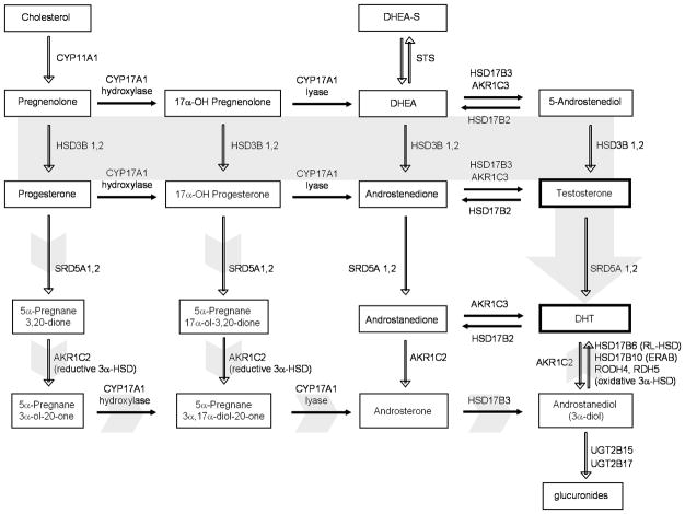
Purpose: Abiraterone is a potent inhibitor of the steroidogenic enzyme CYP17A1 and suppresses tumor growth in patients with castration-resistant prostate cancer (CRPC). The effectiveness of abiraterone in reducing tumor androgens is not known, nor have mechanisms contributing to abiraterone resistance been established.

Experimental design: We treated human CRPC xenografts with abiraterone and measured tumor growth, tissue androgens, androgen receptor (AR) levels, and steroidogenic gene expression versus controls.

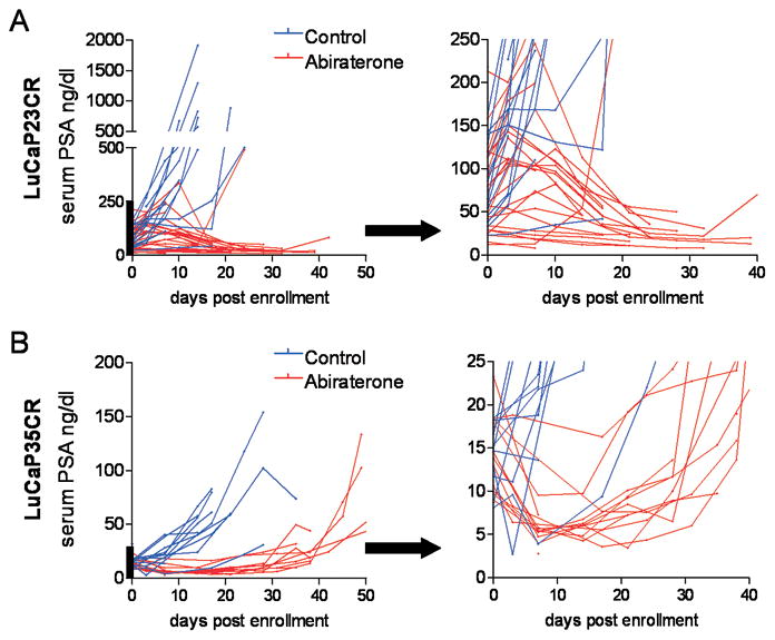
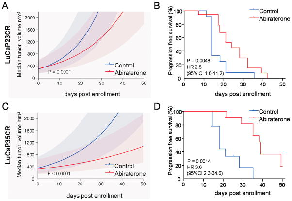
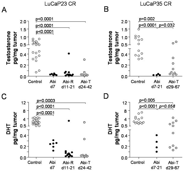
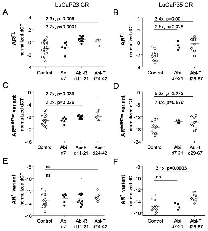
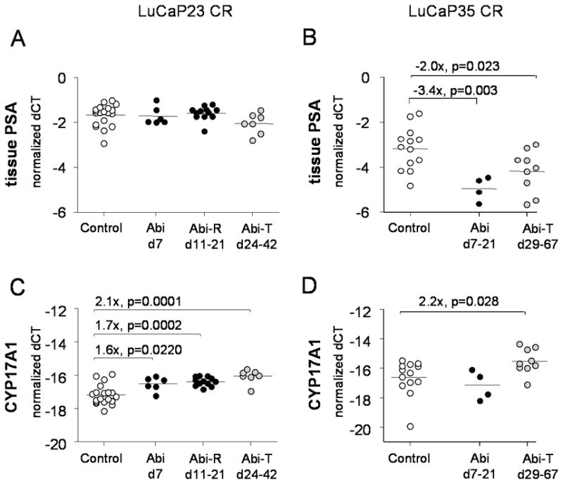
Results: Abiraterone suppressed serum PSA levels and improved survival in two distinct CRPC xenografts: median survival of LuCaP35CR improved from 17 to 39 days (HR = 3.6, P = 0.0014) and LuCaP23CR from 14 to 24 days (HR = 2.5, P = 0.0048). Abiraterone strongly suppressed tumor androgens, with testosterone (T) decreasing from 0.49 ± 0.22 to 0.03 ± 0.01 pg/mg (P < 0.0001), and from 0.69 ± 0.36 to 0.03 ± 0.01 pg/mg (P = 0.002) in abiraterone-treated 23CR and 35CR, respectively, with comparable decreases in tissue DHT. Treatment was associated with increased expression of full-length AR (AR(FL)) and truncated AR variants (AR(FL) 2.3-fold, P = 0.008 and AR(del567es) 2.7-fold, P = 0.036 in 23 CR; AR(FL) 3.4-fold, P = 0.001 and AR(V7) 3.1-fold, P = 0.0003 in 35CR), and increased expression of the abiraterone target CYP17A1 (∼2.1-fold, P = 0.0001 and P = 0.028 in 23CR and 35CR, respectively) and transcript changes in other enzymes modulating steroid metabolism.

Conclusions: These studies indicate that abiraterone reduces CRPC growth via suppression of intratumoral androgens and that resistance to abiraterone may occur through mechanisms that include upregulation of CYP17A1, and/or induction of AR and AR splice variants that confer ligand-independent AR transactivation.


Figures

Figure 1
Figure 1
Changes in serum PSA levels in LuCaP23CR and LuCaP35CR prostate cancer xenografts in response to treatment with abiraterone. Castrate male SCID mice were implanted subcutaneously with LuCaP23CR or LuCaP35CR tumors and randomly assigned to vehicle control or abiraterone treatment for 21 days. Serum PSA curves for control (blue) or abiraterone-treated mice (red) are shown for individual mice bearing Lu-CaP23CR (A) and LuCaP35CR (B) xenografts. The segment of the y-axis denoted by the heavy bar in each graph is enlarged and presented in the adjacent panels on an expanded y-axis in order to more clearly demonstrate the decline in serum PSA over the first ~10–15 days after initiation of treatment.
Figure 2
Figure 2
Tumor growth and survival in LuCaP23CR and LuCaP35CR prostate cancer xenografts treated with abiraterone. Castrate male SCID mice were implanted subcutaneously with LuCaP23CR (A and B) or LuCaP35CR (D and E) tumors and randomly assigned to vehicle control or abiraterone treatment for 21 days. Median tumor volume trajectories with 95% confidence bands for control (blue) or abiraterone-treated mice (red) bearing the LuCaP23CR (A) and LuCaP35CR (D) xenografts. Kaplan-Meier plots of progression-free survival (defined as tumor size <1000mm3) in control or abiraterone-treated mice bearing the LuCaP23CR (B) and LuCaP35CR (E) xenografts. P-values for curve comparisons generated using the Mantel-Haenszel logrank test.
Figure 3
Figure 3
Tumor testosterone (upper panels) and DHT levels (lower panels) in control and abiraterone-treated LuCaP23CR (A,C) and LuCaP35CR (B,D) xenografts. Androgen levels in abiraterone-treated xenografts were evaluated by mass spectrometry in tumors resected at early (days 7–21) or late time points after therapy. Abiraterone-resistant tumors (Abi-R) recurred (defined as progression to >1000mm3) and were resected during the 21-day period of abiraterone treatment. Abiraterone-treated tumors (Abi-T) recurred and were resected after the completion of abiraterone treatment. This occurred between days 24–42 for LuCap23CR tumors, and between days 29–67 for LuCaP35CR (none of which recurred during the abiraterone-treatment period). P values represent unpaired two-sided t-tests between the indicated groups. P values <0.05 were considered significant. One p value in panel D (in italics) is included as trending toward statistical significance (p<0.10).
Figure 4
Figure 4
Expression of full length and truncated AR splice variants in LuCaP23CR and LuCaP35CR tumors treated with abiraterone compared to vehicle control. Transcript levels for full length AR (ARFL; A, B), the AR7 splice variant (C, D), and theARdel567es splice variant (E, F) were measured by qRT-PCR in frozen LuCaP23CR (A, C, E) and LuCaP35CR (B, D, E) tumors. White circle denote vehicle treated controls. Black circles denote tumors resected while on abiraterone treatment, either at early time points (d7–21 or at abiraterone-resistant re-growth (Abi-R). Gray circles denote abiraterone-treated tumors (Abi-T) which recurred and were resected after completion of abiraterone treatment. Fold changes are calculated from the difference in mean delta Ct’s between abiraterone treated and vehicle treated controls (delta-delta CT method; fold = 2ddCt). P values from two sample t-tests. P values < 0.05 were considered significant. The p values in panel D (in italics) are included as trending toward statistical significance (p<0.10). n.s. not significant.
Figure 5
Figure 5
Expression of PSA and CYP17A1 in LuCaP23CR and LuCaP35CR tumors treated with abiraterone compared to vehicle control. Transcript levels for PSA (A, B) and CYP17A1 were measured by qRT-PCR in frozen LuCaP23CR (A, C) and Lu-CaP35CR (B, D) tumors. White circle denote vehicle treated controls. Black circles denote tumors resected while on abiraterone treatment, either at early time points (d7–21 or at abiraterone-resistant re-growth (Abi-R). Gray circles denote abiraterone-treated tumors (Abi-T) which recurred and were resected after completion of abiraterone treatment. Fold changes are calculated from the difference in mean delta Ct’s between abiraterone treated and vehicle treated controls (delta-delta CT method; fold = 2ddCt). P values from two sample t-tests. P values < 0.05 were considered significant.
Figure 6
Figure 6
The pathways of androgen biosynthesis. In the classical pathway (solid gray arrow), C21 precursors (pregnenolone and progesterone) are converted to the C19 adrenal androgens DHEA and androstenedione (AED) by the sequential hydroxylase and lyase activity of CYP17A1. DHEA and AED are converted to testosterone by a series of reactions involving the activity of HSD3B1 and 2, HSD17B3 and AKR1C3 enzymes. Testosterone is converted to the potent androgen DHT by the activity of SRD5A1 and 2. Oxida-tive 3 α-HSD enzymes (including HSD17B6 (RL-HSD), HSD17B10, HSD17B13 (DHRS9), RODH4 and RDH5) can act to inhibit the pre-receptor catabolism of DHT. An alternative pathway (short gray arrows) has also been proposed in which C21 precursors are first acted upon by SRD5A and the reductive activity of AKR1C2, followed by CYP17A1, HSD17B3 and subsequent oxidation to DHT Adapted from Mostaghel EA, Nelson PS. Intracrine androgen metabolism in prostate cancer progression: mechanisms of castration resistance and therapeutic implications. Best Pract Res Clin Endocrinol Me-tab. 2008;22:243, with permission (pending).

Similar articles

Cited by

References

    1. Eisenberger MA, Blumenstein BA, Crawford ED, Miller G, McLeod DG, Loehrer PJ, et al. Bilateral orchiectomy with or without flutamide for metastatic prostate cancer. N Engl J Med. 1998;339:1036–42. - PubMed
    1. Chen CD, Welsbie DS, Tran C, Baek SH, Chen R, Vessella R, et al. Molecular determinants of resistance to antiandrogen therapy. Nat Med. 2004;10:33–9. - PubMed
    1. Stanbrough M, Bubley GJ, Ross K, Golub TR, Rubin MA, Penning TM, et al. Increased expression of genes converting adrenal androgens to testosterone in androgen-independent prostate cancer. Cancer Res. 2006;66:2815–25. - PubMed
    1. Mostaghel EA, Page ST, Lin DW, Fazli L, Coleman IM, True LD, et al. Intra-prostatic Androgens and Androgen-Regulated Gene Expression Persist after Testosterone Suppression: Therapeutic Implications for Castration-Resistant Prostate Cancer. Cancer Res. 2007;67:5033–41. - PubMed
    1. Montgomery RB, Mostaghel EA, Vessella R, Hess DL, Kalhorn TF, Higano CS, et al. Maintenance of intratumoral androgens in metastatic prostate cancer: a mechanism for castration-resistant tumor growth. Cancer Res. 2008;68:4447–54. - PMC - PubMed

Publication types

MeSH terms