. 2011 Nov;90(5):883-95.
doi: 10.1189/jlb.0311134. Epub 2011 Aug 1.

Lung dendritic cells at the innate-adaptive immune interface

Affiliations

Lung dendritic cells at the innate-adaptive immune interface

Tracy Voss Condon et al. J Leukoc Biol. 2011 Nov.

Abstract

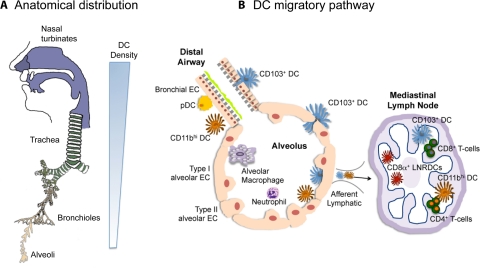
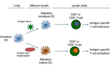
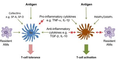
This review updates the basic biology of lung DCs and their functions. Lung DCs have taken center stage as cellular therapeutic targets in new vaccine strategies for the treatment of diverse human disorders, including asthma, allergic lung inflammation, lung cancer, and infectious lung disease. The anatomical distribution of lung DCs, as well as the division of labor between their subsets, aids their ability to recognize and endocytose foreign substances and to process antigens. DCs can induce tolerance in or activate naïve T cells, making lung DCs well-suited to their role as lung sentinels. Lung DCs serve as a functional signaling/sensing unit to maintain lung homeostasis and orchestrate host responses to benign and harmful foreign substances.


Figures

Figure 1.
Figure 1.. Anatomical location and migratory pathway of lung DCs.
(A) DCs are located throughout the respiratory tract. They are most abundant in the nose, nasal turbinates, and trachea. As shown by the sliding scale at right, the density of lung DCs declines as one descends the respiratory tract. (B) Two subsets of cDCs are present: an intraepithelial web of CD103+ cDCs (blue) and CD11bhi cDCs (orange) in the lamina propria, together with pDCs (yellow). The alveoli are lined with type I and type II alveolar ECs (peach). AMs (purple) form the main population of resident alveolar cells, whereas neutrophils can be recruited in response to inflammatory stimulation. Both cDC subsets are present in the alveoli and migrate to the draining LNs (right), where they present antigens to CD4+ and CD8+ T cells. CD103+ cDCs are specialized in cross-priming as CD8α+ LNRDCs (red).
Figure 2.
Figure 2.. Antigen-induced T cell tolerance or antigen-induced T cell activation is controlled by the nature of the inhaled antigen.
In the absence of second signals provided by PAMPs or host endogenous DAMPs, antigen-exposure DCs migrate to the regional draining LNs and promote T cell tolerance (upper pathway). In the presence of PAMPs or DAMPs, antigen-exposed DCs mature and up-regulate cell-surface costimulatory molecules, migrate to regional LNs, and promote antigen-specific CD4+ and CD8+ T cell activation.
Figure 3.
Figure 3.. Complexity in the regulation of lung DC function.
Antigen-specific T cell tolerance (left) or activation (right) is regulated at multiple levels, some of which are mutually antagonistic. SP-A/D, Surfactant protein A/D.

References

    1. Aschoff L. (1924) The reticuloendothelial system. Ergeb. Inn. Med. Kinderheilkd. 26, 1–118
    1. van Furth R., Cohn Z. A. (1968) The origin and kinetics of mononuclear phagocytes. J. Exp. Med. 128, 415–435 - PMC - PubMed
    1. Gordon S., Taylor P. R. (2005) Monocyte and macrophage heterogeneity. Nat. Rev. Immunol. 5, 953–964 - PubMed
    1. Volkman A., Gowans J. L. (1965) The origin of macrophages from bone marrow in the rat. Br. J. Exp. Pathol. 46, 62–70 - PMC - PubMed
    1. Steinman R. M., Cohn Z. A. (1973) Identification of a novel cell type in peripheral lymphoid organs of mice. I. Morphology, quantitation, tissue distribution. J. Exp. Med. 137, 1142–1162 - PMC - PubMed

Publication types