Review
. 2011 Aug 1;3(8):a005090.
doi: 10.1101/cshperspect.a005090.

Angiogenesis

Affiliations
Review

Angiogenesis

Donald R Senger et al. Cold Spring Harb Perspect Biol. .

Abstract

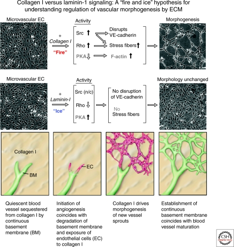
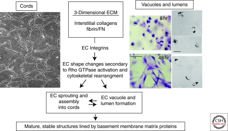
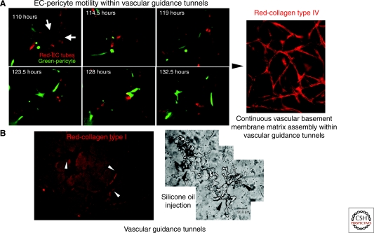
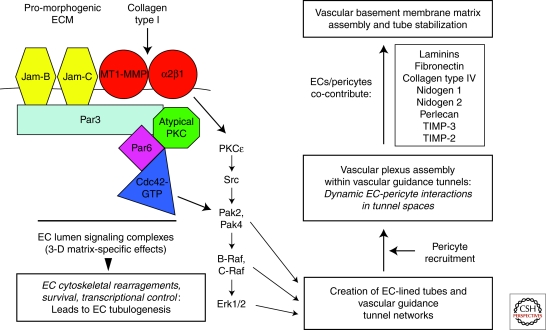
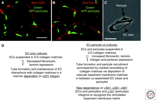
Extracellular matrix (ECM) is essential for all stages of angiogenesis. In the adult, angiogenesis begins with endothelial cell (EC) activation, degradation of vascular basement membrane, and vascular sprouting within interstitial matrix. During this sprouting phase, ECM binding to integrins provides critical signaling support for EC proliferation, survival, and migration. ECM also signals the EC cytoskeleton to initiate blood vessel morphogenesis. Dynamic remodeling of ECM, particularly by membrane-type matrix metalloproteases (MT-MMPs), coordinates formation of vascular tubes with lumens and provides guidance tunnels for pericytes that assist ECs in the assembly of vascular basement membrane. ECM also provides a binding scaffold for a variety of cytokines that exert essential signaling functions during angiogenesis. In the embryo, ECM is equally critical for angiogenesis and vessel stabilization, although there are likely important distinctions from the adult because of differences in composition and abundance of specific ECM components.


Figures

Figure 1.
Figure 1.
Signals transduced by interstitial collagen type I and laminin-1 (laminin-111) in dermal microvascular ECs, and consequences for capillary morphogenesis in vitro. In this proposed model, interstitial collagens, but not laminins, activate Src and Rho and suppress Rac and PKA activities through β1 integrins. Such signaling results in induction of actin stress fibers, disruption of VE-cadherin, and formation of precapillary cords. In contrast, laminins activate Rac and PKA and suppress Rho activity and therefore do not provoke morphogenesis. Although laminin-1 (revised nomenclature = laminin-111) is not one of the laminin isoforms typically associated with vascular basement membranes (see text) and therefore further testing of this model requires additional investigations with vascular laminin isoforms, these marked distinctions in signaling by collagen type I (“fire”) and laminin-1 (“ice”) suggest a mechanism through which degradation of basement membrane and exposure of activated and proliferating ECs to interstitial collagens within the provisional matrix initiates morphogenesis of new capillary sprouts. (n/c) = no change. (This figure is adapted from Liu and Senger [2004] and Davis and Senger [2005] and is reprinted, with permission, from the Federation of American Societies for Experimental Bio (FASEB) ©2004 and Wolters Kluwer Health ©2005, respectively.)
Figure 2.
Figure 2.
Fundamental contribution of the ECM-integrin-cytoskeletal signaling axis to the assembly of EC cords and lumens during vasculogenesis and angiogenesis. In this schematic, two models of EC morphogenesis are illustrated. In the left panel, confluent human microvascular EC monolayers were overlaid with a type I collagen gel and allowed to undergo cord formation, illustrating how collagenous ECM markedly converts an EC monolayer into an interconnecting network of cords. This process is dependent on the α1β1 and α2β1 integrins and the small GTPase, RhoA. In the right panels, human ECs are suspended as individual cells in 3-D collagen matrices and are fixed, stained, and photographed at 8 and 48 hr of culture, illustrating intracellular vacuoles and early lumen formation at 8 hr and interconnecting networks of EC-lined tubes at 48 hr. Bar equals 50 µm. The three panels on the far right side are cross-sections from plastic-embedded collagen gels revealing EC intracellular vacuoles (upper two right panels) or EC lumens (lower right panel). Bar equals 25 µm. The vacuole and lumen formation process shown is completely dependent on the α2β1 integrin and the Cdc42 and Rac1 small GTPases. (This figure is adapted from Davis and Senger [2005] and reprinted, with permission, from Wolters Kluwer Health ©2005.)
Figure 3.
Figure 3.
Vascular guidance tunnels: ECM conduits generated during vessel tubulogenesis that regulate EC-pericyte interactions, ECM remodeling, vascular basement membrane matrix assembly, and vessel stabilization. The process of EC tubular morphogenesis in 3-D collagen matrices leads to the creation of networks of EC-lined tubes, and also networks of vascular guidance tunnels in which ECs reside. The tunnels are formed during lumen and tube formation through EC cell surface proteolysis by MT1-MMP. EC-lined tubes within vascular guidance tunnels recruit pericytes to the tube ablumenal surface; subsequently, pericytes migrate along this surface within the tunnels. (A) Time-lapse imaging of EC tubes (RFP-labeled) and pericytes (GFP-labeled) reveals motility of pericytes along the tube ablumenal surface over time. Arrows indicate EC tube ablumenal surface. Bar equals 25 µm. Active motility of pericytes along tubes leads to EC-pericyte interactions and contributions of basement membrane components, such as collagen type IV, which is deposited continuously along the ablumenal surface (right panel; Bar equals 50 µm). (B) Vascular guidance tunnels are formed during EC tubulogenesis through MT1-MMP-mediated proteolysis and can be shown using anticollagen type I antibodies (left panel; Bar equals 50 µm; the tunnels are the negative stained areas; arrowheads indicate tunnel borders) or by microinjection of tunnels with silicone oil (right panel; Bar equals 20 µm; arrowheads indicate tunnel borders).
Figure 4.
Figure 4.
Molecular signaling events controlling vascular tube morphogenesis, matrix remodeling, and stabilization in 3-D extracellular matrices. Multicomponent signaling complexes control EC lumen and tube formation in 3-D matrices. A key aspect of this mechanism is the integration of Cdc42-mediated signaling with cell surface proteolysis through MT1-MMP. A downstream kinase signaling cascade is activated leading to EC cytoskeletal changes, survival, and transcriptional control that regulates EC tubular morphogenesis. EC tube formation leads to the generation of networks of vascular guidance tunnel spaces within the ECM that are occupied by ECs, thereby allowing for EC motility and remodeling events (illustrated in Fig. 3). Pericytes are recruited to these EC-lined tunnels and EC-pericyte motility within these tunnels and along the tube ablumenal surface leads to ECM remodeling and vascular basement membrane matrix assembly. Both ECs and pericytes have been shown to contribute basement membrane matrix components (illustrated in Fig. 5), as well as TIMP-2 and TIMP-3, which together control the formation and stability of the vascular basement membrane.
Figure 5.
Figure 5.
Pericyte recruitment to EC-lined tubes leads to vascular basement membrane matrix assembly in 3-D matrices. Green-fluorescent protein (GFP)-tagged bovine retinal pericytes were mixed with ECs in 3-D collagen matrices over a period of 5 days, during which EC tubes formed and recruited pericytes. Cultures were fixed at 5 days and immunostained for CD31 (panel A) or for collagen type IV (panel B); bar equals 50 µm. Thin plastic sections were obtained and photographed under transmitted light (panel C); bar equals 25 µm. (Panel D) RT-PCR and protein analyses of EC-only and EC-pericyte cocultures revealed specific changes in ECM and integrin expression, indicating distinctly different regulation in EC-pericyte cocultures in comparison with ECs alone (Stratman et al. 2009a).

References

    1. Akiyama SK, Yamada SS, Chen WT, Yamada KM 1989. Analysis of fibronectin receptor function with monoclonal antibodies: Roles in cell adhesion, migration, matrix assembly, and cytoskeletal organization. J Cell Biol 109: 863–875 - PMC - PubMed
    1. Aloisi M, Schiaffino S 1971. Growth of elementary blood vessels in diffusion chambers: Electron microscopy of capillary morphogenesis. Virchows Arch Abt B Zellpath 8: 328–341 - PubMed
    1. Aoudjit F, Vuori K 2001. Matrix attachment regulates Fas-induced apoptosis in endothelial cells: A role for c-flip and implications for anoikis. J Cell Biol 152: 633–643 - PMC - PubMed
    1. Aplin AE, Short SM, Juliano RL 1999. Anchorage-dependent regulation of the mitogen-activated protein kinase cascade by growth factors is supported by a variety of integrin α chains. J Biol Chem 274: 31223–31228 - PubMed
    1. Aplin AC, Fogel E, Zorzi P, Nicosia RF 2008. The aortic ring model of angiogenesis. Meth Enzymol 443: 119–136 - PubMed

Publication types

LinkOut - more resources