. 2011 Aug 8;194(3):367-75.
doi: 10.1083/jcb.201012132. Epub 2011 Aug 1.

PARP regulates nonhomologous end joining through retention of Ku at double-strand breaks

Affiliations

PARP regulates nonhomologous end joining through retention of Ku at double-strand breaks

C Anne-Marie Couto et al. J Cell Biol. .

Abstract

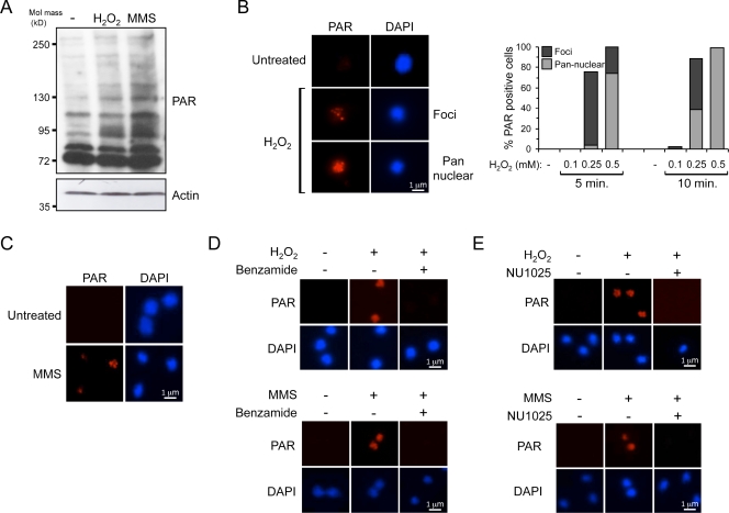
Poly adenosine diphosphate (ADP)-ribosylation (PARylation) by poly ADP-ribose (PAR) polymerases (PARPs) is an early response to DNA double-strand breaks (DSBs). In this paper, we exploit Dictyostelium discoideum to uncover a novel role for PARylation in regulating nonhomologous end joining (NHEJ). PARylation occurred at single-strand breaks, and two PARPs, Adprt1b and Adprt2, were required for resistance to this kind of DNA damage. In contrast, although Adprt1b was dispensable for PARylation at DSBs, Adprt1a and, to a lesser extent, Adprt2 were required for this event. Disruption of adprt2 had a subtle impact on the ability of cells to perform NHEJ. However, disruption of adprt1a decreased the ability of cells to perform end joining with a concomitant increase in homologous recombination. PAR-dependent regulation of NHEJ was achieved through promoting recruitment and/or retention of Ku at DSBs. Furthermore, a PAR interaction motif in Ku70 was required for this regulation and efficient NHEJ. These data illustrate that PARylation at DSBs promotes NHEJ through recruitment or retention of repair factors at sites of DNA damage.


Figures

Figure 1.
Figure 1.
PARylation is induced in D. discoideum after SSBs. (A) Ax2 cells were untreated (−) or exposed to 0.5 mM H2O2 for 10 min or 5 mM MMS for 30 min. Whole-cell extracts were analyzed by Western blotting using the indicated antibodies. (B) Ax2 cells were treated with H2O2 as indicated. Coverslips were subjected to immunofluorescence using PAR antibodies. The percentages of PAR-positive cells were scored from a population of >200 cells. Cells were categorized into those that exhibit pannuclear staining or PAR nuclear foci. Data are representative of three independent experiments. (left) Representative images are shown. (C) Ax2 cells were untreated or treated with 5 mM MMS for 30 min. Coverslips were processed for immunofluorescence and stained with PAR antibodies. (D) Ax2 cells were treated with carrier (ethanol) or 5 mM benzamide and exposed to 0.5 mM H2O2 for 10 min (top) or 5 mM MMS for 30 min (bottom). Coverslips were processed for immunofluorescence using PAR antibodies. (E) Ax2 were treated with carrier (DMSO) or 1 mM NU1025 and analyzed as in D.
Figure 2.
Figure 2.
D. discoideum Adprt1b and Adprt2 are required for tolerance to SSBs. (A) The domain structure of Adprt1a, Adprt1b, and Adprt2 illustrating the PARP catalytic and regulatory domains, putative zinc finger, pADR, WGR, and BRCA1 C-terminal (BRCT) domains. (B) The indicated strains and their respective parental controls, Ax2 and Ax4, were untreated or exposed to 5 mM MMS for 30 min. Cells were subjected to immunofluorescence using PAR antibodies. The percentages of PAR-positive cells were scored from a population of >200 cells. (C) The indicated strains and their respective parental controls, Ax2 and Ax4, were assessed for their ability to survive exposure to MMS. Error bars represent the SEM from three independent experiments.
Figure 3.
Figure 3.
Adprt1a is required for PARylation after DSBs. (A) Ax2 cells were treated with phleomycin (micrograms per milliliter) and subjected to immunofluorescence using PAR antibodies. Data are representative of three independent experiments. Cells were categorized into those that exhibit pannuclear staining or PAR nuclear foci. (B) Ax2 cells were treated with 1 mM NU1025 or DMSO before exposure to 100 µg/ml phleomycin for 30 min. Cells were subjected to immunofluorescence using PAR antibodies. (C) The indicated strains and their respective parental controls, Ax2 and Ax4, were assessed for PARylation after phleomycin treatment as in B. The percentages of PAR-positive cells were scored from a population of >200 cells. Error bars represent the SEM from three independent experiments. *, P < 0.05 compared with the positive control (Ax2 + phleomycin).
Figure 4.
Figure 4.
Adprt1a is required to promote NHEJ. (A) REMI efficiency of the indicated strains was assessed as described in Materials and methods. The numbers analyzed were such that experimental strains were compared with >200 colonies from the positive control (Ax2 plus the restriction enzyme). Data are represented as the percentage of REMI induction relative to parental Ax2 controls. *, P < 0.05 compared with the positive control (Ax2). (B) Ax2, adprt1a, and ku80 spores were germinated before exposure to phleomycin, and cell survival was established as described in Materials and methods. (C) Ax2 and adprt1a cells were assessed for HR efficiency by measuring targeted integration of the blasticidin resistance cassette at the cdk8 locus. The percentage of HR is the proportion of aggregation-deficient colonies against the total number of blasticidin-resistant colonies. The n number represents to total of blasticidin-resistant colonies analyzed from multiple independent transfections. Error bars represent the SEM from three independent experiments.
Figure 5.
Figure 5.
The PBZ domain in D. discoideum Ku70 is required for NHEJ. (A) The indicated strains were exposed to phleomycin as indicated. After 80 min, the indicated cell fractions were analyzed by Western blotting. (B) Alignment of the PBZ domains present in D. discoideum (Dd) Ku70 and vertebrate APLF (APLF1 and APLF2), SNM1, and CHFR. Conserved amino acids are highlighted in black, and similar amino acids are represented in gray. (C) Serial dilutions of GST, GST fused to the C terminus of Ku70 (GST-Ku70C), or GST fused to the same fragment lacking the PBZ domain (GST-Ku70CΔPBZ) were blotted onto nitrocellulose filters. Inclines represent the relative amount of protein blotted onto the filter. After incubation in PAR polymer, Western blotting was performed using the indicated antibodies. (D, left) Whole-cell extracts were prepared from the indicated strains and subjected to Western blotting. The indicated strains were incubated with phleomycin, and Ku was immunoprecipitated (IP) from whole-cell extracts using Ku80 antisera. (right) Myc-Ku70 was confirmed by Western blotting (WB) using Myc antisera. (E) The indicated strains were left untreated or exposed to phleomycin. After 80 min, the indicated cellular fractions were analyzed by Western blotting. (F) REMI efficiency of the indicated strains was assessed as in Fig 4 A. The numbers analyzed were such that experimental strains were compared with >200 colonies from the positive control (ku70 cells expressing Myc-Ku70 plus restriction enzyme). The data are represented as the percentage of REMI induction relative to ku70 cells expressing Myc-Ku70. Error bars represent the SEM from three independent experiments. *, P < 0.05 compared with the positive control (ku70 cells expressing Myc-Ku70). Molecular masses are given in kilodaltons.

References

    1. Ahel I., Ahel D., Matsusaka T., Clark A.J., Pines J., Boulton S.J., West S.C. 2008. Poly(ADP-ribose)-binding zinc finger motifs in DNA repair/checkpoint proteins. Nature. 451:81–85 10.1038/nature06420 - DOI - PubMed
    1. Amé J.C., Rolli V., Schreiber V., Niedergang C., Apiou F., Decker P., Muller S., Höger T., Ménissier-de Murcia J., de Murcia G. 1999. PARP-2, A novel mammalian DNA damage-dependent poly(ADP-ribose) polymerase. J. Biol. Chem. 274:17860–17868 10.1074/jbc.274.25.17860 - DOI - PubMed
    1. Amé J.C., Spenlehauer C., de Murcia G. 2004. The PARP superfamily. Bioessays. 26:882–893 10.1002/bies.20085 - DOI - PubMed
    1. Ariumi Y., Masutani M., Copeland T.D., Mimori T., Sugimura T., Shimotohno K., Ueda K., Hatanaka M., Noda M. 1999. Suppression of the poly(ADP-ribose) polymerase activity by DNA-dependent protein kinase in vitro. Oncogene. 18:4616–4625 10.1038/sj.onc.1202823 - DOI - PubMed
    1. Audebert M., Salles B., Calsou P. 2004. Involvement of poly(ADP-ribose) polymerase-1 and XRCC1/DNA ligase III in an alternative route for DNA double-strand breaks rejoining. J. Biol. Chem. 279:55117–55126 10.1074/jbc.M404524200 - DOI - PubMed

Publication types

LinkOut - more resources