. 2011 Oct;157(2):891-904.
doi: 10.1104/pp.111.181388. Epub 2011 Aug 1.

Low-fluence red light increases the transport and biosynthesis of auxin

Affiliations

Low-fluence red light increases the transport and biosynthesis of auxin

Xing Liu et al. Plant Physiol. 2011 Oct.

Abstract

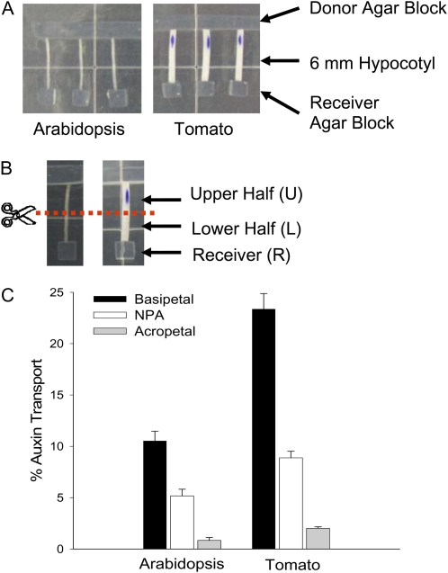
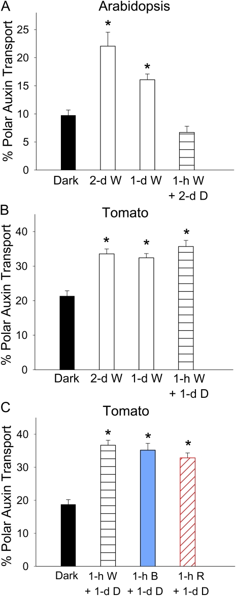
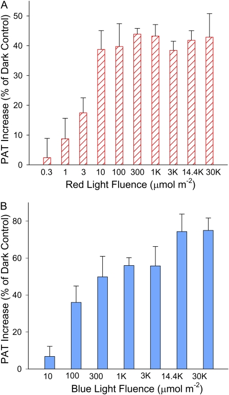
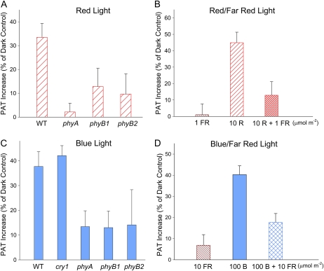
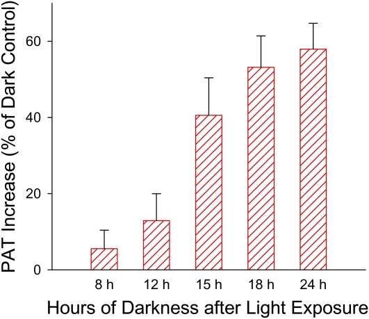
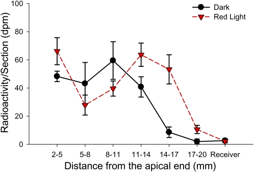
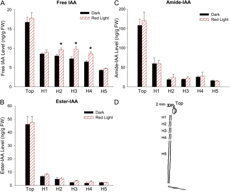
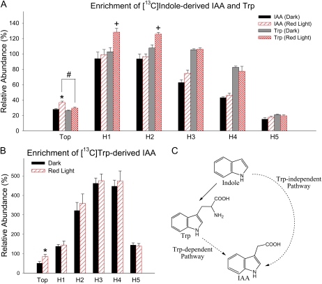
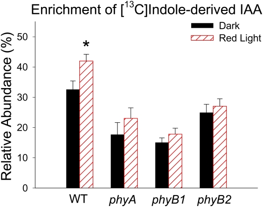
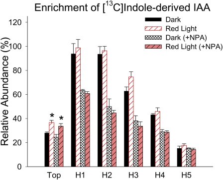
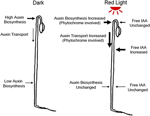
In plants, light is an important environmental signal that induces photomorphogenesis and interacts with endogenous signals, including hormones. We found that light increased polar auxin transport in dark-grown Arabidopsis (Arabidopsis thaliana) and tomato (Solanum lycopersicum) hypocotyls. In tomato, this increase was induced by low-fluence red or blue light followed by 1 d of darkness. It was reduced in phyA, phyB1, and phyB2 tomato mutants and was reversed by far-red light applied immediately after the red or blue light exposure, suggesting that phytochrome is involved in this response. We further found that the free indole-3-acetic acid (IAA) level in hypocotyl regions below the hook was increased by red light, while the level of conjugated IAA was unchanged. Analysis of IAA synthesized from [¹³C]indole or [¹³C]tryptophan (Trp) revealed that both Trp-dependent and Trp-independent IAA biosynthesis were increased by low-fluence red light in the top section (meristem, cotyledons, and hook), and the Trp-independent pathway appears to become the primary route for IAA biosynthesis after red light exposure. IAA biosynthesis in tissues below the top section was not affected by red light, suggesting that the increase of free IAA in this region was due to increased transport of IAA from above. Our study provides a comprehensive view of light effects on the transport and biosynthesis of IAA, showing that red light increases both IAA biosynthesis in the top section and polar auxin transport in hypocotyls, leading to unchanged free IAA levels in the top section and increased free IAA levels in the lower hypocotyl regions.


Figures

Figure 1.
Figure 1.
Etiolated Arabidopsis and tomato (cv Ailsa Craig) seedlings had measurable basipetal PAT in excised hypocotyl sections. A, The PAT assay. The donor agar block contained 10−7 m [3H]IAA; 10 μm NPA was added to the receiver agar block in the NPA control. B, Radioactivity in the upper half (U), lower half (L), and receiver (R) was determined separately at the end of the transport period. C, PAT in 6-d dark-grown Arabidopsis and 4-d dark-grown tomato hypocotyls after a 3-h transport period (n = 10). Similar results were also obtained using 8-d dark-grown Arabidopsis plants. Percentage auxin transport equals the percentage of radioactivity in L and R divided by the sum of L, R, and U. Error bars represent se. [See online article for color version of this figure.]
Figure 2.
Figure 2.
Light increased PAT in etiolated Arabidopsis and tomato seedlings. Percentage PAT equals percentage basipetal auxin transport minus the percentage acropetal auxin transport. Asterisks indicate significant changes. A, PAT in Arabidopsis hypocotyls was significantly increased by 2 d or 1 d of continuous 80 μmol m−2 s−1 W light (P < 0.0005, n = 10, Student’s t test) but not by 1 h of W light followed by 2 d of darkness (D). B, PAT in tomato hypocotyls was significantly increased by 2 d or 1 d of continuous 21 μmol m−2 s−1 W light and by 1 h of W light followed by 1 d of darkness (P < 0.0001, n = 10, Student’s t test). C, PAT in tomato hypocotyls was significantly increased by 1 h of 21 μmol m−2 s−1 W light or by 1 h of 4 μmol m−2 s−1 B or R light followed by 1 d of darkness (P < 0.0001, n = 10, Student’s t test). [See online article for color version of this figure.]
Figure 3.
Figure 3.
Increase of PAT in etiolated tomato hypocotyls in response to different B and R light fluences followed by 1 d of darkness. The increase in PAT is shown as percentage of the dark control. A, Increase of PAT in etiolated tomato seedlings when different R light fluences were applied. The increase was significant when R light fluence was equal to or higher than 3 μmol m−2 (P < 0.05, n = 8, Student’s t test). B, Increase of PAT in etiolated tomato seedlings when different B light fluences were applied. The increase was significant when B light fluence was equal to or higher than 100 μmol m−2 (P < 0.05, n = 8, Student’s t test). [See online article for color version of this figure.]
Figure 4.
Figure 4.
R and B light-induced increases of PAT in etiolated tomato seedlings were mediated by phytochrome. A, The increase in PAT in wild-type (WT) tomato seedlings by 10 μmol m−2 R light exposure was reduced or abolished in phyA, phyB1, or phyB2 tomato seedlings. B, FR light (1 μmol m−2) applied immediately after 10 μmol m−2 R light reversed the increase in PAT induced by 10 μmol m−2 R light (P < 0.05, n = 10, Student’s t test). C, B light (3,000 μmol m−2) exposure significantly increased PAT in wild-type and cry1 tomato seedlings but not in phyA, phyB1, or phyB2 tomato seedlings (P > 0.2, n = 8, Student’s t test). D, FR light (10 μmol m−2) applied immediately after 100 μmol m−2 B light reversed the increase in PAT induced by 100 μmol m−2 B light (P < 0.05, n = 10, Student’s t test). FR light alone (10 μmol m−2) did not show significant effects on PAT (P > 0.3, Student’s t test). In A and C, all tomato plants were in the cv MoneyMaker background. Data shown are increased PAT as a percentage of dark controls in corresponding genotypes. [See online article for color version of this figure.]
Figure 5.
Figure 5.
Greater than 12 h of darkness after the R light exposure was required for the increase in PAT to occur. When the dark period following 10 μmol m−2 R light exposure equaled or exceeded 15 h, PAT was significantly increased in etiolated tomato seedlings (P < 0.005, n = 10, Student’s t test). A similar result was also obtained using 3,000 μmol m−2 B light exposure (Supplemental Fig. S1A). [See online article for color version of this figure.]
Figure 6.
Figure 6.
PAT velocity was increased by R light exposure. In tomato seedlings treated with 100 μmol m−2 R light exposure followed by 1 d of darkness, the pulse of [3H]IAA reached the tissue section 17 mm below the IAA source after a 3-h chase period, while the pulse of [3H]IAA reached the tissue section 14 mm below the IAA source in dark control plants (n = 4). A similar result was obtained using B light exposure (Supplemental Fig. S1B). [See online article for color version of this figure.]
Figure 7.
Figure 7.
Light increased the level of free IAA in specific regions of etiolated tomato hypocotyls. Tissue sections that displayed a significant change in IAA level after 100 μmol m−2 R light exposure followed by 1 d of darkness are indicated by asterisks (P < 0.05, n = 4, Student’s t test). A, Level of free IAA in etiolated tomato tissue sections with or without R light exposure. FW, Fresh weight. B, Level of ester-linked IAA (determined following a 1-h 1 n NaOH hydrolysis at room temperature and then subtracting the level of free IAA) in etiolated tomato tissue sections with or without R light exposure. No significant change in response to R light was found in any tissue sections. C, Level of amide-linked IAA (determined following a 3-h 7 n NaOH hydrolysis at 100°C and then subtracting the level of free + ester IAA) in etiolated tomato tissue sections with or without R light exposure. No significant change in response to R light was found in any tissue section. D, Tissue sections analyzed in A to C. Top, Meristem, cotyledons, and the hook region; H1 to H4, 6-mm hypocotyl sections adjacent to each other; H5, the remaining hypocotyl below H4. [See online article for color version of this figure.]
Figure 8.
Figure 8.
Light increased the biosynthesis of IAA from labeled precursors in the top section of etiolated tomato seedlings. Tissue sections that show significant change in IAA biosynthesis after 100 μmol m−2 R light exposure followed by 1 d of darkness are indicated by asterisks (P < 0.05, n = 4, Student’s t test). Relative abundance is defined as the ratio of the ion abundance ([m/z 131]/[m/z 130] × 100) corrected for the natural abundance of 13C in the unlabeled IAA. A, Relative enrichment of the unlabeled pools by labeled IAA and Trp synthesized from [13C]indole during a 4-h feeding period. The enrichment of label in Trp in H1 and H2 was significantly increased after R light exposure (+; P < 0.05, n = 4, Student’s t test). In the top section after R light exposure, the enrichment of label in IAA was significantly higher than that of Trp (#; P < 0.05, n = 4, Student’s t test). B, Enrichment of labeled IAA from that synthesized from [13C]Trp during a 4-h feeding period. C, A simplified summary of IAA biosynthetic pathways. The solid arrow represents a single-step process; dashed arrows indicate multiple steps; thus, the pathways are shown in abbreviated form. [See online article for color version of this figure.]
Figure 9.
Figure 9.
R light-induced increase in IAA biosynthesis in the top section (meristem, cotyledons, and hook) of tomato seedlings was mediated by phytochrome. IAA biosynthesis was significantly increased after 100 μmol m−2 R light exposure followed by 1 d of darkness in the top section of the wild type (WT [*]; P < 0.05, n = 6, Student’s t test) but not in phyA, phyB1, or phyB2 mutants. In dark-grown seedlings, IAA biosynthesis in phyA and phyB1 was significantly lower than in the wild type (P < 0.01, n = 6, Student’s t test); after R light exposure, IAA biosynthesis in all three mutants was significantly lower than in the wild type (P < 0.001, n = 6, Student’s t test). Relative abundance is defined as described in Figure 8. All tomato plants were in the cv MoneyMaker background, and only the top section as described in Figure 7D was used for analysis. [See online article for color version of this figure.]
Figure 10.
Figure 10.
The effect of NPA on IAA biosynthesis from labeled precursors. Enrichment of label in IAA from that synthesized from [13C]indole during a 4-h feeding period in the presence and absence of NPA is shown. Relative abundance is defined as described in Figure 8. When 10 μm NPA was supplied along with [13C]indole, the level of labeled IAA in H1, H2, H3, and H4 sections was significantly reduced compared with controls without NPA (P < 0.01, n = 4, Student’s t test), as predicted from inhibiting PAT. However, the significant labeling in H5 confirms the localized biosynthesis of IAA in this tissue. R light increased the enrichment of [13C]indole-derived IAA in the top section in the presence and absence of NPA (*; P < 0.05, n = 4, Student’s t test). [See online article for color version of this figure.]
Figure 11.
Figure 11.
A summary of the low-fluence R light effect on the transport and biosynthesis of IAA. In dark-grown seedlings, IAA biosynthesis is high at the top of the hypocotyl and is low in the lower part of the hypocotyl. IAA synthesized in the top section moves basipetally in the PAT stream to supply free IAA to lower hypocotyl regions. When seedlings were exposed to R light, IAA biosynthesis at the top section increased, and IAA transport in the adjacent hypocotyl region also increased to move newly synthesized IAA to the lower hypocotyl regions, which maintained a constant free IAA level at the top section and increased free IAA in the lower portion of the hypocotyl. [See online article for color version of this figure.]

Similar articles

Cited by

References

    1. Aloni R. (2010) The induction of vascular tissues by auxin. Davies P, , Plant Hormones, Ed 3. Springer, Dordrecht, The Netherlands, pp 485–518
    1. Baldi BG, Maher BR, Cohen JD. (1989) Hydrolysis of indole-3-acetic acid esters exposed to mild alkaline conditions. Plant Physiol 91: 9–12 - PMC - PubMed
    1. Bandurski RS, Schulze A, Cohen JD. (1977) Photo-regulation of the ratio of ester to free indole-3-acetic acid. Biochem Biophys Res Commun 79: 1219–1223 - PubMed
    1. Barkawi LS, Tam Y-Y, Tillman JA, Normanly J, Cohen JD. (2010) A high-throughput method for the quantitative analysis of auxins. Nat Protoc 5: 1609–1618 - PubMed
    1. Barker-Bridges M, Ribnicky D, Cohen J, Jones A. (1998) Red light-regulated growth. II. Changes in the abundance of indoleacetic acid in the maize mesocotyl. Planta 204: 207–211

Publication types

LinkOut - more resources