. 2011 Oct;31(19):4107-18.
doi: 10.1128/MCB.05546-11. Epub 2011 Aug 1.

Human CABIN1 is a functional member of the human HIRA/UBN1/ASF1a histone H3.3 chaperone complex

Affiliations

Human CABIN1 is a functional member of the human HIRA/UBN1/ASF1a histone H3.3 chaperone complex

Taranjit Singh Rai et al. Mol Cell Biol. 2011 Oct.

Abstract

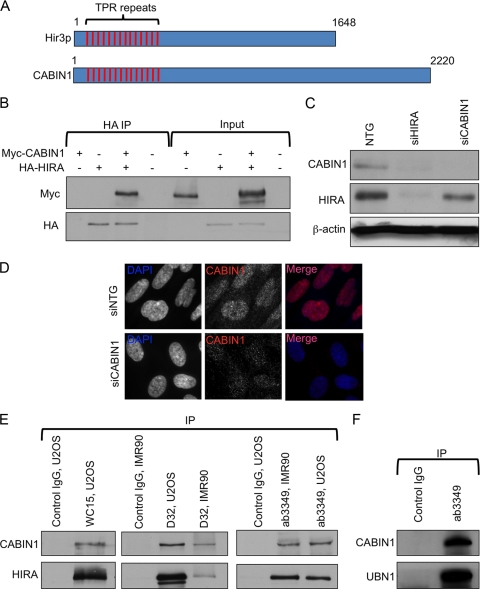
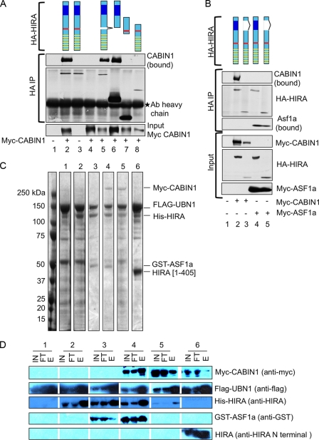
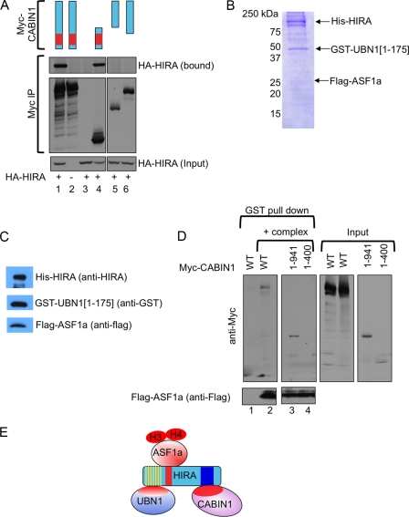
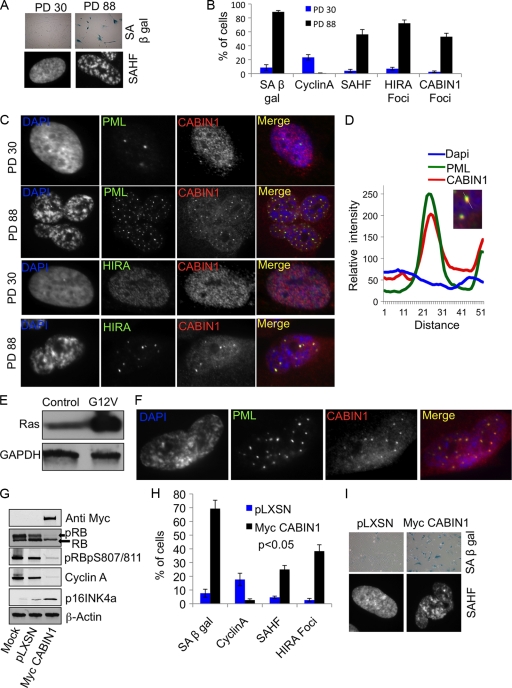
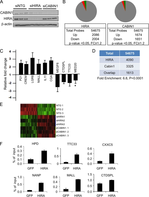
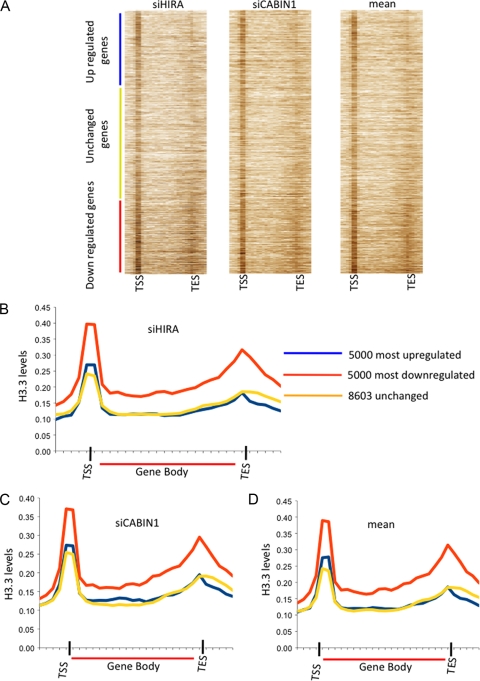
The mammalian HIRA/UBN1/ASF1a complex is a histone chaperone complex that is conserved from yeast (Saccharomyces cerevisiae) to humans. This complex preferentially deposits the histone variant H3.3 into chromatin in a DNA replication-independent manner and is implicated in diverse chromatin regulatory events from gene activation to heterochromatinization. In yeast, the orthologous complex consists of three Hir proteins (Hir1p, Hir2p, and Hir3p), Hpc2p, and Asf1p. Yeast Hir3p has weak homology to CABIN1, a fourth member of the human complex, suggesting that Hir3p and CABIN1 may be orthologs. Here we show that HIRA and CABIN1 interact at ectopic and endogenous levels of expression in cells, and we isolate the quaternary HIRA/UBN1/CABIN1/ASF1a (HUCA) complex, assembled from recombinant proteins. Mutational analyses support the view that HIRA acts as a scaffold to bring together UBN1, ASF1a, and CABIN1 into a quaternary complex. We show that, like HIRA, UBN1, and ASF1a, CABIN1 is involved in heterochromatinization of the genome of senescent human cells. Moreover, in proliferating cells, HIRA and CABIN1 regulate overlapping sets of genes, and these genes are enriched in the histone variant H3.3. In sum, these data demonstrate that CABIN1 is a functional member of the human HUCA complex and so is the likely ortholog of yeast Hir3p.


Figures

Fig. 1.
Fig. 1.
Physical interaction of HIRA, UBN1, and CABIN1. (A) Schematic alignment of CABIN1 and S. cerevisiae Hir3p showing conserved N-terminal TPRs. (B) Ectopically expressed HA-HIRA and myc-CABIN1 interaction, detected by coimmunoprecipitation (IP) with anti-HA antibodies. (C) siRNA-mediated knockdown of endogenous HIRA and CABIN1 confirms specificity of HIRA and CABIN1 antibodies. (D) siRNA-mediated knockdown of endogenous CABIN1 confirms specificity of the CABIN1 antibody by immunofluorescence. (E) Interaction of endogenous HIRA and CABIN1 in osteosarcoma cells (U2OS) and primary fibroblasts (IMR90), detected by coimmunoprecipitation with antibodies to HIRA (WC15 and D32) and CABIN1 (ab3349). Anti-mouse IgG (anti-myc 9E10; Santa Cruz) and anti-rabbit IgG (M7023; Sigma) were used as control IgGs for mouse and rabbit antibodies, respectively. (F) Interaction of endogenous UBN1 and CABIN1, detected by coimmunoprecipitation with antibodies to CABIN1 (ab3349).
Fig. 2.
Fig. 2.
Mapping of CABIN1 interaction domain on HIRA. (A) U2OS cells were transiently transfected with the indicated plasmids and immunoprecipitated (IP) with anti-HA. Lane 1, mock; lane 2, wild-type HA-HIRA and wild-type myc-CABIN1; lane 3, wild-type HA-HIRA only; lane 4, wild-type myc-CABIN1 only; lanes 5 to 8, wild-type myc-CABIN1 and indicated HA-HIRA mutants [lane 5, HA-HIRA(del 439-475); lane 6, HA-HIRA(del520-1017); lane 7, HA-HIRA(421-729); lane 8, HA-HIRA(1-600)]. Schematics are color coded as follows: yellow bars, WD40 repeats; red bars, B domain; blue boxes, C domain. Ab, antibody. (B) U2OS cells were transiently transfected with the indicated plasmids and immunoprecipitated with anti-HA. Lane 1, mock; lane 2, wild-type myc-CABIN1 and wild-type HA-HIRA; lane 3, wild-type myc-CABIN1 and HA-HIRA(del737-963); lane 4, wild-type myc-ASF1a and wild-type HA-HIRA; lane 5, wild-type myc-ASF1a and HA-HIRA(del737-963). Schematics are color coded as in panel A. (C) Insect Sf9 cells were infected with baculoviruses encoding the indicated proteins, and complexes were purified by anti-Flag affinity chromatography. Shown is a Coomassie blue (R250) stain of recombinant proteins. Lanes correspond to following proteins: 1, flag-UBN1; 2, Flag-UBN1 and HisHIRA; 3, Flag-UBN1, HisHIRA, and GST-ASF1a; 4, Flag-UBN1, HisHIRA, GST-ASF1a, and myc-CABIN1; 5, Flag-UBN1, HisHIRA, and myc-CABIN1; 6, Flag-UBN1, HIRA(1-405), and myc-CABIN1. (D) Western blot analysis with indicated antibodies of input lysate (IN), column flowthrough (FT), and elution (E) from panel C.
Fig. 3.
Fig. 3.
Mapping of HIRA interaction domain on CABIN1. (A) U2OS cells were transiently transfected with the indicated plasmids and immunoprecipitated with anti-myc. Lane 1, wild-type myc-CABIN1 and wild-type HA-HIRA; lane 2, wild-type myc-CABIN1; lane 3, wild-type HA-HIRA; lane 4, myc-CABIN1(1-941) and wild-type HA-HIRA; lane 5, myc-CABIN1(942-2220) and wild-type HA-HIRA; lane 6, myc-CABIN1(401-2220) and wild-type HA-HIRA. (B) Insect Sf9 cells were infected with baculoviruses encoding His-HIRA, GST-UBN1(1-175), and Flag-ASF1a, and recombinant complex was purified by nickel affinity chromatography. Shown in the figure is a Coomassie blue (R250) stain of recombinant proteins. (C) Western blot of complex from panel B with indicated antibodies. (D) The trimeric protein complex from panel B was incubated with lysates from U2OS cells transfected with wild-type myc-CABIN1, myc-CABIN1(1-941), or myc-CABIN1(1-400), as indicated. Bound proteins were Western blotted as indicated. (E) Schematic of the HUCA complex based upon binding studies reported here and previously published literature.
Fig. 4.
Fig. 4.
CABIN1 regulates senescence. (A) PD 30 and PD 88 IMR90 fibroblasts stained for markers of senescence, SA β-Gal and SAHF. (B) Cells from panel A were scored for the percentage of cells expressing SA β-Gal, cyclin A, SAHF, HIRA foci, and CABIN1 foci. Values are means ± standard errors of the means (SEM) of three independent experiments. (C) Cells from panel A stained with antibodies to HIRA, PML bodies, and CABIN1 and with DAPI to visualize SAHF. (D) Relative intensity of DAPI, PML body, and CABIN1 fluorescence along a straight line through the CABIN1/PML focus. (E) Western blot showing expression of oncogenic H-RasG12V in IMR90 cells. (F) H-RasG12V-expressing cells stained with antibodies to PML bodies, CABIN1, and with DAPI (4′,6-diamidino-2-phenylindole). (G) IMR90 cells were infected with pLXSN or pLXSN-myc-CABIN1, and lysates were Western blotted with the indicated antibodies. (H) Cells from panel G were scored for SA β-Gal, cyclin A, HIRA foci, and SAHF. Values are means ± SEM of three independent experiments. (I) Cells from panel G stained to detect SA β-Gal and with DAPI to detect SAHF.
Fig. 5.
Fig. 5.
HIRA and CABIN1 regulate overlapping sets of genes. (A) HeLa cells were nucleofected with siRNAs to HIRA, CABIN1, or the nontargeting (NTG) control as indicated. Three independent nucleofections were preformed with Smartpool siRNAs to HIRA or CABIN1. Lysates were Western blotted to detect HIRA, CABIN1, and β-actin as indicated. (B) Pie charts showing significantly upregulated and downregulated probes after HIRA or CABIN1 knockdown. Green indicates upregulated probes, and red indicates downregulated probes. (C) qRT-PCR analysis to confirm expression changes detected by microarray after siHIRA knockdown. Fold change is calculated by dividing normalized (to housekeeping gene) expression in siHIRA cells by normalized (to housekeeping gene) expression in siNTG cells. *, P > 0.05. P < 0.05 for the other 9 genes. Values are means ± SEM of four independent experiments. (D) Table showing overlap of changing probes after HIRA and CABIN1 knockdown. (E) Heat maps showing hierarchical clustering of genes whose expression changes after three independent siHIRA or siCABIN1 knockdowns, compared to three independent siNTG nucleofections. Green indicates upregulated probes, and red indicates downregulated probes. (F) Sonicated chromatin from HeLa cells was immunoprecipitated with antibodies to GFP or HIRA, and the indicated target genes were detected by qPCR. Expression of HPD and MALL increased on HIRA knockdown, while expression of all others decreased. Genes were selected at random from a list of genes with a fold change (FC) of >1.2-fold and P < 0.05 from siHIRA knockdown cells. Values are means ± SEM of three independent ChIP experiments. P < 0.05 for all comparisons.
Fig. 6.
Fig. 6.
Genes activated by HIRA are enriched in histone H3.3. (A) Heat map showing H3.3 enrichment on genes ordered by fold change after HIRA or CABIN1 knockdown versus siNTG and mean fold change of the two independent knockdowns versus siNTG. On the y axis, genes are rank ordered by fold change; on the x axis, positions along the gene are shown in 1-kb windows from 5 kb upstream of the gene to the transcription start site (TSS) and 1-kb windows from the transcription end site (TES) to 5 kb downstream of the gene. Within the gene bodies, windows of 5% of the length of the gene were used. (B) Average histone H3.3 enrichment over three classes of genes, defined according to their response to HIRA knockdown. x axis, position along the gene in 1-kb windows from 5 kb upstream of the gene to the transcription start site (TSS) and 1-kb windows from the TES to 5 kb downstream of the gene. Within the gene bodies, windows of 5% of the length of the gene were used. (C) Average histone H3.3 enrichment over three classes of genes, defined according to their response to CABIN1 knockdown. Results are color coded as in panel B. (D) Average histone H3.3 enrichment over three classes of genes, defined according to their averaged response to HIRA and CABIN1 knockdown. Results are color coded as in panel B.

Similar articles

Cited by

References

    1. Adams P. D. 2009. Healing and hurting: molecular mechanisms, functions, and pathologies of cellular senescence. Mol. Cell 36:2–14 - PubMed
    1. Anderson H. E., et al. 2010. Silencing mediated by the Schizosaccharomyces pombe HIRA complex is dependent upon the Hpc2-like protein, Hip4. PLoS One 5:e13488. - PMC - PubMed
    1. Anderson H. E., et al. 2009. The fission yeast HIRA histone chaperone is required for promoter silencing and the suppression of cryptic antisense transcripts. Mol. Cell. Biol. 29:5158–5167 - PMC - PubMed
    1. Balaji S., Iyer L. M., Aravind L. 2009. HPC2 and ubinuclein define a novel family of histone chaperones conserved throughout eukaryotes. Mol. Biosyst. 5:269–275 - PMC - PubMed
    1. Banumathy G., et al. 2009. Human UBN1 is an ortholog of yeast Hpc2p and has an essential role in the HIRA/ASF1a chromatin-remodeling pathway in senescent cells. Mol. Cell. Biol. 29:758–770 - PMC - PubMed

Publication types

MeSH terms